Prenatal Diagnosis of Galen Vein Aneurysm
Using Ultrasonography and Magnetic
Resonance Imaging and Perinatal and
Long-Term Neurological Outcomes: A Case Series
Diagnóstico do aneurisma de veia de Galeno por meio de
ultrassonogra
fi
a e ressonância magnética e resultados
perinatais e neurológicos: série de casos
Pedro Pires
1Larisse de Brito Aurélio Martins
2Norma Maria Tenório Brito Pires
2Heron Werner
3Adilson Cunha Ferreira
4Edward Araujo Júnior
51Department of Obstetrics and Gynecology, Universidade de Pernambuco, Recife, PE, Brazil
2Real Hospital Português de Beneficência em Pernambuco, Recife, PE, Brazil
3Clínica de Diagnóstico por Imagem, Rio de Janeiro, RJ, Brazil 4Faculdade de Medicina de São José do Rio Preto, São José do Rio
Preto, SP, Brazil
5Department of Obstetrics, Escola Paulista de Medicina, Universidade Federal de São Paulo, São Paulo, SP, Brazil
Rev Bras Ginecol Obstet 2017;39:309–314.
Address for correspondence Edward Araujo Júnior, PhD, Department of Obstetrics, Escola Paulista de Medicina, Universidade Federal de São Paulo, Rua Belchior de Azevedo, 156, apto. 111, Torre Vitória, São Paulo, SP, Brazil (e-mail: [email protected]).
Keywords
►
Galen vein aneurysm
►
prenatal diagnosis
►
ultrasonography
►
magnetic resonance
►
perinatal outcomes
Abstract
Objective
To describe the prenatal diagnosis of Galen vein aneurysm (GVA) based on
ultrasonography and magnetic resonance imaging (MRI) in a series of cases, as well as
its postnatal outcomes and follow-up until 4 years of age.
Methods
A retrospective longitudinal study was performed, analyzing a database
comprising seven cases of prenatal diagnosis of GVA at two Brazilian institutions from
February of 2000 to May of 2012. The following data were evaluated: gestational age at
diagnosis, GVA dimensions on ultrasonography, associated fetal changes,
fi
ndings on
fetal echocardiography, gestational age at delivery, type of delivery, birth weight,
Apgar score at the 1st and 5th minutes, neonatal outcomes, and survival with follow-up
until 4 years of age.
Results
The mean gestational age
standard deviation on the prenatal diagnosis of
GVA based on ultrasonography was 25
4.9 weeks. The mean length of GVA was 3.2
0.4 cm. The mean gestational age at birth was 37.5
0.7 weeks, and a cesarean section
was performed in 85.7% of the cases (6/7). The mean birth weight was 3,070
240.4 g.
The total survival rate was 42.8% (4/7), with three neonatal deaths. Of the four
survivors, three presented with normal neuropsychomotor development until 4 years
received
September 27, 2016
accepted after revision
January 11, 2017
published online
April 18, 2017
DOIhttps://doi.org/ 10.1055/s-0037-1601401.
ISSN 0100-7203.
Introduction
Galen vein aneurysm (GVA) is a rare congenital malformation arising because of the presence of multiple arteriovenous shunts that drain to a median prosencephalic vein.1There is usually only a single malformation corresponding to1% of all vascular cerebral malformations. However, it may be associated with congenital heart disease, hydrops, and cystic hygroma.2Its etiology is unknown, and there is no described familial inheritance. Heart failure is the most common symptom in the neonatal period, but seizures and other neurological signs may also be observed.3,4
Because GVA has a low incidence rate but high morbidity and mortality rates, a prenatal diagnosis is necessary for adequate follow-up, delivery, and parent counseling. In gener-al, the condition is prenatally diagnosed based on conventional ultrasonography when a cystic image that confirms dilation of the vein, located either in the middle region or slightly deviated from the central region, below the third ventricle on the middle supratentorial line is identified.5Color Doppler imaging shows a turbulentflow inside the cyst, which may be associated with secondary ventriculomegaly.6Magnetic reso-nance imaging (MRI) helps to confirm the diagnosis and also reveals complications such as hemorrhagic injury in the white
matter of the brain.7Other prenatal diagnostic methods such as ultrasonography in the 3-dimensional power Doppler mode have been described, but these have shown no advantages over conventional ultrasonography and MRI.8,9Fetal echocardiog-raphy may help detect early signs of heart failure, which, together with hydrops, is the most common consequence of GVA.
Here we present a series of seven cases of prenatal diagnosis of GVA with their mainfindings based on conventional ultra-sonography and MRI as well as their postnatal outcomes.
Methods
A retrospective longitudinal study was performed, analyzing a database of seven cases of prenatal diagnosis of GVA from February of 2000 to May of 2012 at two Brazilian institutions: Centro Integrado de Saúde Amaury de Medeiros da Universi-dade de Pernambuco (UPE) and Clínica de Diagnóstico por Imagem (CDPI). This study was approved by the Committee on Ethics in Research of the Universidade de Pernambuco (UPE). Five cases were from UPE and 2 from CDPI.
The following data were evaluated: gestational age (in weeks) at diagnosis, GVA dimensions on ultrasonography, associated fetal changes,findings on fetal echocardiography,
of age and only one showed serious neurological sequelae. Ultrasonography and MRI
showed similar
fi
ndings for all seven cases.
Conclusions
Galen Vein Aneurysm is associated with a high neonatal death rate.
Therefore, its prenatal diagnosis is essential for parent counseling and follow-up at
tertiary care institutions.
Resumo
Objetivo
Descrever o diagnóstico pré-natal de uma série de casos de aneurisma de
veia de Galeno (AVG) por meio de ultrassonogra
fi
a e ressonância magnética (RM), bem
como os resultados pós-natais e acompanhamento até 4 anos de vida.
Métodos
Realizou-se um estudo retrospectivo longitudinal com análise de banco de
dados de sete casos de diagnóstico pré-natal de AVG em dois serviços brasileiros entre
fevereiro de 2000 e maio de 2012. Foram avaliados a idade gestacional ao diagnóstico,
dimensões do AVG na ultrassonogra
fi
a, alterações fetais associadas, achados da
ecocardiogra
fi
a fetal, idade gestacional ao parto, tipo de parto, peso ao nascimento,
índice de Apgar no 1° e 5° minutos, resultados neonatais, e sobrevida com
acompan-hamento até 4 anos de idade.
Resultados
A idade gestacional média
desvio-padrão ao diagnóstico pré-natal do
AVG pela ultrassonogra
fi
a foi de 25
4,9 semanas. O comprimento médio do AVG foi
3,2
0,4 cm. A idade gestacional média ao nascimento foi 37,5
0,7 semanas, sendo
que, em 85,7% dos casos (6/7) o parto foi cesáreo. O peso médio ao nascimento foi de
3.070
240,4 gramas. A sobrevida total foi de 42,8% (4/7), com três óbitos neonatais.
Dos quatro sobreviventes, três apresentaram desenvolvimento neuropsicomotor
normal até a idade de 4 anos, sendo que apenas um apresentou sequelas neurológicas
graves. Ultrassonogra
fi
a e RM apresentaram achados semelhantes nos sete casos.
Conclusões
O AVG está associado à elevada taxa de óbito neonatal, sendo, portanto,
fundamental o seu diagnóstico pré-natal precoce para aconselhamento dos pais e
seguimento em serviço terciário.
Palavras-chave
►
Aneurisma da veia de
Galeno
►
Diagnóstico pré-natal
►
Ultrassonogra
fi
a
►
Ressonância
magnética
gestational age at delivery, type of delivery, birth weight, Apgar score at the 1st and 5th minutes, neonatal outcomes, and survival with follow-up until 4 years of age. In addition, we have described the mainfindings based on color Doppler ultrasonography and MRI.
Results
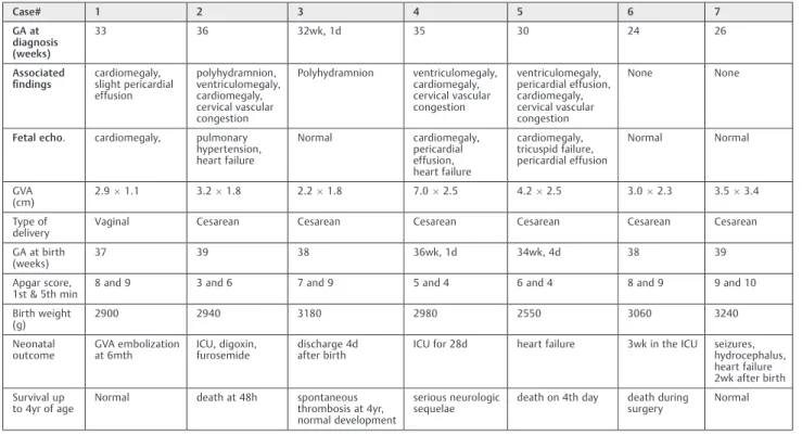
The mean gestational age standard deviation on prenatal diagnosis of GVA by ultrasonography was 254.9 weeks. The mean length and width of the GVA on diagnosis were 3.2 0.4 cm and 2.21.6 cm, respectively. The mean gestational age at birth was 37.50.7 weeks, and a cesarean section was performed in 85.7% of cases (6/7). The average birth weight was 3,070240.4 g. Mean Apgar scores at the 1st and 5th minutes were 8.50.7 and 9.50.7, respectively. The overall survival rate was 42.8% (4/7), with three neonatal deaths. Of the four survivors, three presented with normal neuropsycho-motor development until 4 years of age and only one showed serious neurological sequelae.►Table 1presents the
descrip-tion of the pre- and postnatal data of the seven cases of GVA.►Fig. 1shows the pre- and postnatal imagingfindings
of case #7. ►Table 2 presents the description of the main
findings based on conventional color Doppler ultrasonography and MRI in the seven cases of the prenatal diagnosis of GVA.
Discussion
Here we present a series of cases of prenatal diagnosis of GVA based on ultrasonography at an average gestational age of
25 weeks; our diagnosis agrees with thefindings reported in most publications.8–10Magnetic resonance imaging is used to evaluate associated neurological findings that may be of prognostic value. In our case series, MRI showed no diagnostic advantages over ultrasonography. In a series of 18 cases of GVA, MRI identified 3 cases of neuronal migration abnormalities that had not been identified by ultrasonography.2
In our series, associatedfindings were present in 71% of the cases (5/7) and cardiomegaly was the most frequent finding. However, in only two cases, a therapeutic preterm delivery was performed owing to congestive heart failure in the fetus. The mean gestational age at delivery was 37.5 weeks, and the mean birth weight was adequate in terms of the gestational age. In a series of 21 cases, the mean gesta-tional age at birth was high (38.7 weeks) and the mean birth weight was also adequate in terms of the gestational age (3096 g).2The most frequent type of delivery in our case series was cesarean section (86%), which is in accordance with the high incidence of this type of delivery in Brazil, both in public and private institutions, regardless of fetal malformations.11
Associated anomaly is a proven factor of adverse perinatal outcome in cases of GVA and termination of the pregnancy is indicated in countries where this procedure is legal.2 Re-garding the type of delivery, in the absence of fetal cardiac dysfunction and isolated GVA, normal delivery is the best choice. In the presence of fetal cardiac dysfunction and isolated/associated GVA, there is no consensus in the litera-ture and the choice should be based on the gestational age and the neonatal intensive care.
Table 1 Prenatal outcomes of fetuses diagnosed with Galen vein aneurysm
Case# 1 2 3 4 5 6 7
GA at diagnosis (weeks)
33 36 32wk, 1d 35 30 24 26
Associated
findings cardiomegaly,slight pericardial effusion polyhydramnion, ventriculomegaly, cardiomegaly, cervical vascular congestion Polyhydramnion ventriculomegaly, cardiomegaly, cervical vascular congestion ventriculomegaly, pericardial effusion, cardiomegaly, cervical vascular congestion None None
Fetal echo. cardiomegaly, pulmonary hypertension, heart failure Normal cardiomegaly, pericardial effusion, heart failure cardiomegaly, tricuspid failure, pericardial effusion Normal Normal GVA
(cm) 2.91.1 3.21.8 2.21.8 7.02.5 4.22.5 3.02.3 3.53.4
Type of
delivery Vaginal Cesarean Cesarean Cesarean Cesarean Cesarean Cesarean
GA at birth (weeks)
37 39 38 36wk, 1d 34wk, 4d 38 39
Apgar score, 1st & 5th min
8 and 9 3 and 6 7 and 9 5 and 4 6 and 4 8 and 9 9 and 10
Birth weight
(g) 2900 2940 3180 2980 2550 3060 3240
Neonatal
outcome GVA embolizationat 6mth ICU, digoxin,furosemide discharge 4dafter birth ICU for 28d heart failure 3wk in the ICU seizures,hydrocephalus, heart failure 2wk after birth
Survival up
to 4yr of age Normal death at 48h spontaneousthrombosis at 4yr, normal development
serious neurologic
sequelae death on 4th day death duringsurgery Normal
Despite advancements in prenatal diagnosis, neonatal mor-tality is high in GVA, with three neonatal deaths (43%) ob-served in our case series, as well as a case of serious neurological sequelae in the 4-year follow-up (14%). In all these neonatal deaths, the fetuses showed cardiomegaly in the
In a systematic review of 90 cases of prenatal diagnosis of GVA, the mortality rate was 54%, and serious neurological sequelae were found in 14% of the cases2; this was consis-tent with thefindings of our study. Postnatal treatment of GVA will depend on its size; small GVAs with lowflow may undergo spontaneous thrombosis, as observed in case #3. Patients with neurological and cardiac symptoms must be treated by a radiological or surgical intervention.5 When GVA is not life threatening, the vascular malformation is best embolized after 5 months from birth,12 as per-formed in cases #1 and #7, which showed good postnatal outcomes and normal neurological development in the 4-year follow-up.
In summary, we have presented a series of cases of prenatal diagnosis of GVA based on ultrasonography and MRI. Because GVA is associated with high rates of neonatal
death, its prenatal diagnosis is essential for parent counsel-ing and follow-up at tertiary care institutions.
References
1 Gailloud P, O’Riordan DP, Burger I, et al. Diagnosis and manage-ment of vein of galen aneurysmal malformations. J Perinatol 2005;25(08):542–551
2 Deloison B, Chalouhi GE, Sonigo P, et al. Hidden mortality of prenatally diagnosed vein of Galen aneurysmal malformation: retrospective study and review of the literature. Ultrasound Obstet Gynecol 2012;40(06):652–658
3 Gupta AK, Varma DR. Vein of Galen malformations: review. review Neurol India 2004;52(01):43–53
4 Mai R, Rempen A, Kristen P. Prenatal diagnosis and prognosis of a vein of Galen aneurysm assessed by pulsed and color Doppler sonography. Ultrasound Obstet Gynecol 1996;7(03):228–230 Table 2 Findings on magnetic resonance imaging and ultrasonography in our cases of prenatal diagnosis of Galen vein aneurysm
Case # Magnetic resonance imaging Ultrasonography
1 Ellipsoid expansive formation,2.0 cm in its largest diameter at the level of the middle line in the tentorial region, posterior to the pituitary, hypointense T1 and T2 signals, suggesting aflow void, communicating with the sinus rectus, consistent with aneurysmal dilation of the Galen vein, with no signs of ventriculomegaly.
An elongated cystic image, measuring2.91.1 cm, was observed in a location posterior to the thalamus, continuing on the middle line and spreading superiorly between the hemispheres. On the color Doppler, a low-resistanceflow was observed, with an arterial
pattern prevailing. The echographic aspect and theflow pattern are consistent with Galen vein aneurysm. Enlarged cardiac area and slight pericardial effusion.
2 Slight ventriculomegaly and expansive ellipsoid forma-tion in a middle line locaforma-tion, spreading to the posterior fossa, consistent with arteriovenous malformation (Galen vein aneurysm).
Slight ventriculomegaly and an elongated anechoic image in a middle line location, spreading to the pos-terior fossa. On the color Doppler, an abundantflow of very low resistance, consistent with an arteriovenous malformation (Galen vein aneurysm). Enlarged cardiac area with slight pericardial effusion.
3 Intracranial ellipsoid expansive formation, 3.02.1 cm in size, located on the middle cerebral line (supraten-torial), consistent with Galen vein aneurysm.
A homogeneous intracranial cystic area, measuring 2.2 1.8 cm, localized on the middle line (supratentorial). On color Doppler, intense arteriovenousflow. Normal heart size and shape.
4 Ellipsoid expansive formation measuring 7.02.5 cm, supratentorial, slight dilation of the posterior horn of the brain ventricle and the 3rd ventricle, consistent with Galen vein aneurysm associated with ventriculomegaly.
A cystic area with a tubular aspect, measuring 4.1 3.7 cm, on the middle line (supratentorial), being
con-firmed based on color Doppler as an arteriovenousflow within, consistent with Galen vein aneurysm. Slight ventricular dilation. Slightly enlarged cardiac area. Vascular congestion of the cervical region.
5 Ventricular dilation and ellipsoid expansive formation, measuring6.04.0 cm, on the middle line (supratentorial), consistent with Galen vein aneurysm.
A cystic area with a tubular aspect, measuring5.5 4.0 cm, on the middle line (supratentorial), confirmed based on color Doppler as an arteriovenousflow within, consistent with Galen vein aneurysm. Slight ventricular dilation.
6 Expansive lesion with lobulated contours and
well-de-fined limits, with a hypointense T2 signal and an iso/ hypointense T1 signal, measuring 3.01.82.3 cm, interhemispheric, posterior to the 3rd ventricle, with no compressive effect.
Anechoic image measuring 3428 mm, located pos-teriorly above the thalamus. Slightly dilated lateral ventricles. Color Doppler with turbulentflow. Normal 3rd and 4th ventricles.
7 Expansive lesion with lobulated contours and well-defined limits, with a hypointense T2 signal and an iso/ hypointense T1 signal, measuring 3.53.42.6 cm, interhemispheric, posterior to the 3rd ventricle, no compressive effect.
5 Pilu G, Nicolaides KH. Diagnosis of fetal abnormalities: the 18–23-week scan. New York: Parthenon; 1999. Vein of Galen Aneurysm; 14–7
6 Diebler C, Dulac O, Renier D, Ernest C, Lalande G. Aneurysms of the vein of Galen in infants aged 2 to 15 months. Diagnosis and natural evolution. Neuroradiology 1981;21(04):185–197 7 Wagner MW, Vaught AJ, Poretti A, Blakemore KJ, Huisman TA.
Vein of galen aneurysmal malformation: prognostic markers depicted on fetal MRI. Neuroradiol J 2015;28(01):72–75 8 Rios LT, Araujo Júnior E, Nardozza LM, Moron AF, Martins MdaG.
Prenatal diagnosis of an aneurysm of the vein of galen by three-dimensional power and color Doppler ultrasonography. Clin Med Insights Case Rep 2012;5:77–80
9 Ergenoğlu MA, Yeniel AÖ, Akdemir A, Akercan F, KaradadaşN. Role of 3D power Doppler sonography in early prenatal diagnosis of Galen vein aneurysm. J Turk Ger Gynecol Assoc 2013;14(03):178–181 10 Beucher G, Fossey C, Belloy F, Richter B, Herlicoviez M, Dreyfus M.
[Antenatal diagnosis and management of vein of Galen aneurysm: review illustrated by a case report]. J Gynecol Obstet Biol Reprod (Paris) 2005;34(06):613–619 French.
11 Barros FC, Matijasevich A, Maranhão AG, et al. Cesarean sections in Brazil: will they ever stop increasing? Rev Panam Salud Publica 2015;38(03):217–225