DEPARTAMENTO DE BIOLOGIA VEGETAL
Characterising the Formation and Functional Role of Vesicular
Clustering during Influenza A Virus Infection
Ana Laura Vinagre Costa e Sousa
Mestrado em Microbiologia Aplicada
Dissertação orientada por:
Doutora Maria João Amorim
Professora Doutora Maria Filomena Trabucho Caeiro
2017
ii
“Human life itself may be almost pure chaos, but the work of the artist is to take these handfuls of confusion and disparate things, things that seem to be irreconcilable, and put them together in a frame to give them some kind of shape and meaning” – Katherine Anne Porter
iii
Acknowledgements
“Sometimes the most ordinary things could be made extraordinary, simply by doing them with the right people” – Elizabeth Green
I would like to first say thank-you to my “chefa”, Erin Telley for opening the doors of the Electron Microscopy field to me and supporting my attendance at conferences and courses. Without a doubt, if I did not have established my career at Instituto Gulbenkian de Ciência, I would not have developed this work. Also, if I was not at Instituto Gulbenkian de Ciência, I would not have met my lab mate Sara. Some results described in this thesis were accomplished with her help and support by freezing her hands during several afternoons but also for cheering me up in the hard days – chocolate always make the days brighter - made this work undoubtedly easier. As well, I have to thank my lab colleagues, Joana and Marta, who helped me with the cryostat sections, and Luis, who helped me with the super-resolution acquisitions and reconstructions.
I would like to express my gratitude to my external supervisor, Doctor Maria João Amorim, for accepting me into her group – Cell Biology of Viral Infection (CBV). She gave me intellectual freedom in my work and engaged in me new ideas. To all in CBV group, thank-you for listening all the electron microscopy presentations. Also from the CBV group, I want to highlight all the support and the friendship from Silvia. Her help with experiments when my days were overwhelming but also with the patience necessary for reviewing my reports and thesis, always putting complex ideas into simple terms and demanding a high quality of work from which I greatly benefited as a student but also as a professional in science. I also need to emphasize the support to all the bureaucratic questions from my internal supervisor Professor Filomena Caeiro.
Finally, although this master degree was one of the hardest things I did, I cannot forget the friends that it brought into my life. First my car sharing colleague Alexandra. I cannot thank-you more for all the talks and the messages! Although you still doubt, I can tell you are a great friend and you have a big heart that you hide so well that only the ones that are persistent enough can have access to. But also my “montaditos” team: André, Bia e Jorge. You guys made my time at Faculdade de Ciências lot more fun.
To my family from the heart: Luis, Flávia, Abreu, Vanessa e Gonçalo. Saying sorry for not being present it’s not enough. Last the most important person, my “mamy” which was, is and will be always present in the bad and in the good times.
iv
Characterising the Formation and Functional Role of Vesicular
Clustering during Influenza A Virus Infection
Ana Laura Vinagre Costa e Sousa
2017
This thesis was fully performed at Instituto Gulbenkian de Ciência under the direct supervision of Dr Maria João Amorim in the scope of the Master in Applied Microbiology of the Faculty of Sciences of the University of Lisbon.
v
Resumo
Os vírus Influenza, que pertencem à família Orthomyxoviridae, estão divididos em quatro tipos A, B, C e D (Hause et al. 2013; Hause et al. 2014). Entre estes, o influenza A (IAV) é o vírus que causa maior impacto na saúde pública. O seu reservatório natural são as aves marinhas, no entanto possui a capacidade de gerar infeções zoonóticas encontrando-se disseminado em muitas espécies de aves e mamíferos (Acheson 2011; Taubenberger & Kash 2010). Nos seres humanos, o IAV é o patógeno responsável pela gripe, uma doença aguda respiratória associada a alta morbidade e mortalidade (Lofgren et al. 2007).
O IAV é composto por um invólucro viral derivado da membrana plasmática da célula. Este envelope é revestido por uma camada externa de proteínas virais, designadamente hemaglutinina e neuraminidase. O interior do virião contém o genoma viral, constituído por oito complexos ribonucleoproteicos (vRNPs). Cada vRNP é composto por um dos oito segmentos de ácido ribonucleico viral que codificam para diferentes proteínas virais. Os vRNPs funcionam como unidades independentes sendo necessário um conjunto completo de oito vRNPs diferentes para a montagem de uma partícula infeciosa (Fields et al. 2007).
Os vRNPs são transportadas por piggyback em vesículas destinadas à periferia da célula através da interação entre a polimerase 2 viral e a GTPase Ras related in brain 11 (Rab11) (Amorim et al. 2011; Eisfeld et al. 2011; Momose et al. 2011). Em homeostasia, o Rab11 está envolvido na reciclagem de proteínas endocitadas. Regula a formação, movimento e fusão de vesículas originárias do compartimento de reciclagem de endossomas e encontra-se espalhado pelo citoplasma (Grant & Donaldson 2009). Em células infetadas, o Rab11 acumula no citoplasma formando agregados vesiculares. Sabe-se que estas vesículas são heterogéneas, compostas por membrana simples e dupla (SMV e DMV, respetivamente), o que indica que o IAV conduz não só ao agrupamento vesicular mas também ao rearranjo das suas membranas (Vale-Costa et al. 2016).
Este trabalho permitiu fundamentar um modelo alternativo para a montagem genómica do IAV, em que o agrupamento vesicular pode funcionar como uma plataforma induzida pelo virus para a criação de hotspots de vRNPs. A alta co-localização entre o conjunto de todas as vRNPs permitiria a ocorrência das interações RNA-RNA necessárias à junção do genoma completo composto pelos oito vRNPs. O agrupamento vesicular surge assim como um passo essencial no ciclo replicativo do IAV. Pelo que, a definição da cronologia de eventos que levam à sua maturação e as alterações na composição vesicular tornam-se fundamentais para compreender os mecanismos de montagem genómica do IAV.
Neste trabalho experimental foi aplicada uma multi-metodologia de crio-microscopia eletrónica (EM). A razão da escolha dos métodos de crio-EM baseia-se na capacidade que estas técnicas têm para preservar os detalhes ultra-estruturais sem introduzirem os artefactos morfológicos de volume, forma e desnaturação de epitopos associados aos métodos cuja abordagem é puramente química (Studer et al. 1989). No entanto, para a sua aplicação, estes métodos tiveram que sofrer otimizações. Os mesmos permitiram elucidar o modo como a agregação vesicular se forma em células A549 que expressam constitutivamente GFP-Rab11 durante a infeção e qual a função das vesículas agregadas no ciclo replicativo do IAV.
vi
A metodologia de crio-EM usada incorporou duas técnicas conhecidas na área: a técnica de congelamento de alta pressão e de fixação química por substituição a baixas temperaturas e a técnica de Tokuyasu. A primeira utiliza a baixa temperatura do azoto (-196ºC) e alta pressão (aproximadamente 2000 bar) para crio-imobilizar as amostras. A técnica permitiu a preservação celular para análise morfológica e a quantificação vesicular (sem introdução de artefactos de fixação química). A segunda técnica, Tokuyasu, também tira partido da baixa temperatura do azoto para crio-imobilizar as amostras e permite manter as estruturas hidratadas para uma imunomarcação ótima. Esta técnica foi usada para discernir a origem das estruturas vesiculares em estudo.
As metodologias usadas neste trabalho foram alvo de diversas otimizações, com o intuito de preservar as características ultra-estruturais o mais próximo possível do seu estado nativo (McDonald 2007). Na otimização da técnica de congelamento de alta pressão dois parâmetros foram avaliados: a vitrificação e a preservação morfológica das células GFP-Rab11. A vitrificação refere-se ao processo de transformação de um liquido em sólido sem cristalização. A cristalização induz artefactos nas células, reconhecíveis sob a forma de redes interligadas de espaços vazios onde outrora expandiram os cristais de gelo. Ambos os parâmetros de vitrificação e preservação morfológica são afetados pelo tipo de suporte de amostra utilizado para congelar, bem como pelo tipo de enchimento escolhido para preencher o espaço livre entre a amostra e o suporte. Os tipos de suporte para congelamento de amostras testados foram os discos planos e os suportes côncavos. O primeiro tipo – discos planos permite o congelamento de células em camada única e foi testado em combinação com três tipos de enchimentos (hexadeceno, dextano a 20% (m/v) e albumina sérica bovina a 20% (m/v)). O segundo tipo – suportes côncavos – permite o congelamento em pellet e foram testados em combinação com hexadeceno. Estabeleceu-se que o melhor protocolo para processamento de células GFP-Rab11 por congelamento de alta pressão tendo em conta o objeto de estudo (a ultra-estrutura vesicular) foi alcançada ao usar a combinação de discos planos com hexadeceno.
Para a otimização da técnica de Tokuyasu, quatro marcadores: Golgi Marker (GM) 130, calnexina, nucleoproteína (NP) e GFP-Rab11 foram estudados em células infetadas com IAV para a capacidade de manter sua imunorreatividade em diferentes condições de fixação. Três fixadores foram usados durante o teste: formaldeído a 2% (v/v) com glutaraldeído a 0,2%, 0,1% e 0,05% (v/v). Como controlo foi adicionado o fixador de referência em imunocitoquímica: formaldeído a 4% (v/v). O formaldeído permite a manutenção da imunorreatividade celular, no entanto o glutaraldeído conduz a uma melhor preservação morfológica da ultra-estrutura. O fixador que permitiu a preservação da imunorreatividade de todos os marcadores foi o formaldeído a 4% (v/v) e, como tal, o fixador usado no estudo.
A combinação entre as técnicas de crio-EM, congelação por alta pressão e Tokuyasu, permitiu quantificar o número de vesículas de membrana simples e dupla (SMV e DMV, respetivamente) durante a infeção por IAV. Foi possível determinar que o número de SMVs - utilizadas como veículos para o transporte de vRNPs - aumenta durante a infeção por IAV e que as membranas destas vesiculas de membrana simples são reorganizadas para produzir as DMVs. Desta forma, é possível afirmar que as SMV juntamente com as DMVs parecem constituir os agregados vesiculares formados durante a infeção e que permitem a convergência espacial dos vRNPs.
Para além das técnicas de crio-EM descritas, também a técnica de correlação entre microscopia de luz e eletrónica (CLEM) foi desenvolvida e otimizada para pesquisa da função dos agregados vesiculares. A técnica de CLEM aplicada combinou as técnicas de hibridização in situ fluorescente (FISH), microscopia estocástica de reconstrução ótica (STORM) e EM e contribuiu para uma caracterização
vii
técnica de STORM permitiu a identificação específica de segmentos do genoma do IAV em agregados de Rab11, enquanto a EM forneceu o contexto ultra-estrutural da célula. No entanto, a precisão da correlação alcançada foi baixa, não tendo permitido a identificação do conteúdo genómico transportado por cada vesícula dentro dos agregados. Neste sentido, podemos afirmar que, apesar de poder ser combinada com outras técnicas, a técnica de CLEM integrando FISH, STORM e EM é uma técnica ainda em desenvolvimento e que requer melhorias adicionais.
Em resumo, o trabalho aqui descrito conduziu à conclusão de que as vesículas de membrana simples e dupla, portadoras de vRNPs, são originárias do compartimento de reciclagem de endossomas. Ambos os tipos de vesículas foram encontrados nos agregados e identificadas com os mesmos marcadores imunes. A sua relevância funcional para a montagem dos vRNPs continua por inferir, sendo necessária a contínua otimização da combinação das técnicas de microscopia de luz e de microscopia eletrónica para determinar um método de CLEM funcional.
viii
Abstract
Influenza viruses, from the Orthomyxoviridae family, are divided in four types A, B, C and D (Hause et al. 2013; Hause et al. 2014). Influenza A (IAV) as the capacity to generate zoonotic infections and is widespread in many mammalian and avian species. (Acheson 2011; Taubenberger & Kash 2010). In humans, IAV is the pathogen responsible for flu, a respiratory acute disease associated with high morbidity and mortality (Lofgren et al. 2007).
IAV as a segmented genome divided in eight different viral ribonucleoprotein complexes (vRNP). Each vRNP complex is composed by one of the eight viral ribonucleic acid segments. The vRNPs function as independent units and a complete set of eight different vRNPs is required for assembly of an infectious IAV particle (Fields et al. 2007).
It is known that vRNPs are transported by piggybacking in vesicles destined to the cell periphery via interaction between the viral polymerase basic protein 2 and the host GTPase Ras-related in brain 11 (Rab11) (Amorim et al. 2011; Eisfeld et al. 2011; Momose et al. 2011). Also, it was stated that during infection there is an aggregation of vesicles creating clusters. The clusters were composed by vesicles with single and double membranes (SMV and DMV), indicating that IAV leads not only to clustering but also to the rearrangement of vesicular membranes (Vale-Costa et al. 2016).
This work substantiated an alternative model for IAV genomic assembly in which vesicular clustering can function as a viral induced platform promoting the creation of vRNPs hotspots. The high co-localization among the pool of all vRNPs would allow the establishment (or completion) of the RNA-RNA interactions necessary for assembly of the eight RNA-RNA segments before budding. Vesicular clustering thus emerges as an essential step in IAV lifecycle.
In this work, a multi electron microscopy (EM) cryo-methodology was not just applied but also optimized and developed to elucidate how vesicular clustering is formed in A549 cells constitutively expressing GFP-Rab11 during infection and what the vesicles functional role in the lifecycle of IAV.
The EM cryo-methodology incorporated the High Pressure Freezing – Freeze Substitution (HPF-FS) and Tokuyasu techniques. The first permitted the morphological analysis of cells and vesicular quantification (without introducing chemical fixation artifacts) and the second enabled the immunolabeling of structures to discern their origin. Also, to further investigate the functional role of clusters a correlative light electron microscopy (CLEM) method that combines Fluorescent in situ Hybridization (FISH), Stochastic Optical Reconstruction microscopy (STORM) and EM was developed.
The optimization of HPF-FS technique established that the best vitrification and morphological preservation of GFP-Rab11 cells was achieved when using flat disks, as specimen carriers for the freezing process, filled with 1-hexadecene, as a cryoprotectant. For Tokuyasu, 4% (v/v) formaldehyde was the fixative to enable the best preservation of immunoreactivity.
Together, both techniques, lead to conjecture that the number of SMVs - used as vehicles for vRNP trafficking - increase during IAV infection and are modified to rearrange membranes and produce
ix
enable the spatial convergence of vRNPs.
The CLEM technique incorporating FISH, STORM and EM contributed to a further characterization of SMVs and DMVs role within the clusters. The FISH technique allowed the identification of IAV genome segments on Rab11 clusters imaged by STORM, whereas EM provided the ultrastructural context of the cell. A low accuracy of the achieved correlation did not allow the identification of the segment content of each vesicle within the clusters. This combinatorial but powerful method thus requires further improvement.
In summary, the work herein described led to the conclusion that vesicles of single and double membrane, carrying vRNPs, originate from the endosomal recycling compartment. Both types of vesicles were found in clusters. Their functional relevance for vRNP assembly remains to be determined, as the CLEM method developed for its study needs further optimization.
x
Table of Contents
Acknowledgments……….iii Resumo………..v Abstract……….viii Table of Contents………..xFigures and Tables Index………..xiii
Abbreviation List….. ………...xiv
1. Importance of the Study ... 1
2. Research Approach ... 4
2.1 Study Location ... 4
2.2 Research Question and Hypothesis ... 4
2.3 Objectives... 4 2.3.1 General Objectives ... 4 2.3.2 Specific Objectives ... 4 3. Literature Review ... 5 3.1 Influenza A Virus ... 5 3.1.1 Structure ... 5 3.1.2 Lifecycle ... 6
3.2 Electron Microscopy Cryo-Methods to Study IAV at the Nanoscale ... 9
3.2.1 High Pressure Freezing – Freeze Substitution ... 10
3.2.2 Tokuyasu ... 11
3.2.3 Correlative Light Electron Microscopy ... 12
4. Material and Methods ... 13
4.1 Cell Culture ... 13
4.2 Infection ... 13
xi
4.3.2 Freeze Substitution ... 14
4.3.3 Ultramicrotomy and Post-Staining ... 14
4.3.4 Data Acquisition ... 14
4.3.5 Data Analysis ... 14
4.4 Immunolabeling ... 17
4.4.1 Optimization of Fixation ... 17
4.4.2 Tokuyasu Technique ... 17
4.4.3 Data Acquisition and Analysis ... 18
4.5 Correlative Light Electron Microscopy ... 18
4.5.1 Light Microscopy ... 18
4.5.2 Electron Microscopy ... 19
4.5.3 Data Acquisition and Analysis ... 20
5. Results ... 21
5.1 High Pressure Freezing – Freeze Substitution Technique Optimization ... 21
5.1.1 Vitrification Evaluation ... 21
5.1.2 Morphological Preservation ... 22
5.2 Characterization of IAV induced Vesicular Clustering ... 23
5.2.1 Quantification of Vesicle Number during IAV Infection ... 23
5.2.2 Vesicles Morphological Study ... 25
5.2.3 Clustered Structures Origin and Cellular Context ... 28
5.2.4 CLEM Method Development – FISH, STORM and EM ... 31
6. Discussion ... 36
6.1 Characterization of Vesicular Structures Induced by IAV Infection ... 36
6.1.1 High Pressure Freezing – Freeze Substitution Optimization: Vitrification and Morphological Preservation ... 37
6.1.2 Quantification of Vesicle Number during IAV Infection ... 37
6.1.3 3D Morphological Study ... 38
xii
6.2.1 Immunolabeling Optimization ... 38
6.2.2 Electron Microscopy Analysis ... 39
6.3 CLEM Method Development ... 39
7. Conclusion... 40
7.1 Future Perspectives ... 42
8. Appendix ... 43
8.1 Appendix 1 – Publications Resulting from Work Carried Out during my Masters ... 43
8.2 Appendix 2 – Conferences and Courses Attended during my Masters ... 43
xiii
Figures and Tables Index
Figure 1. IAV infection alters Rab11 distribution ... 2
Figure 2. Ultrastructural features of Rab11 vesicular clustering revealed by CLEM technique ... 3
Figure 3. IAV virion structure ... 5
Figure 4. IAV lifecycle ... 7
Figure 5. Specimen carrier and aclar flat disk example ... 11
Figure 6. Scheme of HPF optimization ... 13
Figure 7. Scheme of immunolabeling fixation optimization ... 17
Figure 8. Scheme of Tokuyasu processing, ultracryosectioning and immunolabeling ... 17
Figure 9. Scheme of FISH on Cryostat Sections ... 19
Figure 10. Scheme of coordinate system for targeted ultramicrotomy ... 20
Figure 11. Vitrification evaluation ... 21
Figure 12. Analysis of morphological preservation of the cell ultrastructure ... 22
Figure 13. Photomontage for the quantification of vesicles number ... 24
Figure 14. Quantification of SMV in mock, 4, 8, 12, 16, 20 and 24 hours PI with IAV ... 24
Figure 15. Quantification of DMV in mock, 4, 8, 12, 16, 20 and 24 hours PI with IAV ... 25
Figure 16. SMV and DMV morphology at 16 hours PI with IAV ... 25
Figure 17. Serial slices through single-axis tomograms of a 16 hours IAV infected cell ... 26
Figure 18. 3D model of SMVs and DMVs of a 16 hours IAV infected cell ... 27
Figure 19. Fixatives testing for GM130, calnexin, NP and GFP-Rab11 detection ... 28
Figure 20. Ultrastructural analysis of a 16 hour IAV infected cell fixed with 4% FA ... 29
Figure 21. Immuno-gold labeling of GM130, calnexin, NP and GFP-Rab11 ... 30
Figure 22. SMVs and DMVs label with GFP-Rab11 and NP ... 31
Figure 23. FISH on cryostat sections of cells uninfected and infected for 8 hours with IAV ... 32
Figure 24. Ultrastructural preservation of an IAV 8 hours infected cell after FISH labeling and STORM acquisition ... 33
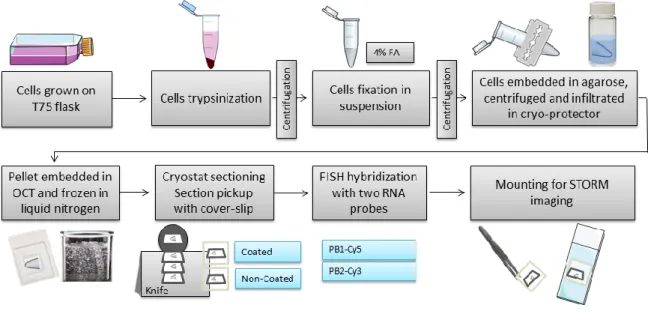
Figure 25. CLEM workflow for an IAV infected cell ... 34
Figure 26. STORM reconstruction and EM alignment ... 35
Figure 27. IAV genome bundling models ... 36
Figure 28. Vesicular collision models ... 40
Figure 29. Recycling impairment model ... 41
xiv
Abbreviation List
APES (3-Aminopropyl)triethoxysilane
BSA Bovine serum albumin
CLEM Correlative light electron microscopy
cRNA Complementary RNA
DMEM Dulbecco's modified eagle medium
DMV Double membrane vesicle
EM Electron microscopy
ER Endoplasmic reticulum
ERC Endosomal recycling compartment
FA Formaldehyde
FISH Fluorescent in situ hybridization
FS Freeze substitution
GA Glutaraldehyde
GM Golgi marker
HA Hemagglutinin
HPF High-pressure freezing
IAV Influenza A virus
LM Light microscopy
M1 Matrix protein 1
M2 Matrix protein 2
MOI Multiplicity of infection
mRNA Messenger ribonucleic acid
MTOC Microtubule-organizing center
NA Neuraminidase
NP Nucleoprotein
NS Non-structural protein
OCT Optimal cutting temperature compound
PA Polymerase acidic protein
PB Phosphate buffer
PB1 Polymerase basic protein 1
PB2 Polymerase basic protein 2
PBS Phosphate-buffered saline
PI Post-infection
Rab11 Ras-related in brain 11
RER Rough endoplasmic reticulum
ROI Region of interest
RT Room temperature
SMV Single membrane vesicle
STORM Stochastic optical reconstruction microscopy
TEM Transmission electron microscopy
vRNA Viral ribonucleic acid
vRNP Viral ribonucleoprotein complexes
1
1. Importance of the Study
“Disease cannot be understood unless it is realized that the ultimate abnormality must lie in the cell” – Rudolf Virchow
Influenza viruses, from the Orthomyxoviridae family, are obligatory intracellular parasites that rely on the host cell chemical energy, protein synthetizing machinery and controlled environment to replicate. They are divided in four types A, B, C and D (Hause et al. 2013; Hause et al. 2014). From these, influenza A (IAV) is a major threat to public health as it evolves exceedingly fast and is a zoonotic virus. The natural reservoirs of IAV are the wild aquatic waterfowl and shorebirds; however, due to stable host switches, the virus is widespread in many mammalian and avian species. (Acheson 2011; Taubenberger & Kash 2010).
There is a substantial diversity of circulating IAV virus subtypes classified by the nature of its surface glycoproteins: neuraminidase (NA) and hemagglutinin (HA). Eighteen HA (H1-H18) and eleven NA (NA1-NA11) types were identified and can be combined to generate the different IAV subtypes denominated HxNx depending on the surface glycoprotein content (Tong et al. 2013).
The subtypes are created by antigenic re-assortment which is made by a mechanism of antigenic shift. The antigenic shift is created when virus from different origins co-infect a host-cell and exchange genetic material. This mechanism have the capacity to created viral subtypes with different antigenic properties from the parental strains which can overcome the immune system and even break the inter-species barrier as reviewed in (Bouvier & Palese 2008).
In humans, IAV is the pathogen responsible for flu, a severe respiratory acute disease associated with high morbidity and mortality (Lofgren et al. 2007). Data from the World Health Organization, estimates that there are approximately 1 billion cases of flu every year, 3 to 5 million severe related illness hospitalizations and 300,000 to 500,000 associated deaths. In addition to yearly epidemics, IAV pandemics can also occur sporadically with associated devastating mortality. The most notable example is the “Spanish flu” originated from avian strains, between 1918 and 1920 caused around 40 million deaths. There are other cases worth mentioning, such as the “Asian flu” in 1957, also from avian origin but establishing in human via complex mixes (reassortment) with human viruses, and, more recently, the “Swine flu” in 2009, from swine origin (Saunders-Hastings & Krewski 2016).
IAV pandemics have up to now, been caused by infections with viruses unknown to the human immune system. In most cases the virus has crossed the host species barrier from avian and swine and established in humans. The ability of IAV to overcome the species barrier resides in the nature of its RNA segmented genome, divided in eight different particles called viral ribonucleoprotein complexes (vRNP). Each vRNP complex is composed by one of the eight viral ribonucleic acid (vRNA) segments individually wrapped with multiple monomers of the RNA-binding nucleoprotein (NP) and a heterotrimeric viral RNA polymerase composed by polymerase acidic protein (PA), polymerase basic protein 1 (PB1) and 2 (PB2) bound at the 3´ and 5´termini hairpin of the vRNA (Lee et al. 2017; Moeller et al. 2012; Arranz et al. 2012).
The vRNPs function as independent units for transcription and replication; however, for viral assembly, a complete set of eight different vRNPs is required increasing the complexity of genome
2
packaging. Identifying the mechanisms sustaining IAV genome is of utmost importance for human health. It has been widely reported that each segment has specific nucleotides located at the 3´ and 5´ termini responsible for RNA-RNA interactions that sustain the cohesion of the eight-segment core as reviewed in (Hutchinson et al. 2010). Much has been done to identify regions that are necessary for inclusion of the segments in virions (packaging signals) and for their binding to each other (bundling signals) (Giese et al. 2016). Recently it was suggested that the host recycling pathway facilitated IAV genome assembly.
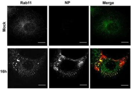
After export from the nucleus, IAV replicated genome is trafficked in a two-step mechanism: 1) vRNPs accumulate nearby the microtubule-organizing center (MTOC) in the endosomal recycling compartment (ERC) and 2) piggyback onto vesicles destined to the cell periphery via interactions between viral PB2 and vesicular GTPase Ras-related in brain 11 (Rab11) (Amorim et al. 2011; Eisfeld et al. 2011; Momose et al. 2011). In homeostasis, Rab11 acts as a molecular switch regulating the steps involved in the processes of vesicle formation from the ERC, movement and fusion towards the plasma membrane being spread in the cytoplasm (Grant & Donaldson 2009). In infected cells, the Rab11 distribution pattern is altered and appears as a puncta indicating that IAV infection altered Rab11-vesicular pathway (figure 1) (Vale-Costa et al. 2016). During this transformation the co-localization of the eight different segments increased in a Rab11 dependent manner, and it was proposed that genome sub-bundling occurred en route from the nucleus to the actual budding site (Lakdawala et al. 2014; Chou et al. 2013).
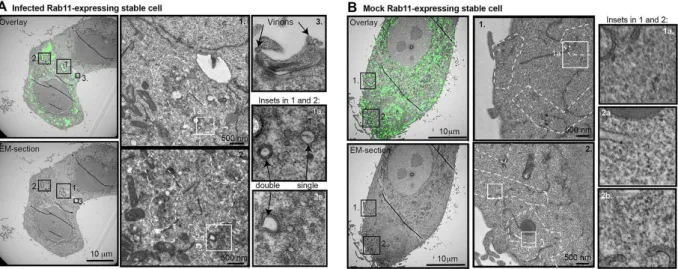
Subsequently, I used correlative light electron microscopy (CLEM) methods to demonstrate the ultrastructural features underneath the Rab11 fluorescent signal (Vale-Costa et al. 2016). In this work it was observed that during infection there is an aggregation of vesicles, without fusion, creating clusters. The clusters were composed by vesicles of heterogeneous sizes with single and double membranes (SMV and DMV), indicating that IAV leads not only to clustering but also to the rearrangement of vesicular membranes (figure 2) (Vale-Costa et al. 2016).
Figure 1. IAV infection alters Rab11 distribution
Immunofluorescence of A549 cells labeled for viral NP and Rab11. Mock cells represent the non-infected control and 16h cells represent the cells infected for 16 hours with influenza A/Puerto Rico/8/34 (PR8) at the multiplicity of infection (MOI) of 3. In mock cells, Rab11 is distributed homogeneously in the cytoplasm of the cell, having an accumulation at the perinuclear region where the MTOC is located. In infected cells, the Rab11 shows aggregation indicating that the infection with IAV leads to an alteration of Rab11 distribution pattern. Bar = 10 µm. Image adapted from our publications Vale-Costa et al. 2016.
3
This work substantiated an alternative model for IAV genomic assembly in which vesicular clustering can function as a viral induced platform promoting the creation of vRNPs hotspots. The high co-localization among the pool of all vRNPs would allow the establishment (or completion) of the RNA-RNA interactions necessary for assembly of the eight RNA-RNA segments before budding. Vesicular clustering thus emerges as an essential step in IAV life cycle. The definition of the chronology of events leading to maturation of vesicular clusters, in particular formation of clusters and alterations in vesicular composition become central to understand the mechanisms of IAV genome assembly.
In the present work, electron microscopy (EM) cryo-methods were applied to further investigate our IAV genome trafficking and assembly model. These methods are crucial in order to define the smallest ultrastructural details hindered by the chemical approaches (Studer et al. 1989). Despite the advantages of using the EM cryo-methods, without the appropriate optimization and development they cannot be used to their full potential (Roingeard 2008; Romero-Brey & Bartenschlager 2015).
In summary, within this work the EM cryo-methodologies were not just applied but also optimized and developed to elucidate how vesicular clustering is formed during infection and what is its functional role in the lifecycle of IAV.
Figure 2. Ultrastructural features of Rab11 vesicular clustering revealed by CLEM technique
Correlative light electron microscopy (CLEM) of A549 cells expressing GFP-Rab11 and processed for Electron Microscopy (EM). A. Cells were infected for 16 hours with PR8 at a MOI of 3. ERC clustered vesicles are scattered in the cytosol. The clusters are composed by a heterogeneous population of vesicles with single and double membranes. B. Mock cells represent the non-infected control. No clusters of Rab11-vesicles were detected in the absence of IAV infection. Bar = 10 µm. Image adapted from our publications Vale-Costa et al. 2016.
4
2. Research Approach
2.1 Study Location
This study was developed in the laboratory of Cell Biology of Viral Infection laboratory and in the Electron Microscopy Facility, at the Instituto Gulbenkian de Ciência located at Rua da Quinta Grande 6, 2780-156 Oeiras, Portugal.
2.2 Research Question and Hypothesis
The vesicular clustering is formed during IAV infection and plays a role in viral lifecycle, promoting the establishment of RNA-RNA interactions among the different segments.
2.3 Objectives
2.3.1 General Objectives
a) Characterise the biogenesis of the vesicles that cluster during IAV infection;
b) Determine the functional relevance of vesicular clustering for IAV genome assembly.
2.3.2 Specific Objectives
a) Optimize an electron microscopy (EM) cryo-method for the preparation of A549 cells constitutively expressing GFP-Rab11 cells evaluating the vitrification rate and morphological preservation;
b) Quantify the number of vesicles of single and double membrane (SMV and DMV) over an IAV infection time-course using the EM cryo-method assessed in specific objective a);
c) Describe the morphological profile of the cellular structures involved in cluster formation during IAV infection using tomography and 3D modeling;
d) Optimize a fixation method that enables immuno-gold labeling and ultrastructural identification to determine the origin of the vesicular content of clusters;
e) Develop a CLEM method to image individual IAV vRNPs in A549 cells constitutively expressing GFP-Rab11 infected cells using super resolution light microscopy and EM.
5
3. Literature Review
“A thorough, sophisticated literature review is the foundation and inspiration for substantial, useful research” – D. Boote and P. Beile
3.1 Influenza A Virus
3.1.1 Structure
IAV is a pleomorphic virus that can assume spherical or filamentous forms. The spherical particles have approximately 100 nm in diameter and the filamentous ones have 300 nm in length (Mosley & Wyckoff 1946). The virion is composed by a host-derived envelope with an external layer of spike-like projections. This layer surrounds the segmented negative-strand RNA genome (figure 3), characteristic of the Orthomyxoviridae family, where IAV belongs (Fields et al. 2007).
3.1.1.1 Viral Envelope
The virion has a host cell derived lipid envelope that contains three transmembrane proteins HA, NA and matrix protein 2 (M2). M2 is a minor component of the virion envelope, whereas HA appears in larger number followed by NA (Compans et al. 1970; Hutchinson et al. 2014). There are 18 genetically different subtypes of HA and 11 of NA (Tong et al. 2013). IAV is classified by the expression of different combinations of HA and NA subtypes at the virion surface (Fields et al. 2007).
HA is synthetized as a precursor protein (HA0) at the rough endoplasmic reticulum (RER), matured at the Golgi complex and transported to the plasma membrane, where is cleaved by cellular proteases to form HA1 and HA2. HA1 and HA2 remain linked through a disulphide bond to form the HA monomer (Rodriguez-Boulan et al. 1984; Braakman et al. 1991; Lamb, Robert and Choppin 1983). HA is presented at the surface of the virion as a homotrimer. It is responsible for: 1) viral recognition and binding to the sialic acid cellular receptor and 2) membrane fusion between the viral envelope and the host cell membrane (Wiley & Skehel 1987).
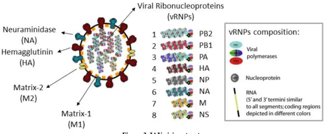
Figure 3. IAV virion structure
IAV is composed of a lipid envelope that contains three structural proteins: hemagglutinin (HA), neuraminidase (NA) and matrix protein 2 (M2). The viral core is coated with the matrix protein 1 (M1), and encloses the viral genome composed by eight vRNPs. Each vRNP encodes for different viral proteins and is composed by the hetero-trimeric viral polymerase (PB1, PB2 and PA) and the negative-sense vRNA genome wrapped by NP. Image produced by Maria João Amorim.
6
NA is presented as a homotetramer at the virion surface (Varghese et al. 1983). It cleaves the sialic acid present in glycoproteins or glycolipids from the cell surface and also on oligosaccharides from mucoproteins. This enzymatic activity mediates the release of newly formed virions from the surface of the host cell and from the abundant mucins present in the respiratory tract (Matrosovich et al. 2004).
The third transmembrane protein, M2, also forms a homotetramer. It functions as an ion channel modulating the pH within the virions. It allows a flow of ions from the endosome across the virion envelop that results in the virion acidification. This process disrupts the connection between the matrix protein 1 (M1) and the vRNPs in the viral core and enables the release of the uncoated vRNPs into the cytoplasm (Holsinger et al. 1994; Pielak & Chou 2011).
3.1.1.2 Viral Core
IAV viral core is enclosed by an internal layer of the matrix protein, M1. M1 covers the inner part of the lipid layer and interacts with the vRNA and the NP holding the vRNP complex structure together. Also, it is involved in the nuclear export of IAV genomic segments together with the nonstructural protein (NS) 2 (Fields et al. 2007). The NS2, also called nuclear export protein, binds to cellular export proteins and nucleoporins recruiting the machinery necessary for nuclear exportation of the complex M1-vRNPs (O’Neill et al. 1998).
There are eight individual vRNPs complexes that together constitute the viral genome (McGeoch et al. 1976). Each composed by a negative-sense single stranded vRNA segment with regions bound to multiple copies of NP and regions NP-free (Lee et al. 2017). Each of the eight genome segments encode for a major viral proteins: PB2, PB1, PA, HA, NP, NA, M and NS (figure 3). However, from this major proteins, ten other viral proteins arise by splicing, ribosomal frameshifts or alternative initiation (Yamayoshi et al. 2014).
The vRNA segment 4, 5 and 6 encode for a single protein each: HA, NP and NA, respectively (Palese & Schulman 1976; Ritchey et al. 1976). The segment 1 encodes for PB2 and PB2-S1 (Yamayoshi et al. 2014), the segment 2 for PB1 and the accessory protein PB1-F2 and PB1-N40 and the segment 3 for PA, PA-N155, PA-N182 and PA-X (Akkina 1990; Chen et al. 2001; Wise et al. 2009; Jagger et al. 2012). Also, the two last segments 7 and 8, encode for the matrix proteins M1, M2 and M42 and for the non-structural proteins, NS1, NS2 and NS3, respectively (Wise et al. 2012; Selman et al. 2012). The vRNPs can be also divided by length in three categories: 90-110 nm (segments 1-3), 60-90 nm (segments 4-6) and 30-50 nm (segments 7-8) (Zheng & Tao 2013).
At the 5´ and 3´ termini of the each segment there is 13 and 12 conserved nucleotides, respectively, that form a hairpin structure to where is connected the heterotrimeric viral RNA-dependent RNA polymerase composed of PA, PB1 and PB2 (Moeller et al. 2012; Lee et al. 2017). There are noncoding regions at both 3´ and 5´ ends with a highly conserved region in the extreme end that functions as a promoter for the replication and transcription by the viral polymerase. These regions also possess a messenger RNA (mRNA) polyadenylation and packaging signals (Lamb, Robert and Choppin 1983; Fields et al. 2007).
3.1.2 Lifecycle
The lifecycle of IAV can be categorized in five major steps: 1) Entry; 2) Uncoating; 3) Replication; 4) Trafficking and 5) Assembly, budding and release (figure 4).
7
3.1.2.1 Entry
The first step of the IAV lifecycle is the entry of the virion in the host cell (figure 4A). The step starts with the recognition and attachment of the virus to the target cell. The viral protein HA (or HA0 for the non-cleaved form) requires activation through cleavage into HA1 and HA2 by host proteases. HA1 recognizes the sialic acid cell receptor located on glycoconjugates at the cellular membrane (Rossmann & Rao 2012).
The sialic acid is connected to a galactose residue by a glycosidic linkage that can assume an α2,3 or α2,6 configuration. The different subtypes of HA have a fine specificity towards the type of configuration. For example, human influenza strains with human adapted HA subtypes, binds to α2,6 linkage configuration rather than α2,3 and avian influenza does the opposite (Connor et al. 1994). After attachment, the influenza virion penetrates the cell via receptor-mediated endocytosis (Matlin et al. 1981; Rust et al. 2004).
3.1.2.2 Uncoating
Uncoating and subsequent release of the vRNPs into the cytosol is mediated by the endosomal acidic environment (pH 5-6). During the process two key steps occur: the fusion between the endosomal membrane and the viral envelope and the release of the vRNPs from the M1 matrix. In the first step, HA conformational rearrangements are triggered. The hydrophobic fusion peptide of the HA2 domain is exposed and is inserted into the endosomal membrane to create a fusion pore (Skehel & Wiley 2000). In the second step, the hydrogen ions present in the endosome are pumped via the viral M2 towards the core of the virion (Kemler et al. 1994). This disrupts the interactions between M1 and vRNPs. The two steps enable the direct release of vRNPs onto the host cytosol (figure 4B).
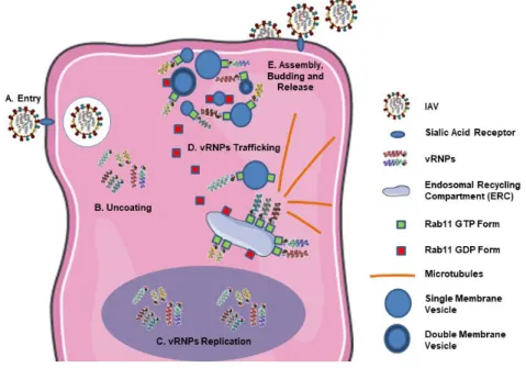
Figure 4. IAV lifecycle
A. IAV binds to host sialic acid receptors at the cell membrane and enters via endocytosis. B. Once inside, the fusion between
the endosomal membrane and the viral envelope is triggered with consequent release of the vRNPs onto the cytosol. C. The vRNPs are transported to the nucleus, where the replication of vRNA happens. D. After replication, the vRNPs are accumulated at the ERC and transported via Rab11-vesicles towards the cell periphery. E. In the periphery occurs the assembly of the complete genomic set and the incorporation onto the viral envelope, before budding and release of the progeny particles.
8
3.1.2.3 Replication
The vRNPs released into the cytosol are transported to the cellular nucleus (figure 4C) by a process that involves the importin α pathway recognition of the vRNPs structural component nucleoprotein (O’Neill et al. 1995; Neumann et al. 1997). There, the process of transcription and replication occurs via a mechanism that is being unraveled by many scientists for many years and is reviewed in (te Velthuis & Fodor 2016). The negative-sense vRNA is used as a template for two positive-sense products: mRNA, for viral protein translation, and complementary RNA (cRNA), the latter serving as a template for synthesis of multiple copies of negative-sense vRNA by the viral RNA-dependent RNA polymerase (te Velthuis & Fodor 2016).
The viral mRNA contains a 5´ cap sequence and a poly-A tail at the 3´ termini (Lamb, Robert and Choppin 1983). The viral RNA-dependent RNA polymerase hijacks the host pre-mRNA cap regions through a process of “cap snatching”. The subunit PB2 recognizes cap structures on host pre-mRNAs (Ulmanen et al. 1981; Peng et al. 1996), PA cleaves these structures (Dias et al. 2009) and PB1 has polymerase activity that elongates transcripts (Braam et al. 1983). The cap regions function as primers for viral mRNA synthesis and are also essential for the efficient binding of ribosomes to the mRNA. Through the cap snatching process, IAV promotes the production of viral components and inhibits host mRNA translation and consecutive protein synthesis. This process disrupts the cellular homeostasis and eventually leads to cell death. The poly-A tail of viral mRNAs is a host independent process that results from stuttering of a poly-U stretch in vRNA templates (Robertson et al. 1981). The viral mRNA is transported to the cytoplasm where translation of both viral envelope and core proteins happens. The viral envelope proteins NA, HA and M2 are synthetized in the cytosol and transported to the endoplasmic reticulum (ER) where they are glycosylated and folded into trimers and tetramers (Braakman et al. 1991; Fields et al. 2007). After folding they are transported through the Golgi apparatus and the trans-Golgi network to the plasma membrane as reviewed in Bouvier & Palese 2008. The viral core proteins, PA, PB1, PB2 and NP are translated and sent to the nucleus to first promote the vRNA replication and second to join the newly replicated vRNA. M1 and NS2 are also translated and imported to the nucleus to assist vRNP export to the cytoplasm (O’Neill et al. 1998; Bui et al. 2000). Part of M1 is transported to the plasma membrane where it interacts with NA and HA C-terminal domains.
3.1.2.4 Trafficking
After nuclear export, vRNPs need to be transported towards the plasma membrane for assembly, budding and release (Fields et al. 2007). The trafficking of the genome is made in a two-step mechanism: first the vRNPs agglomerate at the perinuclear region and second piggyback onto Rab11 vesicles of the endosomal recycling compartment (ERC) to be transported on microtubules towards the cell surface (figure 4D) (Amorim et al. 2011; Eisfeld et al. 2011; Momose et al. 2011).
Rab11 is a GTPase involved in the recycling of endocytosed proteins. It regulates vesicle formation from the ERC, movement and fusion as reviewed in (Grant & Donaldson 2009). It was also described as being implicated in the exocytosis of the ERC vesicles (Takahashi et al. 2012). It acts as a molecular switch with GDP/GTP cycles. Rab11-GDP is the inactive form. It connects to dissociation inhibitors (Rab-GDIs) that delivers Rab11 to target membranes. At the membranes, guanine exchange factors activates Rab11 by changing GDP to GTP. The activated form, Rab11-GTP, can be connected to effectors that mediate the movement and fusion of vesicles with target membranes. The inactivation is made by guanine-activating proteins that hydrolyse GTP. This process leads to a decrease in the
9
(Pasqualato et al. 2004; Vale-Costa & Amorim 2016).
In homeostasis Rab11 is spread throughout the cytoplasm; Whereas in IAV infected cells, Rab11 forms large cytoplasmic aggregates (Vale-Costa et al. 2016). The most recent model proposes that Rab11 aggregates form a viral induced platform for the spatial converging of vRNPs before assembly and budding (Chou et al. 2013; Lakdawala et al. 2014). This platform is composed by clusters of heterogeneous vesicles with single and double membranes (Vale-Costa et al. 2016).
3.1.2.5 Assembly, Budding and Release
Virion assembly comprises the incorporation of viral genome and proteins and occurs at the plasma membrane. Interestingly, in the case of IAV it is possible that viral assembly comprises two steps: the assembly of the viral genome (via a bundling process), composed of eight distinct segments and its packaging (inclusion) into budding virions at the plasma membrane. It is known that to create an infectious particle each virion needs to incorporate a complete genomic set of eight different vRNPs (Hutchinson et al. 2010).
A lot of research was conducted to solve a long standing debate of whether packaging was a selective or a random process, but overwhelming evidence indicates that the packaging process is selective and occurs through RNA-RNA interactions between the different vRNPs that contain specific packaging signals located at the 5´and 3´ termini (Giese et al. 2016)
Regarding assembly of IAV genome, recently exciting data has been published. Using multicolor fluorescence in situ hybridization two groups have shown that several segments co-localize in the cytoplasm and proposed that genome assembly could occur en route to the plasma membrane, favored by kissing and/or fusion events between vRNP-carrying vesicles (Chou et al. 2013; Lakdawala et al. 2014). These studies used confocal microscopy that lacks the resolving power to distinguish between association of vRNPs or simple spatial convergence in the cell cytoplasm.
We have used electron microscopy (EM) to address this question and found that vRNPs converged in space in regions of clustered vesicles that carry vRNPs facing the cytosol (Vale-Costa et al. 2016). Even with EM resolution, we were unable to find proof of association between the distinct vRNPs. This observation did however lead to the proposal of an alternative model for IAV genome assembly: vesicular clustering leads to formation of vRNP hotspots containing the eight segments and might facilitate genome assembly downstream. Doubt remains of where genome assembly occurs, if before reaching the membrane or at the budding site. In budding virions the eight vRNPs were visualized by EM almost pulling the membrane to be accommodated inside a virion (Fournier et al. 2012; Noda et al. 2012).
On the plasma membrane, the vRNPs interact with the M1 viral protein attached to the internal domain of NA and HA (figure 4E). After budding, lAV is released from the cell surface via the action of M2 and NA. M2 is involved in the scission the budded virion and NA cleaves the host sialic acid that attaches viral HA to the cell surface (Fields et al. 2007; Rossman et al. 2010).
3.2 Electron Microscopy Cryo-Methods to Study IAV at the Nanoscale
The visualization of the ultrastructural alterations induced by infection requires the application of methods whose resolution achieves the nanoscale, such as EM. Despite the advantages of the
10
resolution attained by EM, without appropriate methods to prepare samples, it cannot be used to its full potential to the research of viruses and the mechanisms of viral replication (Roingeard 2009; Romero-Brey & Bartenschlager 2015).
The major goal of EM methods is to preserve the ultrastructural features as close as possible to their living or native state (McDonald 2007). For that reason, fixation is one of the most critical steps during sample preparation (Bancroft & Gamble 2002). There are two types of fixation methods: chemical and physical.
The chemical fixation is typically executed with aldehydes that react with basic amino-acid residues on proteins. Such reaction creates methylene bridges between proteins, thus originating a network of crosslinks (Bancroft & Gamble 2002). The aldehydes usually employed are formaldehyde (FA) and glutaraldehyde (GA), composed by one or two aldehyde reactive groups, respectively. The higher cross-linking capacity of GA causes greater loss of protein structure and immunological activity than FA; however, GA preserves the morphology better (Bancroft & Gamble 2002).
Physical fixation, also called cryo-immobilization, uses cryogens instead of chemicals to stabilize cellular components. This process avoids the distortions induced by chemical fixation but it is limited by the water content of biological sample. Water is a poor heat conductor and limits the cooling rates within the sample leading to crystallization (Romero-Brey & Bartenschlager 2015).
Both fixation methods can be combined when handling infectious samples, such as IAV infected cells. The chemical inactivation of the virus is required prior to cryo-immobilization for the safe handling of the specimens. Two cryo-methods where the hybrid fixation can be employed are the High Pressure Freezing (HPF) - Freeze Substitution (FS) and the Tokuyasu techniques.
3.2.1 High Pressure Freezing – Freeze Substitution
The HPF is a freezing technique that cryo-immobilizes the sample by high pressure (around 2000 bar) and low temperature (-196ºC). This combination alters the physical properties of water by lowering its freezing point. The growth rate and nucleation of ice crystals is reduced and enables water vitrification before crystal formation (Moor 1987).
After cryo-immobilization by HPF, the most common way to process the cells is by FS (McDonald 2007). FS replaces the frozen water by an organic solvent, such as acetone, at a low temperature, avoiding the artifacts introduced by dehydration at room temperature (RT) (Morphew 2000). Also, a fixative can be added to the solvent, e.g. uranyl acetate. At -90ºC, the fixatives will penetrate the sample but will be inactive. This means that the fixative is homogenously distributed through the cell and simultaneously fixes all components when the sample is warmed to the fixative reactive temperature (McDonald 2007). After FS, the samples can be embedded in resin, sectioned by ultramicrotomy and analysed by transmission electron microscopy (TEM).
For a successful HPF-FS of cells, one of the most important steps is the loading of samples into the high pressure freezer machine (McDonald 2007). Several types of specimen carriers and fillers, differing in material and geometry, are available depending on the manufacturer (McDonald 2007; Morphew 2000). Two examples are hats and flat disks. The following described carriers belong to the High Pressure Freezer Compact 02 (Wohlwend Engineering Switzerland).
11
& Muller-Reichert 2012). The aluminum hats are round cups with an external diameter of 3 mm and internal diameter of 2 mm (figure 5A). The inner cavity depth can range from 0.025 mm to 0.3 mm, and two hats can be combined in multiple arrangements to form a chamber ranging from 0.025 mm to 0.6 mm in depth. Hats can be used to freeze a high number of cells in a pellet (McDonald 2007).
Flat disks can be made of aclar (figure 5B), that has a thermal conductivity of 0.19-0.22 W/(m.K) (Verkade & Muller-Reichert 2012). For freezing, flat disks need to be combined with a 0.04 mm hat to form a chamber. Flat disks permit growing and freezing cells in a monolayer.
Both types of sample carriers need a filler to occupy the space surrounding the cells and minimize the aqueous material to be frozen (Morphew 2000). The fillers should have good heat transfer properties and bind to water molecules to avoid their rearrangement into crystals (McDonald 2007).
The fillers can be intracellular or extracellular. Extracellular are preferred since they do not penetrate and interfere with the internal structure of cells (McDonald 2007). Some examples of extracellular fillers are: 1-hexadecene, dextran and bovine serum albumin (BSA).
3.2.2 Tokuyasu
Tokuaysu is an EM cryo-method that enables the preparation of frozen-hydrated specimen for cryosectioning and immunolabeling. Cells are primarily fixed in aldehydes, cryo-protected with 2.3 M sucrose and plunge frozen in liquid nitrogen (Griffiths 1993). Sucrose is an intracellular cryo-protector that avoids cellular crystallization and, together with the aldehyde fixation, gives enough consistency to the cells so they can be ultracryosectioned (Tokuyasu 1973).
This technique is the gold-standard for TEM immunogold labeling (Mobius 2009). The sections remain hydrated and the antigens accessibility is not hindered by the presence of a resin. Nevertheless, for high-resolution localization of antigens, the initial fixation must permit a balance between ultrastructure and immunoreactivity preservation. Aldehyde induced cross-linking must be weak enough to maintain epitope accessibility yet preserving ultrastructural morphology. This balance needs to be optimized according to the biological system in study and the sensitivity of the antigens to be detected. Optimization should be done by light microscopy (LM) to avoid lengthy and labor-intensive EM procedures (Morphew 2000).
Figure 5. Specimen carrier and aclar flat disk example
There are several types of specimen carriers available whose design varies between HPF manufacturers. A. Hats made of aluminum with a depth of 0.1 mm. For freezing they need to be combined to form a chamber where the sample resides. B. Flat disks made of aclar. These disks can be used for the growth of monolayers and also need to be combined with a special hat of 0.04 mm for freezing.
12
3.2.3 Correlative Light Electron Microscopy
Correlative light electron microscopy (CLEM) methods emerged as a powerful tool in the field of cellular biology. They allow the imaging of the same structure using two microscopy modalities, LM and EM (Verkade & Muller-Reichert 2012). LM enables the identification of fluorescence/colorimetric tags widely used while EM allows correlating such tags with the ultrastructure within the cellular context (Verkade & Muller-Reichert 2012). These tags can be endogenously expressed by the cell or added externally, for example through immunocytochemistry or fluorescent in situ hybridization (FISH). With the development of new techniques, such as LM super-resolution, a number of different and new CLEM approaches has arisen.
3.2.3.1 STORM, FISH and EM to Study IAV genome
Stochastic optical reconstruction microscopy (STORM) is a type of super-resolution microscopy. It uses a sequential activation and then time-localization of fluorophores with photo-switchable capabilities. At each time-point only a subset of fluorophores is activated to a fluorescent state. Their position is recorded and its centroid position is precisely found. Then this set of fluorophores is inactivated and the cycle is repeated in several acquisitions of different fluorophore sets. Finally, fluorophore positions can be reconstructed to give rise to a high-precision LM image (Rust et al. 2006). STORM can be allied to techniques such as FISH in order to detect fluorescent dyes attached to nucleic acids.
FISH uses complementary genomic sequences attached to fluorescent probes to detect specific parts of the genome (Bancroft & Gamble 2002). For our purpose, it can be used to localize specific viral genome segments in IAV infected cells. For performing the technique, the cells need to be permeabilized so the probes can reach the intracellular targets. There are two types of permeabilization: chemical and physical. The chemical permeabilization cannot be used for EM, since it disrupts the ultrastructure. Instead, cells can be opened through physical permeabilization with sectioning (McDonald 2007).
The sectioning can be done before or after the EM processing. The pre-processing sectioning can be done using a cryostat or vibratome (Vogels et al. 2009). It facilitates the accessibility of the probes to the targets and enables further processing of sections with heavy metals for high-contrast ultrastructural preservation. Within this work, the pre-processing approach was used, with the FISH labeling being done in cryostat sections. The FISH labeling on cryostat sections is a common approach, however some points need to be adapted for the later STORM imaging, EM processing and correlation of techniques.
13
4. Material and Methods
“Setting a goal is not the main thing. It is deciding how you will go about achieving it and staying with that plan” – Tom Landry
4.1 Cell Culture
Human alveolar basal epithelial A549 cells expressing GFP-Rab11 Wild-Type (WT) were cultured with Dulbecco's Modified Eagle Medium (DMEM) (®Life Technologies) supplemented with 2 mM glutamine, 50 μg/ml streptomycin, 50 U/ml penicillin (®Biowest) and 10% (v/v) fetal bovine serum. Cells were maintained in T75 flasks at 37ºC in a humidified atmosphere with 5% CO2. The cells were
sub-cultured every 3-4 days. The procedure consisted in: 1) rinsing cells with phosphate-buffered saline (PBS), 2) trypsinizing with 1 μg/mL solution of trypsin in PBS for 5 minutes at 37oC, 3)
pelleting by centrifugation and 4) resuspending in DMEM accordingly to the desired confluence.
4.2 Infection
Cells were infected with IAV A/Puerto Rico/8/34 (PR8; human subtype H1N1) at a multiplicity of infection (MOI) of 5 in serum-free-DMEM for 30 minutes. Afterwards, infection was stopped by the addition of DMEM and cells were incubated at 37ºC in a humidified atmosphere containing 5% CO2
(Amorim et al. 2011).
4.3 Morphology and Quantification
4.3.1 High Pressure Freezing
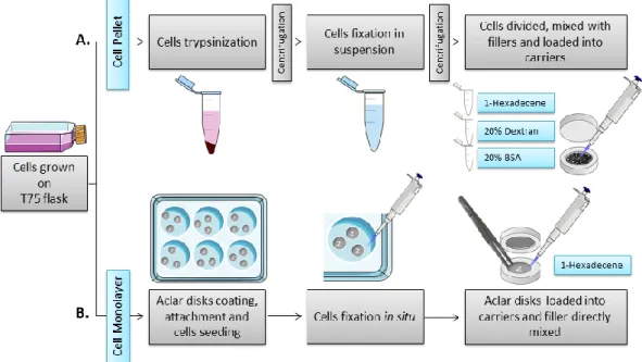
Two types of HPF sample preparation were tested: pellet and monolayer (figure 6 A and B, respectively).
Figure 6. Scheme of HPF optimization
Two types of cell preparations were tested for HPF of cells: pellet or monolayer. A. For pellets, cells were trypsinized and fixed in suspension. In the last step, the pellet was divided to test three fillers: 1-Hexadecene, 20% (w/v) Dextran and 20% (w/v) BSA. The cells mixed with the fillers were pipetted to the carrier. B. For monolayers, cells were seeded in plates previously prepared with aclar disks coated with carbon and engraved with the number “2” for orientation. Cells were fixed in the plate. In the last step, the aclar disks were removed with forceps and loaded into the carrier with the engraved side with the number “2” facing up. The filler, 1-Hexadecene, was added directly to the carrier containing the aclar disk.
14
For the pellet method, cells were seeded in T75 flasks at a density of 5x10⁴ cells/ml, washed with PBS,
trypsinized and pelleted by centrifugation (200 rcf, 5 minutes). For the monolayer preparation, cells were seeded at a density of 4x10⁴ cells/ml onto 3 mm aclar disks attached with dental wax to a 6
well-plate. Prior to seeding, the aclar disks were coated with a layer of carbon and then landmarked with the number “2” using a needle. This was done to track the side of the disk where the cells were seeded – when the number “2” was readable, the cells were on the top. Cell monolayers were fixed in situ, whereas pellets were fixed in suspension both using a mixture of 2% (v/v) formaldehyde (FA) (®EMS) and 0.2% (v/v) glutaraldehyde (GA) (®Polysciences) in 0.1M phosphate buffer (PB), for 2 hours at RT. Cells were washed with PB in between the steps. Pellets were additionally centrifuged and resuspended before loading in carriers. The pellet was loaded into 0.1 mm deep carriers and, due to the thickness of the pellet, three types of fillers were tested: 1-Hexadecene (®Merck), 20% (w/v) Dextran (®Alfa Aesar) and 20% (w/v) BSA (®Sigma Aldrich). The monolayers in the aclar disks were added to a 0.04 mm deep carrier which was filled with 1-hexadecene. Hexadecene was used in this case since it has a low viscosity and surface tension that reduces the air trapped in the carriers (Studer et al. 1989). All samples were frozen using a High Pressure Freezer Compact 02 (®Wohlwend Engineering Switzerland).
4.3.2 Freeze Substitution
For FS the Leica EM AFS2 was used together with a processor Leica EM FSP. The samples were freeze substituted at -90ºC with 0.1% (w/v) uranyl acetate and 0.01% (w/v) tannic acid (®EMS) in acetone for 6 hours. The temperature was raised to -45ºC at a slope of 5ºC/hour. Samples were stabilized at -45ºC for 1.5 hours before washing in acetone three times. Samples were infiltrated and embedded in Lowicryl HM20 (®Polysciences) at -45ºC. Polymerization of the resin was done using ultraviolet light at -25ºC for 48 hours.
4.3.3 Ultramicrotomy and Post-Staining
Sections were taken at 70 nm for 2D imaging. The sections from the flat disks blocks were picked at 1.2 µm starting from the cell bottom. For tomography 120 nm sections were cut. The sectioning was performed on a Leica UC7 and all the sections were picked on palladium-copper grids coated with 1% (w/v) formvar (®Agar Scientific) in chloroform (®VWR). The post-staining was made with 1% (w/v) uranyl acetate and Reynolds lead citrate, for 5 minutes each. For tomography, 15 nm protein A-gold (®UMC, Utrecht) was added to both sides of the sections before staining.
4.3.4 Data Acquisition
The images for technique optimization were acquired on a Hitachi H-7650 operating at 100 keV equipped with a XR41M mid mount AMT digital camera. For quantification and morphology analysis, photomontages and tomograms were acquired on a FEI Tecnai G2 Spirit BioTWIN operating at 120
keV equipped with a Olympus-SIS Veleta CCD Camera.
4.3.5 Data Analysis
4.3.5.1 High Pressure Freezing – Freeze Substitution Technique Optimization
For the optimization of the HPF-FS technique two parameters were studied: vitrification rate and morphology preservation.
15
Tested conditions: Two types of specimen carrier were tested: aluminum hats 0.1 mm deep and aclar flat disks. For the hats, three types of cryo-protectants were tested: 1-hexadecene, 20% (w/v) Dextran and 20% (w/v) BSA. For the flat disks and due to the reduced thickness of sample only 1-hexadecene was used for evaluation.
Sampling: For each condition, five blocks were produced. From each block, one section was taken and one cell was randomly imaged. For each cell, three pictures at the magnifications of 1k, 5k and 10k were acquired on a Hitachi H-7650 equipped with a XR41M mid mount AMT digital camera.
Criteria: Cells with absence of crystallization patterns are vitrified (Studer et al. 1989). Cells with good vitrification were classified with an arbitrary value of 1 and cells with ice damage were categorized as -1. To all images a randomized numerical code was given to allow a non-biased evaluation. The classification was performed by three different evaluators. Results were presented as % of vitrified blocks relative to a total number of blocks without ice damage. The number of blocks was directly translated from the number of vitrified cells, since each cell correspond to one single block.
B. Morphology Preservation
Tested conditions: Aclar flat disk frozen with 1-hexadecene and 0.1mm aluminum hats with 20% (w/v) dextran. The two types of specimen holders involve different cellular preparations that can influence the ultrastructure preservation; however, to reduce the number of conditions, only the cryo-protectant condition from the aluminum hats that obtained the best freezing rate from vitrification (20% dextran) was evaluated.
Sampling: One block with good vitrification of each condition was randomly chosen from the five blocks used for vitrification evaluation. From each block, a total of 10 cells were randomly selected and imaged. Five representative pictures of full cell, mitochondria, nuclei, vesicles and plasma membrane, were acquired at the magnifications of 1.2k, 8k, 10k, 12k and 20k respectively on a Hitachi H-7650 equipped with a XR41M mid mount AMT digital camera.
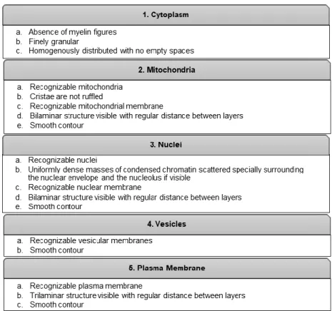
Criteria: Five cellular structures were analysed per cell: cytoplasm, mitochondria, nuclei, plasma membrane and vesicles. For each structure, specific criteria were defined to assess morphological preservation, as described in table 1.
To each criteria, a value of 1 or 0 was attributed, if the cellular structure followed the description or not, respectively. Hence, the maximum score was 18 and the minimum was 0. These scores were transformed in % to facilitate the analysis of the results. To all images a randomized numerical code was given to allow a non-biased evaluation. The evaluation was performed by one evaluator.
Statistical analysis: Non-parametric statistics was used to describe the data with median and interquartile range. The median of morphological preservation scores obtain in each condition were compared by applying the Mann-Whitney test. All the graphs and statistical analysis were done using the software GraphPad Prism 6.
16
4.3.5.2 Quantification
Tested conditions: uninfected (mock) and 4, 8, 12, 16, 20 and 24 hours post-infection (PI) with IAV.
Sampling: two independent infections experiments were done. For each condition, one block was sectioned and five cells were randomly selected and imaged for quantification. To acquire the full cell, the photomontage software from FEI was used.
Criteria: the total number of single-membrane vesicles (SMV) and double-membrane vesicles (DMV) were calculated through Fiji multipoint and measure tool.
Statistical analysis: Non-parametric statistics were used to describe the data with median and interquartile range. Also, the difference of SMV and DMV medians obtained for each infection time-point was tested by applying the Kruskal-Wallis test. The multiple comparisons were tested with Dunn’s test. All the graphs and statistical analysis were done using the software GraphPad Prism 6.
4.3.5.3 3D Morphological Study
Tested conditions: 16 hours PI with IAV.
Sampling: from one block, four serial sections of 120 nm were acquired. Individual single-axis tomograms were done in each section from -62º to +62º at increments of 1. The model was generated from four stacked tomograms. The tomographic reconstructions were made using the program IMOD (Kremer et al. 1996; Mastronarde 1997) and the 3D model using 3Dmod.
Table 1. Criteria for the evaluation of morphological preservation
Criteria used to assess morphological preservation of cellular structures (cytoplasm, mitochondria, nuclei, vesicles and plasma membrane).