Steatosis of indeterminate cause in a pediatric group:
is it a primary mitochondrial hepatopathy?
Esteatose de causa não determinada em grupo pediátrico:
hepatopatia mitocondrial primária?
Gustavo Henrique Silva
I, Gabriel Hessel
II, Kunie Iabiku Rabello Coelho
III, Cecília Amélia Fazzio Escanhoela
IVDepartment of Pathology, Faculdade de Ciências Médicas, Universidade Estadual de Campinas (FCM-Unicamp), Campinas, São Paulo, Brazil
ABSTRACT
CONTEXT AND OBJECTIVE: In children, hepatic steatosis may be related to inborn errors of metabolism (IEMs) or to non-alcoholic fatty liver disease (NAFLD). The aim of this study was to assess and characterize steatosis of indeterminate cause through morphological and morphometric analysis of liver tissue.
DESIGN AND SETTING: Cross-sectional study at the Departments of Pathology of Faculdade de Ciências Médicas, Universidade Estadual de Campinas (FCM-Unicamp) and Faculdade de Medicina de Botucatu, Universidade Estadual Paulista (FMB-Unesp).
METHODS: Eighteen consecutive liver biopsies obtained from 16 patients of ages ranging from 3 months to 12 years and nine months that were inserted in a database in the study period were analyzed using op-tical microscopy and transmission electron microscopy. Through electron microscopy, the mitochondrial density and mean mitochondrial surface area were determined in hepatocytes. Ten patients ranging in age from 1 to 14 years were used as a control group.
RESULTS: “Pure” steatosis was detected, unaccompanied by fibrosis or any other histological alteration. Microvesicular steatosis predominated, with a significant increase in mean mitochondrial surface area.
CONCLUSION: Microvesicular steatosis may be related to primary mitochondrial hepatopathy, especially due to reduction of -oxidation or partial stagnation of oxidative phosphorylation. For these reasons, this form of steatosis (which should not be called “pure”) is likely to represent an initial stage in the broad spectrum of NAFLD. We have drawn attention to cases of steatosis in the pediatric group, in which the microvesicular form predominates, since this may be associated with mitochondrial disorders.
RESUMO
CONTEXTO E OBJETIVO: Em crianças, a esteatose hepática pode se relacionar a erros inatos do metabo-lismo (EIMs) ou à doença hepática gordurosa não-alcoólica (DHGNA). O objetivo deste estudo foi avaliar e caracterizar esteatose de causa indeterminada por meio de análises morfológica e morfométrica em tecido hepático.
TIPO DE ESTUDO E LOCAL: Estudo transversal nos Departamentos de Patologia da Faculdade de Ciências Médicas da Universidade Estadual de Campinas (FCM-Unicamp) e Faculdade de Medicina de Botucatu da Universidade Estadual Paulista (FMB-Unesp).
MÉTODOS: Foram utilizadas 18 biópsias hepáticas consecutivas obtidas de 16 pacientes com idade va-riando de 3 meses a 12 anos e 9 meses, inseridas num banco de dados no período do estudo, que foram analisadas por microscopia óptica e eletrônica. Na microscopia eletrônica, foi realizada determinação da densidade mitocondrial e da área superficial média das mitocôndrias nos hepatócitos. Dez pacientes com idade variando de 1 a 14 anos foram usados como grupo controle.
RESULTADOS: Foi detectada esteatose “pura”, não acompanhada por fibrose ou outra alteração histoló-gica. Foi verificado que, na predominância de esteatose microvesicular, houve aumento significativo da área mitocondrial média.
CONCLUSÃO: A esteatose microvesicular pode estar relacionada à hepatopatia mitocondrial primária, principalmente devido à redução na -oxidação ou parcial estagnação da fosforilação oxidativa. Por essas razões, esta forma de esteatose (que não pode ser chamada de “pura”) possivelmente represente uma fase inicial no amplo espectro da DHGNA. Chamamos a atenção para casos de esteatose no grupo pediátrico com predomínio da forma microvesicular, uma vez que pode haver associação com desor-dens mitocondriais.
IPhD. Postgraduate student, Department of
Pathology, Faculdade de Ciências Médicas, Universidade Estadual de Campinas (FCM-Unicamp), Campinas, São Paulo, Brazil.
IIMD, PhD. Assistant professor, Department
of Pediatrics, Faculdade de Ciências Médicas, Universidade Estadual de Campinas (FCM-Unicamp), Campinas, São Paulo, Brazil.
IIIMD, PhD. Assistant professor, Department of
Pathology, Faculdade de Medicina de Botucatu, Universidade Estadual Paulista (FMB-Unesp), Botucatu, São Paulo, Brazil.
IVMD, PhD. Assistant professor, Department
of Pathology, Faculdade de Ciências Médicas, Universidade Estadual de Campinas (FCM-Unicamp), Campinas, São Paulo, Brazil. KEY WORDS:
Fatty liver. Mitochondria, liver. Anatomy. Infant, newborn. Metabolism, inborn errors. PALAVRAS-CHAVE: Fígado gorduroso. Mitocôndrias hepáticas. Anatomia.
Recém-nascido.
INTRODUCTION
In children, the presence of steatosis is generally related to inborn errors of metabolism (IEMs). he clinical presentation varies and includes changes to general health condition; neurological, diges-tive and respiratory disorders; rapid and progressive neurologi-cal deterioration; myopathy; cardiomyopathy; craniofacial dys-morphism etc. Some forms of the disease present essentially as liver disease with hepatomegaly, jaundice, vomiting, lethargy and abnormalities of liver function.
When these diseases are ruled out ater clinical and laboratory investigation, non-alcoholic fatty liver disease (NAFLD) should be considered. his term, which was initially used for adults, has
become more inclusive.1 It is currently used to describe all age
groups, even children.2,3 In more recent years, several studies on
childhood4 and adolescent hepatic steatosis in cases of NAFLD
have been conducted, especially in relation to obese children,5-13
since the prevalence of NAFLD is higher in these types of chil-dren. In a pediatric group, the risk factors for NAFLD include
obesity, insulin resistance and hypertriglyceridemia.14 he major
features are a higher rate in male children, with serum alanine aminotransferase (ALT) levels that are usually higher than serum aspartate aminotransferase (AST) levels, and with hypertriglyc-eridemia and vague abdominal pain (which is usually the main
reason for seeking clinical evaluation).14-16
According to Mandel et al.17 and Bioulac-Sage et al.,18
child-hood microvesicular steatosis may be associated with
mitochon-drial abnormalities relating to increased numbers of organelles.18,19
here is also a correlation with respiratory chain defects and
con-sequently impaired oxidative phosphorylation.20,21 When this
type of abnormality occurs, transfer of electrons to oxygen mole-cules generates reactive oxygen species, thus producing
superox-ide anions and hydrogen peroxsuperox-ide.22 Furthermore, with
stagna-tion of mitochondrial oxidative capacity, fatty acids accumulate in the cytosol, thus reairming the close relationship between steatosis and mitochondriopathy.
he microvesicular form alone may also be associated with some clinical conditions, such as Reye’s syndrome, Jamaican
vomiting sickness and congenital deiciency of -oxidation.23 In
an experimental study on pigs, it was observed that the regenera-tive capacity of the hepatocytes in macrovesicular steatosis was more efective than in microvesicular steatosis, thereby implying
that the latter has a less favorable prognosis.24
he cases that were studied did not have any clinical associa-tion with any previously cited factor and the indings were cor-related with clinical and laboratory data. here is a continuing debate on the advisability of performing liver biopsy, because of the risk of complications, high costs and lack of efective therapy. However, liver biopsy is the only precise diagnostic method, and it is essential for grading and staging the disease. Furthermore, it
enables patient inclusion in clinical screening programs.25,26
OBJECTIVE
he aim of our study was to evaluate and characterize “pure” ste-atosis in a pediatric group by means of morphological (optical and electron microscopy) and morphometric analysis.
MATERIAL AND METHODS
All pediatric patients who were diagnosed as presenting morpho-logical steatosis by means of optical microscopy and examination of liver fragments processed for transmission electron micros-copy in the Department of Pathology of the School of Medical Sciences of Universidade Estadual de Campinas (FCM-Unicamp) and the Department of Pathology of Faculdade de Medicina de Botucatu (FMB) between 1993 and 2007 were retrospectively selected by means of a database search (speciic records from liver biopsies). Cases in which the cause of steatosis was estab-lished (IEM, obesity, diabetes, inherited metabolic disorders, malnutrition, viral hepatitis, Wilson’s disease, drug use and/or total parenteral nutrition) were subsequently excluded. Sixteen patients whose cause of steatosis could not be identiied were selected. hese patients’ ages ranged from three months to 12 years and nine months. heir heights ranged from 47 to 154.5 cm and their weights ranged from 3.15 to 51.50 kg. A review of all previously selected liver biopsies was undertaken: 18 samples were analyzed, because one patient underwent three biopsies (samples 12, 13 and 14).
he control group was composed of liver biopsies obtained from ten patients aged from one to 14 years who presented nor-mal physical development. In this group, the indications for liver biopsy were similar to those in the study group, i.e. vague abdom-inal pain and/or minimal hepatomegaly associated with slightly and persistently elevated hepatic enzyme levels. he diagnoses for the control individuals were normal, conirmed either from opti-cal or from electron microscopy indings. In all of these patients, the blood glucose levels were always normal. All the clinical data were obtained by consulting patient medical records. he present study was approved by the Research Ethics Committee of FCM-Unicamp (process no. 523/2004).
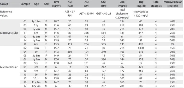
Table 1. Description of some parameters evaluated from the 16 patients studied (18 samples)
Group Sample Age Sex BMI (kg/m2)
AST (U/I)
ALT (U/I)
GGT (U/I)
Chol (mg/dl)
Trig (mg/dl)
Total steatosis
Microvesicular steatosis
Reference values
AST < 37
U/I ALT < 40 U/I GGT < 40 U/I
total cholesterol < 200 mg/dl
triglycerides < 120 mg/dl
Macrovesicular
01 1y 11m F 18.7 20 72 ni 134 46 4 40%
03 11y M 21.6 68 99 28 219 109 3 45%
08 1y 2m M 15.7 73 48 39 216 172 4 20%
11 5m M 14.6 87 386 554 131 347 4 25%
12 4y 6m M 17.5 47 48 20 ni 34 2 40%
14 1y 1m M 15.9 25 25 37 146 51 4 50%
18 6m F 13.5 119 204 585 133 476 4 5%
Microvesicular
02 10m F 15.7 75 71 ni 216 1330 4 55%
04 6y F 14.3 304 104 340 103 516 3 70%
05 3y 8m F 14.9 24 17 15 172 54 1 70%
06 1y 1m M 17.0 75 50 384 144 152 3 70%
07 5m F 12.8 242 151 ni ni ni 3 75%
09 3m M ni 14 19 212 182 421 3 70%
10 4m F 13.7 37 63 197 172 455 3 75%
13 2y M 16.5 26 22 50 156 54 4 60%
15 10 m M 15.8 47 53 31 105 87 4 80%
16 11y 1m M 14.7 20 18 ni 166 75 2 95%
17 12y 9m M ni 39 63 257 281 286 3 75%
BMI = body mass index; ni = nothing included; y = years; m = months; F = female; M = male; AST = aspartate aminotransferase; ALT = alanine aminotransferase; GGT = gamma-glutamyl transpeptidase; Chol = total cholesterol; Trig = triglycerides.
the percentage of hepatocytes afected by microvesicular steato-sis, all cells present in the fragments analyzed were counted (cells with small-droplet steatosis and those with large-droplet steato-sis) and then the percentages of each cell type were calculated. Specimens processed for TEM were ixed in a 2.5% glutaralde-hyde solution, postixed in a 1% osmium tetroxide solution and embedded in araldite resin. Ultrathin sections (70-80 nm) were observed under a LEO 906 (Zeiss) electron microscope. For each patient, a liver tissue block was analyzed to determine the mito-chondrial surface area in 10 microscope ields at a magniication of 6,000 x and the mean mitochondrial density, by counting the number of mitochondria present in 10 hepatocytes. Since this was a retrospective study, identiication of the liver acinus zone at the time of inclusion was not a cause for concern. he acinus zone could not be characterized in this study because of the small dimensions of the TEM blocks. However, hepatocytes were ran-domly selected in all cases, thereby minimizing the error relating to sampling.
he results obtained were compared with the mean values found in the control group of patients. he inclusion criteria for the control group were that these individuals should be under 14 years of age and present a liver biopsy that was diagnosed as normal (n = 10). In both analyses, image capture was ana-lyzed with the aid of the TPS Dig 1.30 sotware. he diference in steatosis between the patients and controls was evaluated using analysis of variance (ANOVA) with log transformation. P val-ues < 0.05 were arbitrarily taken to be signiicant (“acceptable error limit”).
RESULTS
All the samples were described as having fatty liver, without ibrosis and/or deposition in hepatocytes or Kupfer cells. For
each sample evaluated, Table 1 shows the total steatosis values
obtained by means of semiquantitative analysis and the microve-sicular steatosis values obtained by means of estimated analysis. he total steatosis ranged from one to four and the microvesicu-lar steatosis ranged from ive to 95%. Considerable variation was found in the degree of steatosis, as well as in the percentage of microvesicular steatosis compared with macrovesicular steato-sis. Because of this variation, the patients were allocated into two groups: the irst group was composed of patients with predomi-nantly (> 50%) microvesicular steatosis (n = 11); and the second group was composed of patients with predominantly (≤ 50%) macrovesicular steatosis (n = 7).
Marked variation in hepatic enzyme values was observed. he AST levels ranged from 20 to 304 U/I; ALT from 17 to 386 U/I; and gamma-glutamyl transpeptidase (GGT) from 15 to
585 U/I. In Table 1, the biochemical parameters, weight and
height of each patient are shown.
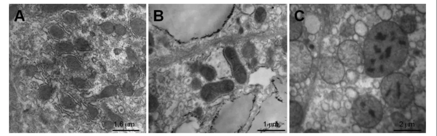
Analysis on the mitochondrial ultrastructure showed that the most frequent morphological abnormalities were megamitochon-dria, with or without crystalline inclusions, and a highly irregular mitochondrial shape, with degenerative changes (decreased elec-tron density and loss of cristae).
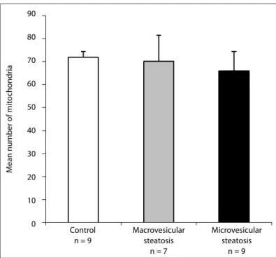
Figure 1. Mean mitochondrial density in the hepatocytes from patients with steatosis, compared with patients in the control group. Analysis of variance (ANOVA) with log transformation.
90
80
70
60
50
40
30
20
10
0
M
ean number of mit
ochondr
ia
Control n = 9
Macrovesicular steatosis
n = 7
Microvesicular steatosis
n = 9
Figure 2. Electron micrographs showing differences in relation to mitochondrial density. A: Hepatocyte from a subject in the control group; B: Hepatocyte from a patient with macrovesicular steatosis; C: Hepatocyte from a patient with microvesicular steatosis.
steatosis group, as demonstrated in Figure 1. Images showing the
diferences in the quantities of mitochondria in the hepatocytes
are presented in Figure 2.
Analysis on the mean mitochondrial surface area showed that there was a signiicant diference between the microvesicu-lar steatosis group and the control and macrovesicumicrovesicu-lar steatosis
groups. hese results are presented in Figure 3 and images are
shown in Figure 4.
DISCUSSION
Since mitochondria are the sites for many metabolic processes
(such as tricarboxylic acid production, fatty acid β-oxidation,
synthesis of urea and other substances, and use of glucose and
fatty acids as fuels for ATP synthesis), a disorder in any of these mechanisms may cause severe damage to the cell and thus to the tissue. Respiratory chain defects are not rare and are known to
cause early liver failure in infancy,21 thereby leading to a change
in fatty acid oxidation.27 Changes to mitochondrial β-oxidation
may result in accumulation of fatty acids in the cytosol, and this
is typically represented by microvesicular steatosis.28,29
In our study, a signiicant elevation in mean mitochondrial surface area was observed, and this proved to be substantial in the patients with predominantly microvesicular steatosis. In con-trast, the mitochondria in the patients with predominantly mac-rovesicular steatosis were considered to be normal in size. hese
results corroborate the reports by Sherlock and Dooley30
indicat-ing that microvesicular steatosis is of greater clinical importance and may progress in a more troubling manner.
Many other reports on the relationship between steatosis and mitochondrial alterations have been published in the literature.
Studies by Sokol and Treem27 have reported that steatosis in
neo-natal liver failure is related to increased mitochondrial density,
with an occasional increase in mitochondrial size. Mandel et al.17
observed mitochondrial abnormalities in the matrix and cris-tae, and also an increase in the total number of mitochondria,
in cases of microvesicular steatosis. Bioulac-Sage et al.18 reported
mitochondrial proliferation (“oncocytic” changes) in patients with microvesicular steatosis.
Figure 4. Electron micrographs showing differences in relation to mean mitochondrial surface area in the hepatocytes. A: Mitochondria in the hepatocytes from a subject in the control group; B: Mitochondria in the hepatocytes from a patient with macrovesicular steatosis; C: Mitochondria in the hepatocytes from a patient with microvesicular steatosis showing considerable dimensions (megamitochondria) and crystalline inclusions.
Figure 3. Mean mitochondrial surface area in the hepatocytes from patients with steatosis compared with patients in the control group. (*) represents a significant difference between the group with microvesicular steatosis, the control group and the group with macrovesicular steatosis. Analysis of variance (ANOVA) with log transformation (P < 0.05).
3.5
3
2.5
2
1.5
1
0.5
0
M
it
ochondr
ial sur
fac
e ar
ea (µm
2)
Control n = 10
Macrovesicular steatosis
n = 7
Microvesicular steatosis
n = 11 In our case study, 64% of the patients with microvesicular
steatosis and 57% of the patients with macrovesicular steatosis showed an elevation in at least one hepatic serum
aminotrans-ferase. Some authors such as Rashid and Roberts15 and Molleston
et al.16 have reported higher rises in AST than in ALT. However,
this relationship is quite typical of alcoholic hepatitis, in which
the AST/ALT ratio is greater than one.2 In our case study, only
39% of the patients with steatosis (of both types) presented ele-vated serum transaminase levels with an AST/ALT ratio greater than one. Elevated serum aminotransferase levels seem to show no direct relationship with either type of steatosis. hus, no rela-tionship with increased mitochondrial volume is found, since the enzyme levels vary widely, both in patients with predominantly microvesicular steatosis and in those with predominantly mac-rovesicular steatosis.
Another indication of compromised mitochondria is the fre-quent observation of megamitochondria, oten showing crys-talline inclusions. Such indings have been correlated with
Wil-son’s disease31 and nonalcoholic steatohepatitis (NASH).32 More
recently, it has been indirectly correlated with oxidative stress in
NAFLD cases.33,34
he mechanisms that induce mitochondrial “hypertrophy” remain obscure. However, changes to the respiratory chain or to other mitochondrial metabolic enzymes, whether inborn or acquired, may lead to an increase in -oxidation and partial stag-nation of oxidative phosphorylation (OxP). We hypothesize that an increase in mitochondrial volume occurs as a consequence. As a result, the volumes of the inner membrane (and cristae) and inner membrane space (sites relating to -oxidation and OxP) increase in an attempt to compensate for metabolic processes that were partially or totally interrupted. Taking into account the
“two-hit” hypothesis proposed by Day and James35 to explain the
patho-genesis of NAFLD, it is important to note that steatosis by itself
does not cause the development of liver disease. However, it is a factor that can sensitize the liver to the damaging efects of a “sec-ond hit.” herefore, factors causing innocuous stress in healthy
livers may lead to the development of NASH in steatotic livers.36
microvesicular steatosis. No similar studies on pediatric groups have been published in the literature, and we would particularly like to highlight that the data on the control group in this study, which has also never previously been published, may be useful as a reference for conducting other electron microscopy studies on pediatric livers.
CONCLUSION
It may be concluded from our results that changes to serum transaminases and the presence of macrovesicular steatosis in this pediatric group did not correlate with changes to mitochon-drial density or size. In addition, increased mitochonmitochon-drial size was probably a morphological expression relating to deranged beta-oxidation and partial stagnation of oxidative phosphoryla-tion. his therefore supports the hypothesis that there may be a mild form of primary mitochondrial hepatopathy, which may then progress in a more worrisome manner, especially as a sec-ond hit.
REFERENCES
1. Browning JD, Horton JD. Molecular mediators of hepatic steatosis
and liver injury. J Clin Invest. 2004;114(2):147-52.
2. Zafrani ES. Non-alcoholic fatty liver disease: an emerging pathological
spectrum. Virchows Arch. 2004;444(1):3-12.
3. Nobili V, Marcellini M, Devito R, et al. NAFLD in children: a prospective
clinical-pathological study and effect of lifestyle advice. Hepatology.
2006;44(2):458-65.
4. Da Silva GH, Coelho KI, Coelho CA, Escanhoela CA. Mitochondrial
alterations in nonalcoholic fatty liver disease. Pediatric case
description of three submitted sequential biopsies. J Gastrointest
Liver Dis. 2009;18(2):215-9.
5. Zhou YJ, Li YY, Nie YQ, et al. Prevalence of fatty liver disease and its
risk factors in the population of South China. World J Gastroenterol.
2007;13(47):6419-24.
6. Imhof A, Kratzer W, Boehm B, et al. Prevalence of non-alcoholic fatty
liver and characteristics in overweight adolescents in the general
population. Eur J Epidemiol. 2007;22(12):889-97.
7. Sagi R, Reif S, Neuman G, et al. Nonalcoholic fatty liver disease in
overweight children and adolescents. Acta Paediatr.
2007;96(8):1209-13.
8. Ciba I, Widhalm K. The association between non-alcoholic fatty liver
disease and insulin resistance in 20 obese children and adolescents.
Acta Paediatr. 2007;96(1):109-12.
9. Radetti G, Kleon W, Stuefer J, Pittschieler K. Non-alcoholic fatty liver
disease in obese children evaluated by magnetic resonance imaging.
Acta Paediatr. 2006;95(7):833-7.
10. Zou CC, Liang L, Hong F, Fu JF, Zhao ZY. Serum adiponectin, resistin
levels and non-alcoholic fatty liver disease in obese children. Endocr
J. 2005;52(5):519-24.
11. Louthan MV, Theriot JA, Zimmerman E, Stutts JT, McClain CJ.
Decreased prevalence of nonalcoholic fatty liver disease in black
obese children. J Pediatr Gastroenterol Nutr. 2005;41(4):426-9.
12. Fishbein M, Mogren J, Mogren C, Cox S, Jennings R. Undetected
hepatomegaly in obese children by primary care physicians: a pitfall
in the diagnosis of pediatric nonalcoholic fatty liver disease. Clin
Pediatr (Phila). 2005;44(2):135-41.
13. Schwimmer JB, Behling C, Newbury R, et al. Histopathology of pediatric
nonalcoholic fatty liver disease. Hepatology. 2005;42(3):641-9.
14. Marion AW, Baker AJ, Dhawan A. Fatty liver disease in children. Arch
Dis Child. 2004;89(7):648-52.
15. Rashid M, Roberts EA. Nonalcoholic steatohepatitis in children. J
Pediatr Gastroenterol Nutr. 2000;30(1):48-53.
16. Molleston JP, White F, Teckman J, Fitzgerald JF. Obese children with
steatohepatitis can develop cirrhosis in childhood. Am J Gastroenterol.
2002;97(9):2460-2.
17. Mandel H, Hartman C, Berkowitz D, et al. The hepatic mitochondrial
DNA depletion syndrome: ultrastructural changes in liver biopsies.
Hepatology. 2001;34(4 Pt 1):776-84.
18. Bioulac-Sage P, Parrot-Roulaud F, Mazat JP, et al. Fatal neonatal liver
failure and mitochondrial cytopathy (oxidative phosphorylation
deficiency): a light and electron microscopic study of the liver.
Hepatology. 1993;18(4):839-46.
19. Ghadially FN. Ultrastuctural pathology of the cell and matrix. 4th ed.
Boston: Butterworth-Heinemann; 1997.
20. Pérez-Carreras M, Del Hoyo P, Martín MA, et al. Defective hepatic
mitochondrial respiratory chain in patients with nonalcoholic
steatohepatitis. Hepatology. 2003;38(4):999-1007.
21. Morris AA. Mitochondrial respiratory chain disorders and the liver.
Liver. 1999;19(5):357-68.
22. Hensley K, Kotake Y, Sang H, et al. Dietary choline restriction causes
complex I dysfunction and increased H(2)O(2) generation in liver
mitochondria. Carcinogenesis. 2000;21(5):983-9.
23. Pessayre D, Mansouri A, Haouzi D, Fromenty B. Hepatotoxicity due to
mitochondrial dysfunction. Cell Biol Toxicol. 1999;15(6):367-73.
24. Oleszczuk A, Spannbauer M, Tannapfel A, et al. Regenerative capacity
differs between micro- and macrovesicular hepatic steatosis. Exp
Toxicol Pathol. 2007;59(3-4):205-13.
25. Reid AE. Nonalcoholic steatohepatitis. Gastroenterology.
2001;121(3):710-23.
26. Sanyal AJ; American Gastroenterological Association. AGA technical
review on nonalcoholic fatty liver disease. Gastroenterology.
2002;123(5):1705-25.
27. Sokol RJ, Treem WR. Mitochondria and childhood liver diseases. J
Pediat Gastroenterol Nut. 1999;28(1):4-16.
28. Natarajan SK, Eapen CE, Pullimood AB, Balasubramanian KA.
Oxidative stress in experimental liver microvesicular steatosis:
role of mitochondria and peroxisomes. J Gastroenterol Hepatol.
2006;21(8):1240-9.
29. Fromenty B, Pessayre D. Inhibition of mitochondrial beta-oxidation as
30. Sherlock S, Dooley J. Nutritional and metabolic liver diseases. In:
Sherlock S, Dooley J eds. Diseases of the liver and biliary system. 11th
ed. Oxford: Blackwell Science; 2002. p. 423-52.
31. Sternlieb I, Berger JE. Optical diffraction studies of crystalline structures
in electron micrographs. II. Crystalline inclusions in mitochondria of
human hepatocytes. J Cell Biol. 1969;43(3):448-55.
32. Caldwell SH, Swerdlow RH, Khan EM, et al. Mitochondrial abnormalities
in non-alcoholic steatohepatitis. J Hepatol. 1999;31(3):430-4.
33. Sanyal AJ, Campbell-Sargent C, Mirshahi F, et al. Nonalcoholic
steatohepatitis: association of insulin resistance and mitochondrial
abnormalities. Gastroenterology. 2001;120(5):1183-92.
34. Le TH, Caldwell SH, Redick JA, et al. The zonal distribution of
megamitochondria with crystalline inclusions in nonalcoholic
steatohepatitis. Hepatology. 2004;39(5):1423-9.
35. Day CP, James OF. Steatohepatitis: a tale of two “hits”? Gastroenterology.
1998;114(4):842-5.
36. Gentile CL, Pagliassotti MJ. The role of fatty acids in the development
and progression of nonalcoholic fatty liver disease. J Nutr Biochem.
2008;19(9):567-76.
Acknowledgements: The authors would like to thank Eduardo Henrique da Silva and Arneth Rodrigues Ribeiro for reviewing the English
translation
Conflict of interest: None
Sources of funding: None
Date of first submission: August 30, 2010
Last received: March 23, 2011
Accepted: March 28, 2011
Address for correspondence:
Gustavo Henrique Silva
Rua Alberto Jackson Brington, 187
Jardim Chapadão — Campinas (SP) — Brasil
CEP 13070-063
Tel. (+55 19) 3241-2137