RESEARCH ARTICLE / ARTIGO
Activity of extracellular compounds of
Pseudomonas
sp.
against
Xanthomonas axonopodis
in vitro
and bacterial leaf
blight in eucalyptus
Lucilene P. Lopes1, Admiltom G. Oliveira Jr.1, Jamile P.O. Beranger1, Cíntia G. Góis1, Fernanda C.S.
Vasconcellos1, Juca A.B. San Martin2, Célia G.T.J. Andrade2, João C.P. Mello3 & Galdino Andrade1
1Departamento de Microbiologia, Universidade Estadual de Londrina, 86051-990, Londrina PR, Brazil; 2Departamento de Biologia Geral, Universidade Estadual de Londrina, 86051-990, Londrina PR, Brazil; 3Departamento de Farmácia e Farmacologia, Universidade Estadual de Maringá, 87020-900, Maringá PR, Brazil
Author for correspondence: Galdino Andrade, e-mail: [email protected]
ABSTRACT
Bacterial leaf blight caused by Xanthomonas axonopodis is a major problem in the production of eucalyptus seedlings. The pathogen causes leaf lesions that reduce the photosynthetic area and limit plant growth. The objective of this study was to evaluate the antibiotic activity of secondary metabolic compounds of a Pseudomonas sp. against X. axonopodis, following extraction with dichloromethane and fractionation by vacuum liquid chromatography. The antibiotic activity of an ethyl acetate phase (F3 fraction) was determined by agar diffusion tests and bioautography. The minimum inhibitory concentration of F3 fraction and the action of F3 on the cell morphology of X. axonopodis was determined by scanning electron microscopy. Electron microscopy revealed changes in exopolysaccharides and morphology of treated cells. When eucalyptus seedlings were sprayed with the F3 fraction before or after inoculation with X. axonopodis in a greenhouse experiment the number of leaf lesions was respectively 93.9% and 89.7% lower than in untreated control plants. High antibiotic activity of the F3 fraction against X. axonopodis and leaf blight suggests that it has potential to control the disease in eucalyptus seedlings.
Key words: Antibiotic activity, antimicrobial, biological control.
INTRODUCTION
Bacterial leaf blight caused by Xanthomonas axonopodis is one of the main foliar diseases that affect
Eucalyptus spp. cultivation (Gonçalves et al., 2008; Alfenas et al., 2009). In the initial stage of the disease, affected plants show water soaked angular lesions concentrated along the main vein, at the leaf edges or scattered on the leaf. When the disease progresses the lesions become necrotic, the photosynthetic area and plant growth are reduced, and the leaves often fall (Gonçalves et al., 2008). Between 2003 and 2008, around 105,500 mini-stumps used for cutting production and 16.5 million infected rooted cuttings were discarded in the Brazilian states of Espírito Santo, Bahia, Goiás, Minas Gerais, and Rio Grande do Sul, with losses of about US $ 10,000,000.00 (Gonçalves et al., 2008).
Management of bacterial leaf blight involves different options that reduce contamination sources and conditions that favor infection. The most common control practices are the use of disease-free seedlings and planting tools free from contamination, and the avoidance of susceptible clones and sprinkler irrigation (Alfenas et al., 2009). Bacterial leaf blight is often severe on seedlings irrigated by means of sprinklers because water films formed
on the leaves facilitate the dissemination, multiplication and host penetration of the pathogen (Beattie & Lindow, 1999; Hirano & Upper, 1983).
Pesticide intervention in eucalyptus cultivation is uncommon compared with other agricultural crops. Hence, few herbicides, insecticides, fungicides and bactericides are registered for eucalyptus (Alfenas et al., 2009). The search for new management options is necessary since the current forms of prevention do not adequately reduce the risk of infection and, as a result, millions of seedlings are discarded annually because of high disease index (Alfenas & Ferreira, 2008). Selection of an alternative option to chemical pesticides for control of bacterial leaf blight is recommended to ensure biological balance and maintain biodiversity in forest areas.
The objective of this study was to evaluate the effects of the dichloromethane phase (DP) fraction produced by Pseudomonas sp. (strain LV) , and of other F fractions obtained from fractionated DP, by vacuum liquid chromatography (VLC), on the severity of bacterial leaf blight of eucalyptus, and on the ultrastructure of X.
axonopodis cells as determined by scanning electron microscopy (SEM).
MATERIALS AND METHODS
Bacterial strains
The pathogen X. axonopodis, strain BSC475a (GenBank, EF101967), was stored in 30% glycerol (v:v) at -20°C and renewed every six months from cells produced on nutrient agar (NA) at 28°C for 48 h. The antagonistic bacterium Pseudomonas sp. LV strain was isolated from an old citrus canker lesion on an orange leaf (Citrus sinensis
cv. Valence) that was collected in Astorga, PR, Brazil [23°13’29.11” S ; 51°39’47.20” W] (Rampazo, 2004). The procedure of storage and cell maintenance was the same as described for X. axonopodis, except that the culture medium was nutrient agar (NA) plus copper chloride (100 mg L-1) as described by Cain et al. (2000).
Production and separation of dichloromethane phase (DP)
The method of production has been patented (Patent #PI0803350-1; www.inpi.gov.br). The initial inoculum of the LV strain was obtained from a culture stored in glycerol and grown as described above. 150 μL of a suspension of log phase cells (108 CFUmL-1, O.D 0.09, λ = 590 nm) were inoculated in 1.5 L of nutrient broth (NB) plus 100 mg.L-1 CuCl2.2H2O and maintained on a shaker operated at 100 rpm for 15 d at 28ºC. Subsequently, the culture was centrifuged (20 min at 4ºC; 9,000 rpm), and 500 mL aliquots of the cell-free supernatant were treated with dichloromethane 1:1 (v:v) in a separation funnel, shaken in an orbital motion for 30 s and left to rest for 15 min; the process was repeated 10 times per aliquot. The dichloromethane phase obtained was concentrated in a rotary evaporator (R 215, Büchi®) under
vacuum system of 50 kpa at 45°C. The sample was then frozen in liquid nitrogen and lyophilised for 24 h. The final amount obtained was approximately 0.5 g of DP per 15 L of supernatant.
Purification of the DP by vacuum liquid chromatography
(VLC)
VLC was carried out in a glass column (20 mm diameter x 350 mm H) filled with 30 g of silica gel 60 (0.063-0.200 mm, Merck) coupled to a vacuum pump with 51 kPa. The DP (2 g mixed with 5 g of silica gel 60) was fractionated using the following mobile phase (v/v): hexane (100; F1), dichloromethane (100; F2), ethyl acetate (100; F3), methanol (100; F4), methanol: water (1:1; F5) and water (100; F6). Forty mL of each organic solvent was
passed through the column 10 times, concentrated in the rotary evaporator under vacuum at 45ºC, frozen in liquid nitrogen and lyophilized for 24 h.
Thin-layer chromatography (TLC)
Using TLC plates of silica gel on aluminum support 60 F254 (Merck) the TLC was performed to assess qualitatively the components of the DP and other fractions obtained by separation processes of VLC. The system eluent (mobile phase) was a mixture of petroleum ether / dichloromethane / ethyl acetate / methanol. Ultraviolet light (254 nm and 366 nm) and TLC bioautography were used for developing the TLC plates. The migration velocity of the mixture was characterized by numerical value Rf, which expresses the relationship between the distance traveled by the compound from the origin (∆x) and the solvent front from origin (∆y).
Evaluation of antibiotic activity TLC bioautography method
The TLC bioautography test (Rahalison et al., 1991) was performed to determine qualitatively the antibiotic activity of all fractions. The TLC plates were each placed in a Petri dish 90 mm in diameter containing 20 mL of NA plus 1 mL cell suspension of X. axonopodis (108 CFU mL–1, D.O 0.1, λ= 590 nm) in the log phase. The plates were incubated at 28°C for 24 h and were read by measuring the diameter of inhibition zones (mm) as revealed by means of a 1% solution 2,3,5-triphenyl-1H-tetrazolium chloride (TTC).
Agar diffusion method
The experimental design was in randomized blocks with seven fractions × one concentration (1000 μg) × two replicates (7 x 1 x 2, n = 14). The antibiotic effect of fractions was investigated by agar diffusion in Petri dishes 90 mm with 20 mL of NA plus 1 mL cell suspensions of
X. axonopodis (108 CFU mL-1) in the log phase, by pour plate method. Five μL aliquots of each fraction were added to antibiotic disks of filter paper 6 mm of diameter, placed on the plates and incubated at 28°C for 24 h. Dried disks plus solvent used in each fraction were also tested to examine the influence of solvent residues on the growth of X. axonopodis.. Plates inoculated with X. axonopodis
were also incubated to assess cell viability. Readings were performed by measuring the diameter of inhibition zones (mm) and calculating the average for each fraction.
Determination of minimum inhibitory concentration (MIC)
The experimental design was in block with six concentrations of F3 fraction (3.12; 6.25; 12.5; 25; 50 and 100μg mL-1) × four repetitions (6 x 4, n = 24). The MIC test was carried out in cell culture plates each with twenty four wells. The non-inoculated NB and cell suspensions of
0.9 mL of NB plus 50 µL of suspensions of X. axonopodis
and 50 µL of the respective concentration of F3 fraction. After incubation at 28°C for 24 h 10 µL of a 1% solution of 2,3,5-triphenyl-1H-tetrazolium chloride (TTC) were added to each well and the plates were incubated at 28°C for 20 min. The wells were then assessed for the presence or absence of pink color, which respectively indicated that the cells were viable or not viable. After the MIC determination, cell suspensions of
X.axonopodis (108 CFU mL-1), with and without the F3 fraction (100 μg mL-1), were inoculated in NB. After 0, 0.5,1, 2, 4, 6, 8, 10, 12, 14, 16 and 20 h of incubation at 28°C, triplicate ten-µL aliquots of the inoculated NB were plated on NA medium, incubated at 28°C for 24 h, and the CFU of each time was calculated to determine the bactericidal or bacteriostatic effect of the different concentrations of F3.
Evaluation of the action of the F3 fraction on the control of bacterial leaf blight in seedlings of eucalyptus
The seedlings of E. urograndis 1404 (hybrid of
E. urophylla and E. grandis) used in the experiment, are characterized by high susceptibility to bacterial leaf blight caused by X. axonopodis. The seedlings were grown in plastic bags each containing 2 L of non-sterile soil (Rhodic Ferrasol; FAO, 1994) and sand (1:1, v:v) in a greenhouse (28°C/22°C and 10 h/14 h day/night, relative humidity of 80%) and watered as needed with tap water. The experiment was performed with 90-day-old seedlings each with 15 leaves, in a completely random block design comprised of two application times of the F3 fraction (pre- and post-application in relation to bacterial inoculation) at 1000 µg mL-1, and 5 replicates each. Seedlings were sprayed with sterile distilled water as the negative control and with a suspension of X. axonopodis (108 CFU mL-1) as the positive control. Before inoculation, the seedlings were watered and covered for 24 h with a polyethylene plastic bag as a moisture chamber. When treated before inoculation, each plant was sprayed with eight mL of F3 fraction, incubated for 24 h, inoculated with a suspension of log phase
X. axonopodis (108 CFU mL–1) and incubated in a moisture chamber for 24 h. When treated after inoculation, the plants were inoculated with the cell suspension of X. axonopodis
and sprayed with the F3 fraction 24 h later. The methods of spraying and maintenance in a moisture chamber were the same as described in the pre-treatment. Lesions were counted on each plant at 15, 20 and 30 days after inoculation. The data were statistically analyzed by the Tukey test (p < 0.05).
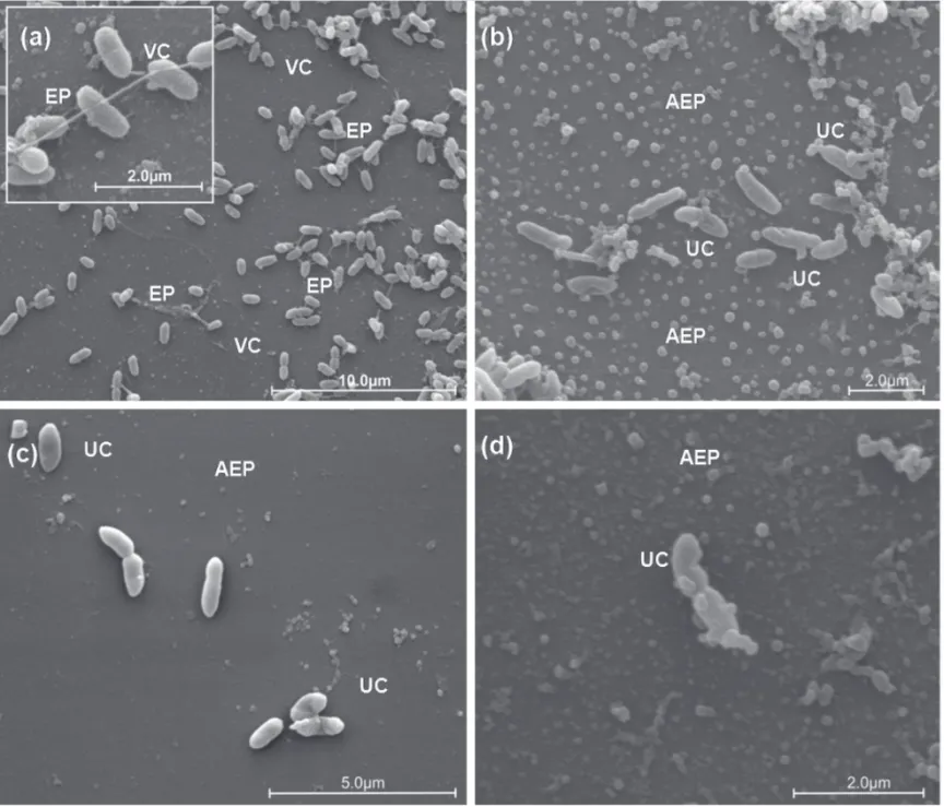
Scanning electron microscope (SEM) study
Suspensions of log-phase cells of X. axonopodis
(108 CFU mL-1) grown in tubes with NB (24 h, 28°C; 100 rpm) were used for the SEM study. Aliquots of 10 mL of
X. axonopodis cultured as described above were sampled from twelve tubes containing 200 μg mL-1 of F3 fraction and from five control tubes without F3, and were incubated at 6 h, 28°C; 100 rpm. After 1, 3 and 6 h, four aliquots of 20 μL of all tubes were collected and spotted onto glass
slides previously coated with a thin layer of polylysine. Afterwards, each slide with X. axonopodis was fixed by immersion in 1 mL of 2.5% glutaraldehyde, 2% paraformaldehyde in 0.1 M sodium cacodylate buffer (pH 7.2) solution for 12 h, following a post-fixation in OsO4 1% for 2 h. The fixed material was dehydrated in an ethanol gradient (70, 80, 90 and 100ºGL), and the sample critical point dried in CO2 (BALTEC CPD 030 Critical Point Dryer). Finally, the slides were taped onto stubs, coated with gold (BALTEC SDC 050 Sputter Coater) and observed under a FEI Quanta 200 scanning electron microscope.
RESULTS
TLC bioautography method
TLC bioautography showed a band of antibiotic activity in the DP fraction and in the F3 fraction of DP, with each band exhibiting the same Rf value (0.66). No band of antibiotic activity against X. axonopodis was found for the other DP fractions.
Agar diffusion method
In the evaluation of antibiotic action by agar diffusion, inhibition zones against X. axonopodis with average diameters of 23.5 and 30.5 mm were observed for the DP and F3 fractions, respectively. No inhibition zones were found for the F1, F2, F4, F5 and F6 fractions (Table 1).
Determination of minimum inhibitory concentration (MIC)
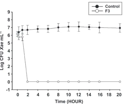
The MIC of F3 fraction against X. axonopodis was of 100 μg mL-1. The cell suspension of X. axonopodis when treated with MIC of F3 fraction significantly decreased the number of CFU after 1 h of incubation. No colonies of the pathogen were found after 2 h of incubation (Figure 1).
Evaluation of the action of the F3 faction on development of bacterial leaf blight in seedlings of eucalyptus
No lesions appeared in the negative control plants that were untreated and not inoculated. On the other hand, increasing numbers of lesions were found in the positive control (untreated and inoculated with X. axonopodis) with an average of 9.4, 32.0 and 75.4 after 15, 20 and 30 days of inoculation, respectively. In the pre and post-inoculation treatments the numbers of lesions were significantly lower than in the positive control. However, no significant differences were found between the times of application in any of the assessments (Figure 2). Application of the F3 fraction 24 h before and 24 h after inoculation, respectively, reduced the number of lesions at 30 days by 93.9% and 89.7% compared the positive control.
Scanning electron microscope (SEM) study
Diameter of inhibition halo in mm
Fractions PD F1 F2 F3 F4 F5 F6 NC
Average 23.5±0.7 - - 30.5±2.1 - - -
-TABLE 1 - Evaluation of antibiotic activity of phase dichloromethane (PD) and its purified fractions by liquid chromatography vacuum (CLV) using six mobile phases with increasing polarity (F1 100% hexane; F2 100% dichloromethane; F3 100 % ethyl acetate; F4 100% methanol; F5 methanol / water (1:1, v / v) and F6 100% water) against Xanthomonas axonopodis
(-) no inhibition zone; NC negative control.
(EPS) (Figure 3A). After 1 h of treatment with F3 fraction, the cells of X. axonopodis showed morphological changes in cell walls and absence of exopolysaccharide (Figure 3B). After 3 h of treatment, the cell walls were severely altered and cell lysis was frequent (Figures 3C and 3D).
DISCUSSION
Many authors reported the use of antagonistic microorganisms such as fungi, rhizobacteria and especially bacteria of genus Bacillus and Pseudomonas as biocontrol agents for plant diseases (Shoda, 2000; Byrne et al., 2005; Lemessa & Zeller, 2007; Mafia, 2009; Todorova & Kozhuharova, 2010). However, there are few reports about the use of microorganisms or secondary metabolic compounds in the control of X. axonopodis ofeucalyptus. Despite of the importance of bacterial leaf blight caused by X. axonopodis and the damage caused by high levels of this disease, information on the infection process and its development in eucalyptus is scanty (Alfenas & Ferreira, 2008; Alfenas et al., 2009).
Among the DP fractions, only the F3 fraction showed antibiotic activity against X. axonopodis. The antibiotic effect actually increased for F3 in comparison to DP. This effect could be due to the purification process which concentrates the active molecule(s). Moreover, F3 showed high solubility in ethyl acetate solvent.
The F3 fraction did not exhibit any phytotoxic effect and was effective in controlling bacterial leaf blight in eucalyptus plants in the greenhouse experiments, in which it significantly reduced the number of leaf lesions whether applied before or after inoculation. These findings are comparable with results obtained by Oliveira et al. (2011) who observed that the same F3 fraction also had high antibiotic activity against X. axonopodis pv. citri
(Xcc), decreasing by 93.5% the number of lesions of citrus canker in orange leaves. Vasconcellos (2009) reported a reduction of 80% in the number of leaf lesions caused by
Xanthomonasarboricola pv. pruni (Xap) in peach seedlings by using same F3 fraction. The F3 fraction was as effective in controlling X. axonopodis in eucalyptus as in the control of Xcc and Xap, indicating that the bacteria of the genus
Xanthomonas have high sensitivity to fraction F3.
Similar results were found by Ji et al. (2008), who observed that strains of Lysobacter antibioticus isolated from rhizosphere of rice were effective in controlling 9
8
7
6
5
4
3
2
1
0
-1
0 2 4 6 8 10 12 14 16 18 20
Time (HOUR)
Control
Log CFU
mL
Xae
-1
F3
FIGURE 1 - Growth curve of Xanthomonas axonopodis treated and not treated with F3 (ethyl acetate phase 100%) at 100 ug mL-1,
obtained from phase dichloromethane (PD) by vacuum liquid chromatography (CLV).
100
80
60
40
20
0
15 20 30
Time (DAYS)
No Lesions Plant
-1
Control
Pre-treatment
Post-treatment
FIGURE 2 - Evaluation of antibiotic activity of F3 (ethyl acetate phase 100%) at 1000 mg ml-1, obtained from phase dichloromethane
FIGURE 3 - Scanning electron microscopy of cells of Xanthomonasaxonopodis untreated and treated with fraction F3 (ethyl acetate phase 100%) at 200 mg mL-1, obtained from phase dichloromethane (PD) by vacuum liquid chromatography (CLV). (A) control cells, untreated
(5,000 x; 20,000 x), (B) Cells of X. axonopodis after 1hour of treatment (10,000 x), (C) Cells of X. axonopodis after 3 hours of treatment (10,000 x), (D) cells of X. axonopodis after 6 hours of treatment (20,000 x). Viable cells (VC), nonviable cells (UC), exopolysaccharides (EP), absence of exopolysaccharide (AEP).
This absence could be related to degradation of EPS or to reduced biosynthesis of EPS. However, further studies are needed to understand these observations.
Considering the importance of EPS in the bacterial infection process in plants, it is possible to relate the observed absence of EPS with the reduced lesion formation by
X. axonopodis in plants treated with F3 fraction. Dharmapuri & Sonti (1999) reported that the production of EPS is an important virulence factor in the infection process of Xoo in rice. Kemp et al. (2004) reported that mutant strains of X. axonopodis pv. manihotis with deficiency in the biosynthesis of EPS, when infiltrated into the plant, were unable to multiply and cause the typical symptoms of bacterial blight in cassava.
Xanthomonas oryzae pv. oryzae (Xoo), which causes bacterial blight in rice. Plants inoculated with supernatant from a cell culture of L. antibioticus had reduced disease incidence when compared with the control plants inoculated with water. In agar diffusion experiments, Ji et al. (2008) reported that five fractions, obtained by supernatant extraction with ethyl acetate and fractionated by TLC, also showed inhibitory zones against Xoo. The compounds with antibiotic activity obtained from L. antibioticus were produced during the cultivation of antagonist cells and exhibited solubility in ethyl acetate.
Besides the absence of EPS, SEM revealed severe morphological changes in the cell walls. Although the mechanism of F3 fraction action is not yet established, these results suggest that the deformities in the cell wall can have caused cell lyses. Zhang et al. (2008) reported similar morphological changes in the cell wall of gram-negative
Xoo and Escherichia coli, and gram-positive Micrococus subtilis and Bacillus subtilis treated with supernatant of Hpa2 proteins cloned from Xoo. Further, the Hpa2 protein has lytic activity causing deformities in the wall and leading to the leakage of cellular contents.
The results of the present study suggest that the F3 fraction obtained by VLC from PD, produced by
Pseudomonas sp. may be a new alternative for the control of X. axonopodis ineucalyptus. However, further studies should be performed to purify, determine and effectively identify the molecules involved in antibiotic activity.
ACKNOWLEDGEMENTS
The Conselho Nacional de Desenvolvimento Científico e Tecnológico - CNPq is acknowledged for providing research grants and scholarships to the authors. The company Klabin – Telêmaco Borba, state of Paraná – is acknowledged for providing the authors with seedlings to be used in the experiments.
REFERENCES
Alfenas AC, Ferreire EM (2008) Emerging diseases in eucalyptus plantations. Fitopatologia Brasileira 33:25-26.
Alfenas AC, Zauza EA, Mafia RG, Assis TF (2009) Clonagem e doenças do eucalipto. 2ª Ed.Viçosa MG. Editora UFV.
Beattie GA, Lindow SE (1999) Bacterial colonization of leaves: A spectrum of strategies. Phytopathology89:353-359.
Byrne JM, Dianese AC, Jia P, Campbell HL, Cuppels DA, Louws FJ, Miller SA, Jones JB, Wilson M (2005) Biological control of bacterial spot of tomato under field conditions at several locations in North America. Biological Control 32:408-418.
Cain CC, Henry AT, Waldo RH, Casida LJ, Falkinham JO (2000) Identification and characteristics of a novel Burkholderia
strain with broad spectrum antimicrobial activity. Applied and Environmental Microbiology 66:4139-4141.
Dharmapuri S, Sonti RV (1999) A transposon insertion in the gumG homologue of Xanthomonas oryzae pv. oryzae causes loss of extracellular polysaccharide production and virulence. FEMS Microbiology Letters 179:53-59.
Gonçalves RC, Lau D, Oliveira JR, Maffia LA, Cascardo JCM, Alfenas AC (2008) Etiology of bacterial leaf blight of eucalyptus in Brazil. Tropical Plant Pathology 33:180-188.
Harman GE (2000) Myths and dogmas of biocontrol changes in perceptions derived from research on Trichoderma harzianum
T-22. Plant Disease 84:377-393.
Hirano SS, Upper CD (1983) Ecology and epidemiology of foliar bacterial plant pathogens. Annual Review of Phytopathology 21:243-270.
Ji GH, Wei LF, He YQ, Wu YP, Bai XH (2008) Biological control of rice bacterial blight by Lysobacter antibioticus strain 13-1. Biological Control 45:288-296.
Kemp BP, Horne J, Bryant A, Cooper RM (2004) Xanthomonas axonopodis pv. manihotis gum D gene is essential for EPS production and pathogenicity and enhances epiphytic survival on cassava (Manihot esculenta). Physiological and Molecular Plant Pathology 64:209-218.
Lemessa F, Zeller W (2007) Screening rhizobacteria for biological control of Ralstonia solanacearum in Ethiopia. Biological Control 42:336-344.
Mafia RG, Alfenas AC, Maffia LA, Ferreira EM, Binoti DHB, Mafia GMV (2009) Plant growth promoting rhizobacteria as agents in the biocontrol of eucalyptus mini-cutting rot. Tropical Plant Pathology 34:10-17.
Oliveira AG, Murate LS, Spago FR, Lopes LP, Beranger JPO, San Martin JAB, Nogueira MA, Andrade CGTJ, Mello JCP, Andrade G (2011) Evaluation of the antibiotic activity of extracellular compounds produced by the Pseudomonas strain against the Xanthomonas citri
pv. citri 306 strain. Biological Control 56:125-131.
Rahalison L, Hamburger M, Hostettmann K, Monod M, Frenk E (1991) Bioautographic agar overlay method for the detection of antifungal compounds from higher plants. Phytochemical Analysis
2:199-203.
Ran LX, Li ZN, Wu GJ, Loon LC, Bakker PAHM (2005) Induction of systemic resistance against bacterial wilt in Eucalyptus urophylla by fluorescent Pseudomonas spp. European Journal of Plant Pathology 113:59-70.
Rampazo LGL (2004) Avaliação de agentes biológicos e seus produtos na incidência de lesões foliares do cancro cítrico. Dissertação MS. Universidade Estadual de Londrina. Londrina PR.
Shoda M (2000) Bacterial control of plant diseases. Journal of Bioscience and Bioengineering 89:515-521.
Todorova S, Kozhuharova L (2010) Characteristics and antimicrobial activity of Bacillus subtilis strains isolated from soil. World Journal of Microbiology and Biotechnology 26:1207-1216.
Vasconcellos FCS (2009) Ação antibiótica de metabólitos de
Pseudomonas sp. no controle da Xanthomonas arboricola pv.
pruni. Dissertação MS. Universidade Estadual de Londrina.
Londrina PR.
Zhang J, Wang X, Zhang Y, Zhang G, Wang J (2008) A conserved Hpa2 protein has lytic activity against the bacterial cell wall in phytopathogenic Xanthomonas oryzae. Applied Microbiology and Biotechnology 79:605-616.