1. Department of Rheumatology & Clinical Immunology, University Medical Center Utrecht, The Netherlands
2. Clínica Universitária de Reumatologia. Faculty of Medicine. University of Coimbra. Portugal
Hypermobility syndromes from
the clinician’s perspective: an overview
ACTA REUMATOL PORT. 2014:39;124-136
abstract
Symptomatic generalized hypermobility is a frequent occurring condition among patients referred to the rheumatologist or other medical specialist. In a subset of patients, a further classifying diagnosis of a specific syndrome can (and should) be made, based on pattern recognition and knowledge of the spectrum of hyper-mobility syndromes. Diagnostic clues are the patient’s and family history and signs at physical examination, including skin abnormalities. It is especially important to recognize hypermobility syndromes with potential-ly life threatening complications.
Genetic testing is only available for some syndromes; is only indicated if there is a reasonable pretest proba-bility regarding a specific syndrome, especially if this syndrome can have life-threatening complications.
The therapy is for the major part of syndromes only symptomatic; key features of management are educa-tion and physical exercises; joint surgery is to be avoided.
Keywords: Hypermobility; Ehlers-Danlos; Marfan;
Loeys-Dietz; Deconditioning
introduction
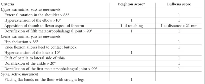
Hypermobility is defined as an increased range of mo-vement in one or more joints. When many joints are hy-permobile in an individual, we speak of generalized hypermobility, a relatively common condition. There are several classification criteria and scores sets for this condition - those of Beigthon and Bulbena are
presen-Johannes WG Jacobs1, José António P. da Silva2
ted in Table I1,2.
Individuals with generalized hypermobility may be asymptomatic. Often, however, joint hypermobility is accompanied by musculoskeletal symptoms such as pain in joints, ligaments, entheses (attachment sites of tendons or ligaments to the bone) and muscles - this is usually referred to as symptomatic hypermobility. In addition, joint hypermobility can be associated with a wider spectrum of symptoms and signs reflecting in-volvement of other organs and organ systems, attribu-table to defective and lax connective tissues. In such cases the term “hypermobility syndrome” is appro-priate.
Most individuals with generalized hypermobility do not stand out clinically and do not meet criteria of a specific syndrome. Others with generalized hypermo-bility may meet criteria of a specific defined syndrome, some of which may have life-threatening complica-tions, making it important not to overlook these diag-noses.
The aim of this paper is to provide the rheumatolo-gist and other medical specialists an overview of the broad spectrum of symptomatic hypermobility syn-dromes they may be confronted with, as a guidance in daily practice. Clinical aspects, with emphasis on mus-culoskeletal problems, are described and clues for diag-nosis and management are provided.
EpidEmiological aspEcts
Generalized hypermobility may actually be a variant of normal mobility, along the upper tail of the Gaussian curve describing the range of motion of normal joints in the population, just as individuals can be (very) short or tall. In general, joint mobility and, thus, hypermobility, decreases with increasing age, is more common in fe-males than fe-males and occurs more often in some racial groups, such as Asian when compared to Caucasians3.
Many young individuals, especially among those performing ballet or gymnastics, would probably meet the criteria of generalized hypermobility if they were screened. In some sports, hypermobility may be an advantage, but it may also be a liability due to the in-creased risk of injuries4,5. Reassuringly, a study in pro-fessional dancers concluded that joint hypermobility was not associated with a higher risk of injuries when assessed prospectively6. However, this observation does not rule out an increased risk of injury associa-ted with hypermobility in the (less trained) general population. In fact, those more prone to injuries may ha -ve fallen short of a desired professional sports career.
People presenting for medical care with sympto-matic hypermobility are most probably a subgroup of those with hypermobility in the population. Further-more, not all cases of this subgroup will meet criteria for generalized hypermobility. An even smaller num-ber will meet criteria for a specific hypermobility syn-drome, such as benign joint hypermobility syndrome (BJHS; for criteria see Table II) or Ehlers Danlos syn-drome (EDS), a group of hypermobility synsyn-dromes with as key features generalized hypermobility and lax skin (Table III). A description of all EDS-types is beyond the scope of this paper.
Exact prevalence estimates for EDS and BJHS are difficult to make for two main reasons. First, there is
the problem of recognition: not all individuals with ge-neralized hypermobility have relevant symptoms and reach medical attention. If joint hypermobility and skin manifestations are mild, they might stay unrecognized. As a consequence, estimates on the prevalence of EDS types and BJHS depend on whether they are based on clinical reports of the syndromes, or on screening stu-dies in populations. In the latter case, the prevalence es-timates will be probably more accurate and higher. A study in a general dermatology population revealed that mild variants of EDS were present in 9% of this population7. Second, there is the problem of current classifications: an almost complete overlap exists bet-ween the signs, symptoms and classification criteria of EDS hypermobility type and those of the more fre-quently occurring BJHS (Table IV)8 and both show a similar familial hereditary pattern.
The prevalence of BJHS is estimated to range from 10–30% in adults, and from 10-15% in male youngs-ters between 11–17 years and up to 20–40% in girls of this age group9. The prevalence of EDS, of which the hypermobility type is the most frequent, is most pro-bably higher than the current estimate of 1:5,00010. Marfan syndrome is an autosomal dominant heredita-ry connective tissue disorder with a prevalence of about 1:5000; 25% of the cases seems to be caused by a new mutation11,12.
tablE i. bEigthon and bulbEna critEria and scorEs to assEss gEnEralizEd hypErmobility
Criteria Beighton score* Bulbena score
Upper extremities, passive movements
External rotation in the shoulder > 85° 1 Hyperextension of the elbow >10° 1 1
Apposition of thumb to flexor aspect of forearm 1, if touching 1 at distance < 21 mm Dorsiflexion of fifth metacarpophalangeal joint > 90° 1 1
Lower extremities, passive movements
Hip abduction > 85° 1 Knee flexion allows heel to contact buttock 1 Hyperextension of the knee > 10° 1
Shift of patella to lateral side of tibia 1 Dorsiflexion of the ankle > 20° 1 Dorsiflexion of the first metatarsophalangeal joint > 90° 1
Spine, active movement
Placing flat hands on the floor with straight legs 1
Beighton score: range 0-9, *each criterion scores 1 point for each side of the body, if present, with exception of placing flat hands on the floor; generalized hypermobility if total score ≥ 5.
Bulbena score: each criterion scores 1 point if bilaterally present; range 0-9; generalized hypermobility if total score ≥ 5 in males and ≥ 6 in females.
BJHS is diagnosed in the presence of either two major criteria, one major and two minor criteria, or four minor criteria. Two minor criteria will suffice where there is an unequivocally affected first-degree relative. BJHS is excluded by presence of Marfan syndrome or Ehlers–Danlos syndrome (other than the Ehlers–Danlos syndrome hypermobility type). Criteria major 1 and minor 1 are mutually exclusive, as are major 2 and minor 2.
*Other, neurophysiological signs include impairment of joint proprioception, lack of efficacy of local anesthetics, and autonomic dysfunction2,3.
tablE ii. thE rEvisEd brighton 1998 critEria for bEnign joint hypErmobility syndromE (bjhs)1
Major criteria
1. A Beighton score ≥ 4/9 (either currently or historically) 2. Arthralgia for >3 months in four or more joints
Minor criteria*
1. A Beighton score of 1–3/9 (0–3 if aged ≥ 50 year)
2. Arthralgia (≥ 3 month) in one to three joints, or back pain (≥ 3 month), or spondylosis, spondylolysis/spondylolisthesis 3. Dislocation/subluxation in more than one joint or in one joint on more than one occasion
4. Three or more soft tissue lesions (e.g. epicondylitis, tenosynovitis, bursitis)
5. Marfanoid habitus (tall, slim, arm span to total height ratio >1.03; upper segment to lower segment ratio <0.89, arachnodactily (+Steinberg/wrist signs))
6. Abnormal skin: striae or hyperextensibility, thin cutis, or papyraceous scarring 7. Eye signs: drooping eyelids or myopia, or antimongoloid slant
8. Varicose veins or hernia or uterine/rectal prolapse
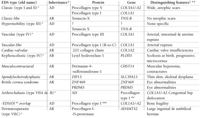
tablE iii. ghEnt updatEd classification -according to gEnEtic rEsEarch of EhlErs–danlos syndromE (Eds)4
EDS type (old name) Inheritance* Protein Gene Distinguishing features5**
Classic (type I and II)# AD Procollagen type V COL5A1/-A2 Wide, atrophic scars
Procollagen type I COL1A1
Classic-like AR Tenascin-X TNX-B No atrophic scars
Hypermobility (type III)# AD ? ? None specific
Tenascin X TNX-B
Vascular (type IV)# AD Procollagen type III COL3A1 Arterial, intestinal & uterine
rupture
Vascular-like AD Procollagen type I (R-to-C) COL1A1 Arterial rupture
Cardiac-valvular AR 2(I) collagen chain COL1A2 Cardiac valve insufficiencies Kyphoscoliotic (type IV)# AR Lysyl hydroxylase-1 PLOD1 Scoliosis at birth, progressive;
microcornea
Musculocontractural AR Dermatan-4- CHST14 Muscular hypotonia,
-sulfotransferase-1 contractures
Spondylocheirodysplastic AR ZIP13 SLC39A13 Thin skin, skeletal dysplasia
Brittle cornea syndrome AR ZNF469 ZNF469 Eye abnormalities
PRDM5 PRDM5 Eye abnormalities
Arthrochalasis (type VIIA & -B)# AD Procollagen COL1A1/-A2 Congenital hip
type I ### dislocation -EDS/OI ##overlap AD Procollagen type I #### COL1A1/-A2 Bone fragility
Dermatosparaxis AR Procollagen-I- ADAMTS2 Large inguinal & umbilical (type VIIC)# -N-proteinase hernias
*AD, autosomal dominant; AR, autosomal recessive; ** all types characterized more or less by generalized hypermobility and lax skin; distinguishing features vary between patients;
#types of Villefranche classification (old term); other types: updated; ##OI: osteogenesis imperfecta; ###deletion of N-propeptide cleavage site; ####delay in N-propeptide cleavage
gEnEral symptoms, signs and complications of hypErmobility syndromEs
Patients with symptomatic hypermobility syndromes may share many clinical characteristics, typically in-cluding chronic musculoskeletal pain, fatigue, signs of autonomic dysfunction, and joint (sub)luxation. Pain and fatigue are the dominant symptoms. In the follow -ing section we discuss the most prevalent symptoms and signs the clinician is confronted with and their as-sociations
pain
There is an increased prevalence of arthralgia (joint pain), chronic generalized myalgia and fibromyalgia (chronic generalized pain in muscles and joints) in hy-permobile children and adults3,8,13-19. In adults, hyper-mobility is also associated with back pain17,20. Perhaps surprisingly, the basis of the association of hypermo-bility and pain is not established. The most consen-sual view is that pain is due to repetitive strain, sprain and microtraumata of muscles and ligaments by the abnormal range-of-motion permitted by hypermobile joints, aggravated by diminished joint proprioception, position sense21-23, and decreased passive muscle ten-sion24. Pain is also related with anxiety and depression, both of which seem to be more prevalent in EDS as well as in BJHS25-27. It is commonly believed that chro-nic pain elicits depressive feelings and that depression has an amplifying effect on chronic pain and fatigue, leading to a vicious circle. These latter symptoms could lead to reduced physical activity and thus physical
de-conditioning (reduced physical condition), aggravating liability to injury, chronic pain and fatigue and initia-ting a downward negative spiral.
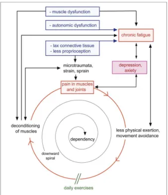
Furthermore, there seems to be a primary involve-ment of muscle in EDS hypermobility type: muscle weakness has been found in the absence of reduced muscles mass, which would have been present if mus-cle weakness was the results of reduced physical activi-ty only28. A hypothetic model on pain, including fatigue and physical deconditioning is depicted in Figure 1.
I
NSUFFICIENT EFFECT OF LOCAL ANALGESICSAn insufficient effect of local analgesics either by intra-dermal injection or as topical cream application has been reported in patients with EDS hypermobility type. This was thought to be due to the lax connective tissues in the skin allowing too much dispersal of the analge-sic29,30. However, the dispersal of a radioisotope labeled solution following deep dermal injection did not differ between EDS patients and healthy controls in a small study31. So, the reason for the insufficient effect of lo-cal analgesics is not yet known.
It was suggested that this phenomenon could be used as a diagnostic test to discriminate between EDS hypermobility type and BJHS30, but further study is warranted.
fatiguE
Hypermobility syndromes are associated with increa-sed fatigue32-34. A hypothesis is that fatigue is a symp-tom of autonomic dysfunction or dysautonomia, des-cribed below33,34. Others suggest that the hyperlaxity of joint ligaments demands increased vigilance,
mus-tablE iv. similarity of villEfranchE critEria for Eds hypErmobility typE and brighton critEria for bjhs*
EDS hypermobility type BJHS
1997 Villefranche criteria6 1998 Brighton criteria1
Major criteria Generalized joint hypermobility (score 5/9) Generalized joint hypermobility: Beighton score of 4/9 or greater (either currently or historically) Skin involvement (hyperextensibility and/or Arthralgia for longer than 3 months in 4 or smooth velvety skin) more joints
Minor criteria Recurring joint dislocations Dislocation/subluxation in > 1 joint, or in 1 joint > 1 time
Chronic joint/limb pain Abnormal skin: striae, hyperextensibility, or thin skin, or papyraceous scarring
* General joint hypermobility is a major criterion for both sets; (sub)luxations an identical minor criterion. The other signs and symptoms appear as major criteria in one set and as minor sign in the other
cle tension and coordination to maintain adequate joint position and body balance23,35,36 thus leading to fatigue. In addition, these patients often show muscle weakness28, reduced exercise tolerance, physical de-conditioning and pulmonary symptoms (see below)37 commonly attributed to reduced exercise because of chronic pain (Figure 1). Finally, fatigue could be asso-ciated with depression and other psychological pro-blems associated with generalized hypermobility34,38. Most probably, fatigue is caused by a constellation of these factors, of which the individual impact will vary between patients.
Fatigue and generalized pain are paramount among the symptoms that establish a clinical similarity and diagnostic confusion between hypermobility syndro-mes and fibromyalgia13-16,39. Some authors even argue whether fibromyalgia is merely a description of the symptoms of EDS or BMJS or a separate disease entity in these patients13-16.
autonomic dysfunction
Autonomic dysfunction comprises disturbances in a variety of functions dependent on the autonomic ner-vous system, leading for instance to postural hypo-tension and tachycardia, (pre)syncope and palpita-tions. Such symptoms have been reported in higher than expected frequency in patients with hypermobi-lity syndromes.33,34Lower urinary tract dysfunction associated with generalized hypermobility of joints may also be related to autonomic dysfunction, along side laxity of the connective tissue of the pelvic floor and the sphincter40,41. Gastro-intestinal distur-bances common in hypermobility syndromes, such as gastro-esophageal reflux, constipation and irritable bo-wel syndrome or malabsorption, are more common in hypermobility syndromes as well and may share a si-milar pathophysiology42. The prevalence of both uri-nary and fecal incontinence has been described as significantly higher in women with hypermobility syndromes than in women without these condi-tions40,41,43-46.
joint (sub)luxation
Joint luxation or subluxation are not specific features of generalized hypermobility and hypermobility syn-dromes. Probably (sub)luxation reflects the severity of the joint laxity and impaired local muscle strength and coordination. If (sub)luxation occurs frequently in a specific joint, this often becomes less painful and so-metimes (sub)luxation can be demonstrated by the pa-tient on request.
ostEoporosis and fracturEs
In EDS and Marfan syndrome, a higher prevalence of low bone mineral density, osteopenia or osteoporosis are reported in most studies47-51but not all. In a study with 23 patients with EDS - hypermobility type and 23 matched controls, EDS subjects had a significantly low er bone mineral density at the femoral neck, but this difference disappeared after adjustment for body height, weight and physical activity levels52. Thus, re-duced exercise or immobility inre-duced by the hyper-mobility syndrome may be important in determining osteopenia, probably in association with the inherited structural deficit. Furthermore, an increased inciden-ce of falls (and thus increased risk of fractures) has been reported in EDS - hypermobility type36, due to impaired balance and muscle weakness. This might be at least partially preventable by appropriate exercise programs.
figurE 1.Vicious circle in generalized hypermobility of chronic pain and fatigue
Via several mechanisms, pain and chronic fatigue may ensue, leading to less physical exercise. This leads to physical deconditioning, associated with chronic pain and fatigue, and a vicious circle. The way to break this vicious circle and often downward spiral is to prevent or treat (further) physical deconditioning by daily physical training (exercises). Not all features have to be clearly present in all patients.
ostEoarthritis
A relationship between hypermobility syndromes and osteoarthritis would be expected especially as long-term complication in patients with frequent (sub)lu-xations. However, the literature data are equivocal: while some papers describe a relation between hyper-mobility and osteoarthritis53,54, others even indicate an inverse relation55-57.
lifE-thrEatEning manifEstations and complications
Some specific hypermobility syndromes may have life-threatening manifestations such as aneurisms and ar-terial ruptures in EDS vascular type, Marfan syndrome, Loeys-Dietz syndrome and the aneurism-osteoarthri-tis syndrome. Next to vascular complications, life-threatening ruptures of the bowel and of the pregnant uterus are also manifestations of EDS vascular type58,59. Luckily, these manifestations or complications do not happen in the more frequently occurring hypermobi-lity syndromes.
classification into a spEcific hypErmobility syndromE
For some of the hypermobility syndromes (e.g. EDS kyphoscoliotic type), the diagnosis can be made very early based on evident signs and symptoms. However, more frequently, the clinician will be faced with pre-viously undiagnosed generalized hypermobility in a patient presenting with symptoms. In such cases, it most often concerns BJHS and EDS hypermobility type. More rarely, such patients may present with ano -ther syndrome, such as EDS classic type, Marfan syn-drome (with mild phenotype) or EDS vascular type (still without clear vascular complications).
So, the first and most important step in eliciting such diagnosis is awareness of the broad spectrum of symptomatic hypermobility syndromes
Would classification of such cases into a specific syndrome be clinically important? And, if so, how could we best do that?
why classify gEnEralizEd hypErmobility?
It is important to identify and classify generalized hy-permobility because some syndromes are associated with life-threatening risks outside the musculoskeletal system, as described above. Although these diseases cannot be cured and complications cannot be totally prevented, awareness and appropriate measures will diminish the risks of such events.
how to go about classification?
Clinical diagnosis is, for most of the syndromes, based on clinical recognition of reported symptoms and signs at physical exam. For most syndromes, there are no (fully) discriminatory lab tests. Luckily, there are use-ful diagnostic tests for the majority of the rarer syn-dromes with life-threatening risks, like EDS vascular type, Marfan syndrome and Loeys-Dietz syndrome. However, the clinician must be aware that not all pa-tients suffering from one of these syndromes have cha-racteristic symptoms, which, in the absence of clinical awareness, will delay recognition often until the first severe vascular complication occurs. This is especial-ly true in young adults. On the other hand, screening all patients with generalized hypermobility with gene-tic testing for these potentially life-threatening syn-dromes would not be a sensible option.
In practice, clinicians should first try to classify pa-tients on the basis of the medical history (including the family) and the physical examination; if a specific syndrome is suspected, appropriate genetic testing may be considered, if available.
For nomenclature, it is recommended that specific hypermobility syndromes are only diagnosed or refer-red to if published classification criteria are satisfied. Patients who do not satisfy such classification criteria should be described simply as having generalized
hy-permobility or an unclassified hyhy-permobility syndrome.
Clinicians should refrain from using supposed syno-nyms for specific syndromes, such those employed for BJHS, composed of various combinations of terms ‘be-nign’, ‘familial’, ‘generalized’, ‘articular’, ‘joint’, ‘hyper-mobility’ and ‘syndrome’, leading to abbreviations like AHS, BFHS, BHS, BHJS, BJFHS, FAH, FGAH, FHS, JHS and HS. Such terms do not correspond to dis-tinctive features and their use adds to confusion.
spEcific hypErmobility syndromEs prEsEnting to thE clinician
It can be challenging to classify a patient with genera-lized hypermobility. Note that specific hypermobility syndromes differ most in the non-musculoskeletal symptoms and signs. In fact, if such non-musculoske-letal symptoms and signs are absent or scarce, clinical classification is difficult given that the skeletal mani-festations are very similar.
The clinician must be aware that frequently the signs and symptoms do not allow a clear
discrimina-tion. In the face of such uncertainty, it is important to remind what the key objective is: to make sure that syndromes with a high risk of life threatening, espe-cially vascular complications, are not overlooked.
Pattern recognition and evaluation of discrimina-ting features (Table V) help making the right diagno-sis. Important clues come from the patient’s history. For example, uncomplicated bowel and vascular sur-gery and uncomplicated vaginal delivery are argu-ments against EDS vascular type, even if they do not exclude this diagnosis completely60,61. Specific discri-minating signs or symptoms include the specific phy-sique (phenotype) and lens dislocations in Marfan syn-drome and the appearance of the skin62,63, for instan-ce in EDS. A family history of people dying relatively young of cardio-vascular complications, especially vas-cular ruptures, are clues for EDS vasvas-cular type, the re-lated Loeys-Dietz syndrome, Marfan syndrome and the aneurism-osteoarthritis syndrome.
Eds-hypErmobility typE / bjhs
Diagnosis of these syndromes are based on clinical cri-teria, including generalized joint hypermobility, re-current joint dislocations, chronic joint/limb pain, and skin involvement, although these features are neither fully discriminating nor pathognomonic. Muscle weakness, muscle pain, and muscle cramps, have also been suggested to be associated with EDS hypermobi-lity type28.
Symptoms of EDS hypermobility type (1997 Ville-franche criteria1) and of BJHS (1998 Brighton crite-ria64) overlap considerably (Table IV). In both sets of criteria, general joint hypermobility is a key/major criterion, with some minor distinction regarding the required number of positive tests for qualifying posi-tive for this criterion. In addition, recurrent joint (sub)luxation is an identical minor criterion for both BJHS and EDS hypermobility type. Other signs and symptoms serve as major criterion in one set and ap-pear as minor sign in the other set of criteria. Specific laboratory (genetic) tests lack for both. So it comes as no surprise that there is discussion whether the EDS hypermobility type and the BJHS are two separate cli-nical syndromes or are in fact manifestations of one clinical condition65,66. However, in a survey, most Bri-tish rheumatologists considered EDS hypermobility type and BJHS to be two different entities, not one sin-gle syndrome67. Also in a study on respiratory disor-ders, patients with EDS hypermobility type and BJHS were / could be discriminated37, using the Villefranche
and Brighton criteria, respectively1,64. The Brighton cri-teria seem to have an acceptable degree of reproduci-bility66,68. One could discuss however which clinical purpose is served by the discrimination between EDS hypermobility type and BJHS as the therapeutic ap-proach is the same.
Neither of these criteria sets include data on tenas-cin-X serum levels, which have been advocated to dis-criminate between the two diagnoses69. Some studies describe reduced tenascin-X serum levels in 5-10% of EDS hypermobility type / BJHS patients, due to tenas-cin-X mutations,70 which does not suggest any dis-crimination. Furthermore, the testing of tenascin-X is still challenging and not routinely available and dis-criminatory serum level cut-offs and test characteris-tics such as sensitivity, specificity and discriminating value are not yet known.
hypErmobility syndromEs a clinician should not ovErlook
These - mostly rare - hypermobility syndromes have potentially life threatening complications, such as rup-ture of arteries: EDS vascular type, the Loeys-Dietz syn-drome71,72Marfan syndrome and the aneurism-os-teoarthritis syndrome.
EDS vascular type is associated with a bad
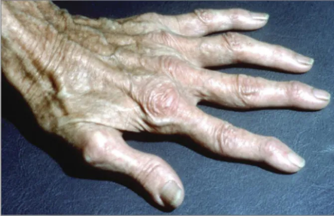
prog-nosis: patients often die relatively young from ruptu-re of arteries and/or hollow organs, such as intestines and uterus during pregnancy73. The typical phenoty-pe consists of thin and translucent skin, showing un-derling veins, giving especially the hands an aged ap-pearance (‘acrogeria’, see Figure 2) and nonspecific di-morphic features of the face. However, all these
typi-figurE 2.Acrogeria in a 32-year-old woman with EDS vascular type. The hand looks much older than the patient really is.
cal features may be absent.
The family history may reveal cases of ruptures of arteries and/or hollow organs, but the present patient could also be the only one in the family with EDS vas-cular type, due to a novel point-mutation in the COL3A1 gene. So, making a clinical diagnosis is not always easy; luckily there are genetic tests for this EDS--type (Table III).
Preventive measures are possible, including life sty-le measures: avoiding sports with risk of trauma and with elevations of blood pressure, stop smoking, meti-culous monitoring and control of blood pressure to low-normal values. In a study of 5-years duration, therapy with the beta-blocker celiprolol prevented major com-plications in patients with EDS vascular type, compared to EDS patients randomly assigned no drug therapy74.
The phenotype of Loeys–Dietz syndrome overlaps with that of EDS vascular type (vascular ruptures) and of Marfan syndrome (aortic aneurysm / dissection and arachnodactyly). The syndrome is caused by TGFBR1
or TGFBR2 mutations, in the genes encoding for
trans-forming growth factor b receptor type 1 or type 2, res-pectively. Transforming growth factor b is a cytokine that exerts diverse roles in cell proliferation and diffe-rentiation, apoptosis (programmed cell death), and ex-tracellular matrix formation11. Patients with TGFBR1 or
TGFBR2 tend to have aortic dissections at smaller
aor-tic-root diameters compared to patients with Marfan syndrome, but compared to patients with EDS vascu-lar type, outcomes after aortic surgery are better72,75.
The criteria for diagnosis of Marfan syndrome are presented in Table VI76. They include involvement of the skeletal system (generalized hypermobility and marfanoid habitus as characterized by the major and
minor skeletal criteria of Table VI) the ocular system (lens dislocation), the cardiovascular system (aortic dissection) and the skin77. However, among individual patients, considerable heterogeneity of phenotype, signs and symptoms is present11.
Genetic testing of Marfan is directed at the many mutations in the FBN1 gene, encoding the structural protein fibrillin-1. About 10% of patients with clini-cally typical Marfan syndrome have no mutation in this gene. In such cases, TGFBR1 and TGFBR2 muta-tions may be detected. To complicate things, TGFBR1 and TGFBR2 mutations are also found in Loeys–Dietz syndrome and may be detected in familial thoracic aor-tic aneurysms and dissections syndrome too78. The an-giotensin II receptor blocker losartan seems to inhibit progressive aortic root dilation in patients with Marfan syndrome12,79. Stickler syndrome (hereditary progres-sive arthro-ophthalmopathy) may cause confusion with Marfan syndrome based on its possible clinical features hypermobility and skeletal abnormalities, such as pectus excavatum or carinatum and highly ar-ched palate. However, skin laxity and vascular com-plications are not features of this syndrome (Table VII)80,81. Although typical cases may be recognized at a young age, the diagnosis often is delayed by variabi-lity of the phenotype82. Genetic testing of the collagen type II gene locus COL2A1 can confirm the clinical diagnosis83.
The aneurism-osteoarthritis syndrome has features similar to Marfan syndrome, but lens dislocation does not occur and generalized hypermobility is less fre-quently present. Next to vascular complications these patients have osteoarthritis at a relatively young age. The specific genetic defect is a mutation of the SMAD3 gene84.
tablE v. discrimination of gEnEralizEd joint hypErmobility syndromEs*
Classification Distinguishing feature
Idiopathic (end of distribution curve)** none Benign joint hypermobility syndrome** none
Ehlers-Danlos types (table 5)** lax skin; specific features of each type Osteogenesis imperfecta bone fragility
Loeys-Dietz syndrome vascular complications
Stickler syndrome eye problems, facial abnormalities
Marfan syndrome skeletal habitus, lens dislocations, aortic arch dissections
*not comprehensive;
**possible overlap of generalized hypermobility as end of normal distribution curve of mobility of joints, benign joint hypermobility syndrome and Ehlers-Danlos hypermobility type
principlEs of managEmEnt of hypErmobility syndromEs
Although there are no randomized controlled studies regarding the effects of existing treatments for muscu-loskeletal problems in patients with symptomatic hy-permobility, this does not mean that certain therapeu-tic strategies could not be helpful85. They are
mentio-ned only briefly below.
• Education is the first step after the diagnosis. It should tackle the feelings of frustration with misun-derstanding of the complaints by the medical profes-sion and social entourage, often during many years86; and the frustration about the absence of clear physical signs and laboratory abnormalities. It also comprises life style advices (e.g. on rest, sleep hygiene, activities,
tablE vi. ghEnt 1996 diagnostic critEria for marfan syndromE7
System Major Criteria Minor Criteria
Skeletal At least 4 of the following features: Two of the major features, or 1 major feature and 2 of the following:
• Pectus carinatum • Joint hypermobility • Pectus excavatum requiring surgery • Moderate pectus excavatum • Wrist and thumb signs • High palate with dental crowding
• Upper-to-lower segment ratio <0.86 or • Characteristic face (dolichocephaly, malar hypoplasia, span:height >1.05 enophthalmos, retrognathia, down-slanting palpebral • Scoliosis >20° or spondylolisthesis fissures)
• Reduced elbow extension (<170°) • Pes planus
• Acetabular protrusion
Ocular • Lens dislocation (ectopia lentis) • Flat cornea
• Increased axial length of globe (causing myopia) • Hypoplastic iris or ciliary muscle (causing decreased
miosis)
Cardiovascular • Dilatation of aortic root involving the • Mitral valve prolapse
sinuses of Valsalva • Dilatation of the pulmonary artery and age <40 y • Dissection of the ascending aorta • Calcified mitral annulus and age <40 y
• Dilatation or dissection of the descending thoracic or abdominal aorta and age < 50 y
Pulmonary None • Spontaneous pneumothorax
• Apical blebs on chest X-ray
Skin None • Striae atrophicae
• Recurrent or incisional hernia
Dura • Lumbosacral dural ectasia None
Genetic • Family history: parent, child, or sibling None meets these criteria independently
• Test: fibrillin-1 mutation known to cause Marfan
• Test: inheritance in the family of DNA marker haplotype linked to Marfan
For all systems, having 1 of the features listed constitutes a major criterion or minor criterion except for the skeletal system, where >1 feature is needed.
The diagnosis of Marfan syndrome requires major criteria in 1 organ and minor criteria (involvement) of another when a positive family history or positive genetic testing (one of the two) is present. In the absence of a positive family history and positive genetic testing, meeting major criteria in at least 2 organ systems and minor criteria in at least a third organ system are required.
prevention of trauma), directions on self-help, redi-recting false cognitions and other cognitive-behavioral strategies, coping, improving patient self-esteem and self-efficacy, genetic counseling.
• Physical therapy and daily exercises at home should be performed: toning exercises for stabilization of joints, exercises improving proprioception, exerci-ses diminishing physical deconditioning (Figure 1) and improving posture. In patients with osteoarthritis of the hip or knee (without a hypermobility syndrome) exercise therapy has demonstrated efficacy in reducing pain and disability87; in hypermobile children also a beneficial effect of physiotherapy on pain was found88. It seems prudent to advice exercises to improve mus-cle strength and proprioception of joints, although in patients with hypermobility syndromes there is lack of data89. The prospective study showing no relations-hip between joint hypermobility and injury in
dan-cers6, who often have a trained body, could also be an argument for exercises. There seems to be no contra-indication against prudent stretching exercises, but long during hyperextension of joint, e.g. standing with hyperextended knees, should be avoided.
Modalities like heat or cold application, electrical stimulation to alleviate pain, acupuncture, acupressu-re, biofeedback, yoga and conscious relaxation are not evidence-based, but might have a beneficial effect on pain in some patients.
It seems prudent to advise weight-bearing exerci-ses for the long bones and spine and an adequate in-take of calcium and vitamin D in patients with a hy-permobility syndrome, to prevent fractures.
• Adaptations and assistive devices (braces, grasps, waterbed, mattress, wheelchair, electric scooter), adap-ted shoes, adaptations to living and working environ-ment could all have a place in the manageenviron-ment strate-gy. Care should be put in avoiding that adaptations and assistive devices lead to less physical exercises or activities, as this could potentially be harmful by in-creasing physical deconditioning and ensuing com-plaints. However, adaptations and assistive devices may lead to increased activities and there can be me-dical and social reasons to prescribe them.
• Drugs, such as medications for pain, disturbed sleep, depression and fatigue could have a place in the management of selected patients with hypermobility syndromes. However, in chronic diseases, these drugs usually only have a mild, often temporarily sympto-matic effect. Apart from treating hypertension, which is always necessary, in Marfan syndrome, losartan has a place and celiprolol in EDS vascular type, to diminish the risk of vascular complications.
• Surgical procedures on joints should be avoided, if possible. For example, in patients with hypermobi-lity and repetitive luxation of the shoulder, surgery of-ten is ineffective, according to clinical experience.
arEas of uncErtainty
The exact incidence of most of the hypermobility syn-dromes is not known. Possibly specific (types of) hy-permobility syndromes are genetically based on diffe-rent genetic aberrations; several new types of EDS have been recognized on the basis of genetic defects over the past years. Although specific drugs have been shown beneficial in Marfan syndrome and EDS vascu-lar type; it has not been investigated whether (other) antihypertensive medications would have a similar ef-fect. Although scarce literature data indicate a
benefi-tablE vii. involvEd organ systEms with thEir manifEstations in sticklEr syndromE*8
Orofacial abnormalities:
• cleft palate (open cleft, submucous cleft, or bifid uvula), highly arched palate
• characteristic face (malar / midfacial hypoplasia, broad and/ or flat nasal bridge, micro / retrognathia)
Ocular abnormalities: vitreous and retinal degeneration (lattice degeneration, retinal hole, detachment or tear)
• Auditory abnormalities:
• high frequency (4–8 kHz) sensorineural hearing loss: at age<20: ≥ 20 dB, at age 20–40: ≥ 30 dB and at age >40: ≥ 40 dB
• hypermobile tympanic membranes
Skeletal abnormalities and symptoms:
• generalized joint hypermobility • chronic musculoskeletal pain
• femoral head disorders (slipped epiphysis or Legg–Perthes-like disease)
• radiographically demonstrated osteoarthritis before age 40
• scoliosis, spondylolisthesis, or Scheuermann-like kyphotic deformity
• mild spondyloepiphyseal dysplasia • pectus excavatum or carinatum
* This list is by no means exhaustive; there is variable intra- and interfamilial heterogeneity in the involvement of these organ systems
cial effect of physiotherapy and exercises, the real long-term effect is not known, nor the effects, pros and cons of specific physiotherapeutic modalities.
corrEspondEncE to
José Antonio Pereira da Silva Rua Carlos Noronha, Nº1. 3140-401 Santo Varão, Portugal E-mail: [email protected]
rEfErEncEs
1. Beighton P, De Paepe A, Steinmann B, Tsipouras P,.Wenstrup RJ. Ehlers-Danlos syndromes: revised nosology, Villefranche, 1997. Ehlers- Danlos National Foundation (USA) and Ehlers-Danlos Support Group (UK). Am J Med Genet 1998; 77: 31-37. 2. Bulbena A, Duro JC, Porta M, et al. Clinical assessment of
hy-permobility of joints: assembling criteria. J Rheumatol 1992; 19: 115-122.
3. Remvig L, Jensen DV,.Ward RC. Epidemiology of general joint hypermobility and basis for the proposed criteria for benign joint hypermobility syndrome: review of the literature. J Rheu-matol 2007; 34: 804-809.
4. Klemp P, Stevens JE,.Isaacs S. A hypermobility study in ballet dancers. J Rheumatol 1984; 11: 692-696.
5. Collinge R, Simmonds JV. Hypermobility, injury rate and reha-bilitation in a professional football squad—a preliminary stu-dy. Phys Ther Sport 2009; 10: 91-96.
6. Roussel NA, Nijs J, Mottram S, et al. Altered lumbopelvic mo-vement control but not generalized joint hypermobility is as-sociated with increased injury in dancers. A prospective study. Man Ther 2009; 14: 630-635.
7. Holzberg M, Hewan-Lowe KO,.Olansky AJ. The Ehlers-Danlos syndrome: recognition, characterization, and importance of a milder variant of the classic form. A preliminary study. J Am Acad Dermatol 1988; 19: 656-666.
8. Bridges AJ, Smith E,.Reid J. Joint hypermobility in adults re-ferred to rheumatology clinics. Ann Rheum Dis 1992; 51: 793--796.
9. Hakim AJ, Grahame R. Joint hypermobility syndrome: an up-date for clinicians. Intern J Adv Rheumatol 2003; 1: 131-138. 10. Steinmann B, Royce PM,.Superti-Furga A. The Ehlers-Danlos
syndrome. 1993351-1993407.
11. Bird HA. Lessons from Marfan syndrome. Rheumatology (Ox-ford) 2007; 46: 902-903.
12. Brooke BS, Habashi JP, Judge DP, et al. Angiotensin II blocka-de and aortic-root dilation in Marfan’s syndrome. N Engl J Med 2008; 358: 2787-2795.
13. Gedalia A, Press J, Klein M,.Buskila D. Joint hypermobility and fibromyalgia in schoolchildren. Ann Rheum Dis 1993; 52: 494--496.
14. Ofluoglu D, Gunduz OH, Kul-Panza E,.Guven Z. Hypermobi-lity in women with fibromyalgia syndrome. Clin Rheumatol 2006; 25: 291-293.
15. Karaaslan Y, Haznedaroglu S,.Ozturk M. Joint hypermobility and primary fibromyalgia: a clinical enigma. J Rheumatol 2000; 27: 1774-1776.
16. Acasuso-Diaz M, Collantes-Estevez E. Joint hypermobility in patients with fibromyalgia syndrome. Arthritis Care Res 1998; 11: 39-42.
17. Larsson LG, Baum J, Mudholkar GS,.Kollia GD. Benefits and
di-sadvantages of joint hypermobility among musicians. N Engl J Med 1993; 329: 1079-1082.
18. Sacheti A, Szemere J, Bernstein B, et al. Chronic pain is a ma-nifestation of the Ehlers-Danlos syndrome. J Pain Symptom Manage 1997; 14: 88-93.
19. Rombaut L, Malfait F, Cools A, De Paepe A,.Calders P. Muscu-loskeletal complaints, physical activity and health-related qua-lity of life among patients with the Ehlers-Danlos syndrome hypermobility type. Disabil Rehabil 2010.
20. Larsson LG, Mudholkar GS, Baum J,.Srivastava DK. Benefits and liabilities of hypermobility in the back pain disorders of in-dustrial workers. J Intern Med 1995; 238: 461-467. 21. Hall MG, Ferrell WR, Sturrock RD, Hamblen DL,.Baxendale
RH. The effect of the hypermobility syndrome on knee joint proprioception. Br J Rheumatol 1995; 34: 121-125. 22. Mallik AK, Ferrell WR, McDonald AG,.Sturrock RD. Impaired
proprioceptive acuity at the proximal interphalangeal joint in patients with the hypermobility syndrome. Br J Rheumatol 1994; 33: 631-637.
23. O’Connor BL, Vilensky JA. Peripheral and central nervous sys-tem mechanisms of joint protection. Am J Orthop 2003; 32: 330-336.
24. Rombaut L, Malfait F, De W, I, et al. Muscle-tendon tissue pro-perties in the hypermobility type of Ehlers-Danlos syndrome. Arthritis Care Res (Hoboken ) 2012; 64: 766-772.
25. Lumley MA, Jordan M, Rubenstein R, Tsipouras P,.Evans MI. Psychosocial functioning in the Ehlers-Danlos syndrome. Am J Med Genet 1994; 53: 149-52.
26. Bulbena A, Duro JC, Porta M, et al. Anxiety disorders in the joint hypermobility syndrome. Psychiatry Res 1993; 46: 59--68.
27. Martin-Santos R, Bulbena A, Porta M, et al. Association bet-ween joint hypermobility syndrome and panic disorder. Am J Psychiatry 1998; 155: 1578-1583.
28. Rombaut L, Malfait F, De W, I, et al. Muscle mass, muscle strength, functional performance, and physical impairment in women with the hypermobility type of Ehlers-Danlos syndro-me. Arthritis Care Res (Hoboken ) 2012; 64: 1584-1592. 29. Arendt-Nielsen L, Kaalund S, Bjerring P,.Hogsaa B. Insufficient
effect of local analgesics in Ehlers Danlos type III patients (con-nective tissue disorder). Acta Anaesthesiol Scand 1990; 34: 358-361.
30. Arendt-Nielsen L, Kaalund S, Hogsaa B, Bjerring P,.Grevy C. The response to local anaesthetics (EMLA-cream) as a clinical test to diagnose between hypermobility and Ehlers Danlos type III syndrome. Scand J Rheumatol 1991; 20: 190-195. 31. Oliver DW, Balan KK, Burrows NP,.Hall PN. Dispersal of
ra-dioisotope labelled solution following deep dermal injection in Ehlers-Danlos syndrome. Br J Plast Surg 2000; 53: 308-312. 32. Barron DF, Cohen BA, Geraghty MT, Violand R,.Rowe PC. Joint hypermobility is more common in children with chronic fati-gue syndrome than in healthy controls. J Pediatr 2002; 141: 421-425.
33. Gazit Y, Nahir AM, Grahame R,.Jacob G. Dysautonomia in the joint hypermobility syndrome. Am J Med 2003; 115: 33-40. 34. Rowe PC, Barron DF, Calkins H, et al. Orthostatic intolerance
and chronic fatigue syndrome associated with Ehlers-Danlos syndrome. J Pediatr 1999; 135: 494-499.
35. Rombaut L, De Paepe A, Malfait F, Cools A,.Calders P. Joint po-sition sense and vibratory perception sense in patients with Eh-lers-Danlos syndrome type III (hypermobility type). Clin
Rheu-matol 2010; 29: 289-295.
36. Rombaut L, Malfait F, De W, I, et al. Balance, gait, falls, and fear of falling in women with the hypermobility type of Ehlers-Dan-los syndrome. Arthritis Care Res (Hoboken ) 2011; 63: 1432--1439.
37. Morgan AW, Pearson SB, Davies S, Gooi HC,.Bird HA. Asthma and airways collapse in two heritable disorders of connective tissue. Ann Rheum Dis 2007; 66: 1369-1673.
38. Voermans NC, Knoop H, van de KN, et al. Fatigue Is a Fre-quent and Clinically Relevant Problem in Ehlers-Danlos Syn-drome. Semin Arthritis Rheum 2009.
39. Rombaut L, Malfait F, De Paepe A, et al. Impairment and im-pact of pain in female patients with Ehlers-Danlos syndrome: a comparative study with fibromyalgia and rheumatoid arthri-tis. Arthritis Rheum 2011; 63: 1979-1987.
40. de Kort LM, Verhulst JA, Engelbert RH, Uiterwaal CS,.de Jong TP. Lower urinary tract dysfunction in children with generali-zed hypermobility of joints. J Urol 2003; 170: 1971-1874. 41. McIntosh LJ, Stanitski DF, Mallett VT, et al. Ehlers-Danlos
syn-drome: relationship between joint hypermobility, urinary in-continence, and pelvic floor prolapse. Gynecol Obstet Invest 1996; 41: 135-139.
42. McLean AM, Paul RE, Jr., Kritzman J,.Farthing MJ. Malab-sorption in Marfan (Ehlers-Danlos) syndrome. J Clin Gas-troenterol 1985; 7: 304-308.
43. Arunkalaivanan AS, Morrison A, Jha S,.Blann A. Prevalence of urinary and faecal incontinence among female members of the Hypermobility Syndrome Association (HMSA). J Obstet Gy-naecol 2009; 29: 126-128.
44. Carley ME, Schaffer J. Urinary incontinence and pelvic organ prolapse in women with Marfan or Ehlers Danlos syndrome. Am J Obstet Gynecol 2000; 182: 1021-1023.
45. Manning J, Korda A, Benness C,.Solomon M. The association of obstructive defecation, lower urinary tract dysfunction and the benign joint hypermobility syndrome: a case-control stu-dy. Int Urogynecol J Pelvic Floor Dysfunct 2003; 14: 128-132. 46. ter Meulen PH, Berghmans LC, Nieman FH,.van Kerrebroeck PE. Effects of Macroplastique Implantation System for stress urinary incontinence and urethral hypermobility in women. Int Urogynecol J Pelvic Floor Dysfunct 2009; 20: 177-183. 47. Dolan AL, Arden NK, Grahame R,.Spector TD. Assessment of
bone in Ehlers Danlos syndrome by ultrasound and densito-metry. Ann Rheum Dis 1998; 57: 630-653.
48. Le Parc JM, Plantin P, Jondeau G, et al. Bone mineral density in sixty adult patients with Marfan syndrome. Osteoporos Int 1999; 10: 475-479.
49. Giampietro PF, Peterson M, Schneider R, et al. Assessment of bone mineral density in adults and children with Marfan syn-drome. Osteoporos Int 2003; 14: 559-563.
50. Moura B, Tubach F, Sulpice M, et al. Bone mineral density in Marfan syndrome. A large case-control study. Joint Bone Spine 2006; 73: 733-735.
51. Yen JL, Lin SP, Chen MR,.Niu DM. Clinical features of Ehlers-Danlos syndrome. J Formos Med Assoc 2006; 105: 475-480. 52. Carbone L, Tylavsky FA, Bush AJ, et al. Bone density in
Ehlers-Danlos syndrome. Osteoporos Int 2000; 11: 388-392. 53. Moriatis WJ, Cameron KL,.Owens BD. Impact of joint laxity
and hypermobility on the musculoskeletal system. J Am Acad Orthop Surg 2011; 19: 463-471.
54. Jonsson H, Eliasson GJ, Jonsson A, et al. High hand joint mo-bility is associated with radiological CMC1 osteoarthritis: the
AGES-Reykjavik study. Osteoarthritis Cartilage 2009; 17: 592--595.
55. Kraus VB, Li YJ, Martin ER, et al. Articular hypermobility is a protective factor for hand osteoarthritis. Arthritis Rheum 2004; 50: 2178-2183.
56. Dolan AL, Hart DJ, Doyle DV, Grahame R,.Spector TD. The re-lationship of joint hypermobility, bone mineral density, and os-teoarthritis in the general population: the Chingford Study. J Rheumatol 2003; 30: 799-803.
57. Chen HC, Shah SH, Li YJ, et al. Inverse association of general joint hypermobility with hand and knee osteoarthritis and se-rum cartilage oligomeric matrix protein levels. Arthritis Rheum 2008; 58: 3854-3864.
58. Yamashita M, Narita M, Ishihara H, Matsuki A,.Oyama T. Ute-rine rupture in a case with Ehlers-Danlos syndrome type IV— anesthetic considerations. Middle East J Anesthesiol 1987; 9: 277-2781.
59. Fuchs JR, Fishman SJ. Management of spontaneous colonic perforation in Ehlers-Danlos syndrome type IV. J Pediatr Surg 2004; 39: e1-e3.
60. Palmquist M, Pappas JG, Petrikovsky B, Blakemore K,.Roshan D. Successful pregnancy outcome in Ehlers-Danlos syndrome, vascular type. J Matern Fetal Neonatal Med 2009; 22: 924-927. 61. Lind J, Wallenburg HC. Pregnancy and the Ehlers-Danlos syn-drome: a retrospective study in a Dutch population. Acta Obs-tet Gynecol Scand 2002; 81: 293-300.
62. Remvig L, Duhn PH, Ullman S, et al. Skin extensibility and consistency in patients with Ehlers-Danlos syndrome and be-nign joint hypermobility syndrome. Scand J Rheumatol 2009; 38: 227-230.
63. Catala-Petavy C, Machet L, Georgesco G, et al. Contribution of skin biometrology to the diagnosis of the Ehlers-Danlos syn-drome in a prospective series of 41 patients. Skin Res Technol 2009; 15: 412-417.
64. Grahame R, Bird HA,.Child A. The revised (Brighton 1998) cri-teria for the diagnosis of benign joint hypermobility syndrome (BJHS). J Rheumatol 2000; 27: 1777-1779.
65. Grahame R. The need to take a fresh look at criteria for hyper-mobility. J Rheumatol 2007; 34: 664-665.
66. Remvig L, Jensen DV,.Ward RC. Are diagnostic criteria for ge-neral joint hypermobility and benign joint hypermobility syn-drome based on reproducible and valid tests? A review of the literature. J Rheumatol 2007; 34: 798-803.
67. Grahame R, Bird H. British consultant rheumatologists’ per-ceptions about the hypermobility syndrome: a national survey. Rheumatology (Oxford) 2001; 40: 559-562.
68. Juul-Kristensen B, Rogind H, Jensen DV,.Remvig L. Inter-exa-miner reproducibility of tests and criteria for generalized joint hypermobility and benign joint hypermobility syndrome. Rheumatology (Oxford) 2007; 46: 1835-1841.
69. Zweers MC, Kucharekova M,.Schalkwijk J. Tenascin-X: a can-didate gene for benign joint hypermobility syndrome and hy-permobility type Ehlers-Danlos syndrome? Ann Rheum Dis 2005; 64: 504-505.
70. Zweers MC, Bristow J, Steijlen PM, et al. Haploinsufficiency of TNXB is associated with hypermobility type of Ehlers-Danlos syndrome. Am J Hum Genet 2003; 73: 214-217.
71. Drera B, Tadini G, Barlati S,.Colombi M. Identification of a no-vel TGFBR1 mutation in a Loeys-Dietz syndrome type II patient with vascular Ehlers-Danlos syndrome phenotype. Clin Genet 2008; 73: 290-293.
72. Loeys BL, Schwarze U, Holm T, et al. Aneurysm syndromes caused by mutations in the TGF-beta receptor. N Engl J Med 2006; 355: 788-798.
73. Pepin M, Schwarze U, Superti-Furga A,.Byers PH. Clinical and genetic features of Ehlers-Danlos syndrome type IV, the vascu-lar type. N Engl J Med 2000; 342: 673-680.
74. Ong KT, Perdu J, De Backer J, et al. Effect of celiprolol on pre-vention of cardiovascular events in vascular Ehlers-Danlos syn-drome: a prospective randomised, open, blinded-endpoints trial. Lancet 2010; 376: 1476-1484.
75. Gelb BD. Marfan’s syndrome and related disorders—more tigh-tly connected than we thought. N Engl J Med 2006; 355: 841--844.
76. De Paepe A, Devereux RB, Dietz HC, Hennekam RC,.Pyeritz RE. Revised diagnostic criteria for the Marfan syndrome. Am J Med Genet 1996; 62: 417-426.
77. Grahame R, Pyeritz RE. The Marfan syndrome: joint and skin manifestations are prevalent and correlated. Br J Rheumatol 1995; 34: 126-131.
78. Mizuguchi T, Matsumoto N. Recent progress in genetics of Mar-fan syndrome and MarMar-fan-associated disorders. J Hum Genet 2007; 52: 1-12.
79. Chiu HH, Wu MH, Wang JK, et al. Losartan Added to beta-Blockade Therapy for Aortic Root Dilation in Marfan Syndro-me: A Randomized, Open-Label Pilot Study. Mayo Clin Proc 2013.
80. Stickler GB, Belau PG, Farrell FJ, et al. Hereditary progressive arthro-ophtalmopathy. Mayo Clin Proc 1965; 40: 433-455. 81. Rose PS, Levy HP, Liberfarb RM, et al. Stickler syndrome:
cli-nical characteristics and diagnostic criteria. Am J Med Genet A 2005; 138: 199-207.
82. Bowling EL, Brown MD,.Trundle TV. The Stickler syndrome: case reports and literature review. Optometry 2000; 71: 177--182.
83. Liberfarb RM, Levy HP, Rose PS, et al. The Stickler syndrome: Genotype/phenotype correlation in 10 families with Stickler syndrome resulting from seven mutations in the type II colla-gen colla-gene locus COL2A1. Genet Med 2003; 5: 21-27. 84. van de Laar IM, van der Linde D, Oei EH, et al. Phenotypic
spectrum of the SMAD3-related aneurysms-osteoarthritis syn-drome. J Med Genet 2012; 49: 47-57.
85. Castori M, Morlino S, Celletti C, et al. Management of pain and fatigue in the joint hypermobility syndrome (a.k.a. Ehlers-Dan-los syndrome, hypermobility type): principles and proposal for a multidisciplinary approach. Am J Med Genet A 2012; 158A: 2055-2070.
86. Berglund B, Anne-Cathrine M,.Randers I. Dignity not fully up-held when seeking health care: Experiences expressed by indi-viduals suffering from Ehlers-Danlos syndrome. Disabil Reha-bil 2010; 32: 1-7.
87. van Baar ME, Dekker J, Oostendorp RA, et al. The effective-ness of exercise therapy in patients with osteoarthritis of the hip or knee: a randomized clinical trial. J Rheumatol 1998; 25: 2432-2439.
88. Kemp S, Roberts I, Gamble C, et al. A randomized comparati-ve trial of generalized vs targeted physiotherapy in the mana-gement of childhood hypermobility. Rheumatology (Oxford) 2010; 49: 315-325.
89. Sahin N, Baskent A, Cakmak A, et al. Evaluation of knee pro-prioception and effects of propro-prioception exercise in patients with benign joint hypermobility syndrome. Rheumatol Int 2008; 28: 995-1000.
90. De Paepe A, Malfait F. The Ehlers-Danlos syndrome, a disorder with many faces. Clin Genet 2012; 82: 1-11.
91. Tofts LJ, Elliott EJ, Munns C, Pacey V,Sillence DO. The diffe-rential diagnosis of children with joint hypermobility: a review of the literature. Pediatr Rheumatol Online J 2009; 7: 1.