ORIGINAL
RES
EAR
CH
Correspondence to: Jessica Caroliny de Jesus Neves − Rua Getúlio Vargas, 109 – CEP: 86620-000 – Guaraci (PR), Brasil. – E-mail: [email protected] – Presentation: Oct.
Study conducted at the Universidade Estadual de Londrina (UEL) – Londrina (PR), Brasil. 1Physical Therapy Student at UEL – Londrina (PR), Brasil.
2Master´s Student at the Associated Master´s Program in Rehabilitation Sciences at UEL - Universidade Norte do Paraná (UNOPAR) – Londrina (PR), Brasil.
3PhD in General Bases of Experimental Surgery at Universidade Estadual Paulista “Júlio de Mesquita Filho” (UNESP) – Botucatu (SP), Brasil; Assistant Professor in the Physical Therapy Department at UEL – Londrina (PR), Brasil.
4PhD, Master´s Degree in Education at UNESP – Marília (SP), Brasil; Associate Professor in the Physical Therapy Department at UEL – Londrina (PR), Brasil.
in preschool children
Prevalência de hipermobilidade articular em crianças pré-escolares
Prevalencia de hipermovilidad articular en niños pre-escolares
Jessica Caroliny de Jesus Neves1, Fabíola Unbehaun Cibinello1, Leonardo George Vitorio Vitor2,
Débora Beckner2, Claudia Patrícia Cardoso Martins Siqueira3, Dirce Shizuko Fujisawa4
ABSTRACT | Joint hypermobility is the ability to per-form movements with a range of motion that is wid-er than normal. This study aimed at establishing the prevalence of joint hypermobility in junior kindergarten and senior kindergarten children from the Municipal Education System of Londrina, Paraná, Brasil. The study was cross- sectional, with a sample of 366 children aged between 5 and 6 years. The detection of joint hypermo-bility was based on the criteria proposed in literature. It was found that 198 (54.1%) of the children evaluated had joint hypermobility, 96 (59.6%) were girls and 102 (49.8%) were boys. Joint hypermobility was found more frequently in the elbow and knee joints, where hyperex-tension occurred. There was no statistically significant difference between the male and female groups in re-lation to joint hypermobility (χ2=3.539, p=0.072). We can
conclude that joint hypermobility found in the junior kindergarten and senior kindergarten children evaluat-ed was common and compatible with the age of the population evaluated in this study.
Keywords | joint hypermobility; joint instability; JK and SK children.
RESUMO | A hipermobilidade articular é a capacidade de realizar movimentos em amplitudes maiores que a normal. O presente estudo teve como objetivo estabelecer a pre-valência de hipermobilidade articular em crianças pré-es-colares da Rede Municipal de Educação de Londrina/PR.
O estudo foi do tipo transversal com amostra constituída de 366 crianças, de 5 e 6 anos de idade. Verificou-se que, das crianças pré-escolares avaliadas, 198 (54,1%) apresen-taram hipermobilidade articular, sendo 96 (59,6%) do sexo feminino e 102 (49,8%) do masculino. A hipermobilidade articular foi encontrada com maior frequência nas arti-culações de cotovelo e joelho, onde ocorreu a hiperex-tensão. Não houve diferença estatisticamente significante entre os grupos masculino e feminino em relação à hiper-mobilidade articular (χ2=3,539, p=0,072). Pode-se concluir
que a hipermobilidade articular nas crianças pré-escolares foi achado comum, compatível com a faixa etária da po-pulação avaliada no estudo.
Descritores | hipermobilidade articular; instabilidade articular; crianças pré-escolares.
Neves et al. Hypermobility in JK and SK children
INTRODUCTION
Joint hypermobility was irst mentioned by Hippocrates as the Celts’ inability to pull a bow string or throw darts because they had “loose limbs”, according to Egri1. Joint hypermobility is caused by tissue loosening or excessive length, which leads to an increase in motion range beyond what is considered normal, and it might be followed, in children, by complaints about pain voiced in the afternoon or at night2,3. We highlight that complaints about musculoskeletal pain is common in pediatric practice, especially in relation to joint hyper-mobility syndrome4.
The British Rheumatology Society (1992) defines joint hypermobility based on the presence of althralgia or musculoskeletal pain with an evolution of at least 3 months, associated with hypermobility5. Con-nective tissue hereditary diseases might be asso-ciated with the syndromes of Down, Marfan, and Ehlers-Danlos6. More frequent in girls, joint mobil-ity7 decreases with age8,9. Besides these conditions, the prevalence of hypermobility varies according to age, sex, ethnicity, and genetic factors, and it is, therefore, multifactorial.
It is believed that the primary cause of hypermobility is ligament loosening associated with musculoskeletal dysfunction, given that the maximal excursion of each joint is inluenced by ligament tension. Ligament loos-ening is determined by genes that codify collagen, elas-tin, and ibrin, making the individual more susceptible to trauma10. For Hall11, individuals with joint hypermo-bility present lower proprioceptive response, and might adopt positions that are biomechanically unfavorable to the limbs, thus predisposing themselves precociously to degenerative conditions.
Hypermobility must be diferentiated from joint instability. he irst is the ability to perform a series of movements in a range that is wider than normal2,3; the latter refers to the amplitude of excessive move-ments performed without protective muscle control11. Excessive hypermobility can provoke joint instability, which might generate sprains, osteoarthritis, pain, diiculties in controlling the body12, and decreased body perception, predisposing the individual to
lesions in soft tissues13. It is believed that there is an alteration in the structure of type 3 collagen1, char-acterized by a higher ratio in relation to type 1 colla-gen14, and proteoglycan alteration, generating distur-bance in proprioception.
he irst quantitative system created to ana-lyze joint hypermobility was devised by Carter and Wilkinson15, who conducted a comparative study between healthy children and children with hip con-genital dislocation, both in school age; they found a prevalence of joint hypermobility of 7% in normal children of both sexes, and in 29.2% of the girls and 71.1% of the boys with hip congenital dislocation. Araújo16 pointed a prevalence of joint hypermobility of 36.31% among children in public schools of São Paulo. he parameters provided by Beighton, Solo-mon, and Soskolne17 are the modiication of Carter and Wilkinson’s theory15, with criteria established by the British Rheumatology Society (1992) to iden-tify hypermobility, currently the most disseminated method in the world.
Joint hypermobility is an important variable to be associated with the postural assessment of children. he individual with an excessive increase of joint mobility sufers alterations in muscle and body pos-ture18, which causes degenerative processes prompted by the use of irregular strength, that is, the exces-sive lengthening of soft tissues that compromise joint integrity might occur. herefore, the detection of joint hypermobility in school children is necessary, consid-ering that it might cause, on a long-term basis, pos-tural alterations, pain, and joint instability. Another aspect that involves the necessity of establishing the prevalence of hypermobility among children results from the lack of studies in the country. We also highlight that the involvement in physical activities through playing has changed in our current soci-ety, that is, children spend more time sitting down, and are, consequently, more sedentary, which might cause alterations in mobility. hus, this study aimed at investigating the prevalence of joint hypermobility in junior kindergarten ( JK) senior kindergarten (SK) children of the Municipal Education System of Lon-drina/PR.
hiperextensión. No hubo diferencia estadísticamente significativa entre los grupos masculino y femenino en relación a la hipermo-vilidad articular (χ2=3,539, p=0,072). Se puede concluir que la
hi-permovilidad articular en los niños pre-escolares fue encontrada
normal, compatible con la línea etaria de la población evaluada en el estudio.
METHODOLOGY
A transversal study was developed in the schools of the Municipal School System of Londrina/PR, in partner-ship with the research project “Detecção precoce de alter-ações porturais em crianças na idade pré-escolar: diagnóstico e orientações” (“Early detection of postural alterations in JK and SK children: diagnosis and guidelines”), approved by the University Hospital/UEL’s Ethics Committee (report number 240/09), and authorized by the Secretaria Municipal de Educação (Municipal Edu-cation Secretariat). he parents and/or responsible for the children were informed about the study and signed the free and informed consent form. he study was con-ducted between February 2011 and May 2012.
In 2011, there were 3,666 children enrolled in the JK and SK programs of Londrina’s Municipal Edu-cation System, according to data from the Municipal Education Secretariat. Considering 0.05 a tolerable sampling error, the sample size (N) was estimated in 360 participants from schools of the northern, south-ern, westsouth-ern, eastsouth-ern, and central regions of the city, with an equitable number of students enrolled. he inclusion criteria were JK and SK children enrolled in the municipal system, who were 5 and 6 years of age, of both sexes. Children with chronic or acute diseases in convalescent stage, with Down, Marfan or Ehlers-Danlos19 syndromes, who presented dii-culty to remain in the orthostatic position during the tests, and those recovering from recent surgeries were excluded. In order to detect joint hypermobility, we used a goniometer to measure the angles, and the cri-teria proposed by Beighton, Solomon, and Soskolne17. hese criteria bilaterally evaluate the joints in difer-ent parts of the body, described below, considering the individuals that present three positive maneuvers as hypermobile without specifying, however, whether the maneuvers must be unilateral or bilateral. Unilateral maximal amplitude was scored with one point, and bilateral with two points20. hus, the children with a minimum score of six and a maximum score of nine points in the sum of the ive criteria were considered hypermobile, according to Fórleo20.
1. Passive approximation of the thumbs over the fo-rearm anterior region;
2. Passive extension of the ifth inger (>90°); 3. Hyperextension of the elbows beyond 10°; 4. Hyperextension of the knees over 10°;
5. Spine lexion while keeping the knees straight until touching the loor with the palms.
he procedures for data collection were initiated
scheduling the days for assessment. he teachers of each classroom were informed about the study and helped explaining the evaluation to the children. Before starting the joint hypermobility assessment, all test items were demonstrated to the children with the purpose of facilitating comprehension and execution. he tests were actively performed by the children in the orthostatic position while they were barefoot on an inlexible mat, with feet parallel in line with the width of the hips. he exam was conducted by a previously trained evaluator.
For the evaluation of musculoskeletal pain in the areas analyzed, and for the detection of joint hyper-mobility, we used a face scale with Maurício de Sousa’s characters (Cebolinha and Mônica), composed of ive facial expressions that vary from no pain to unbear-able pain (0=no pain, 1=light pain, 2=moderate pain, 3=severe pain, 4=unbearable pain)21. A human body scale was also used, on which the child was oriented to indicate the painful spot and its origin22. hese scales had the purpose of verifying the presence and localiza-tion of musculoskeletal pain in the children evaluated; depending on the spot indicated, the pain could be related to joint hypermobility.
he statistical analysis of the variables was per-formed using the program SPSS version 20.0. First, we performed descriptive data analysis, and veriication of normal data distribution through Shapiro-Wilk’s test. he results are presented on graphs, tables, and abso-lute and relative values. Sampling error and conidence interval were also calculated. he categorization of the variables allowed an associative analysis among them through the chi-square test (χ2). A signiicance level of 5% was applied to all analyses.
RESULTS
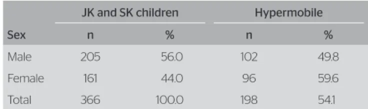
We assessed 366 children, 161 (44.0%) girls and 205 (56.0%) boys. With regard to age 194 (53.0%) children were 5 years old and 172 (47.0%) were 6 years old (Table 1).
Joint hypermobility was identiied in 198 (54.1%) children, 96 (59.6%) girls and 102 (49.8%) boys.
Table 1. Prevalence of hypermobility
JK and SK children Hypermobile
Sex n % n %
Male 205 56.0 102 49.8
Female 161 44.0 96 59.6
Neves et al. Hypermobility in JK and SK children
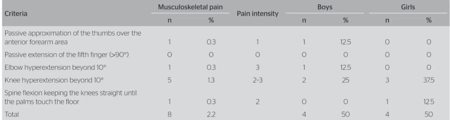
he male and female groups did not present statisti-cally signiicant diference in relation to hypermobil-ity (p=0.072 and χ2=3.539). Out of the 366 children, only 8 (2.2%) children, 4 boys and 4 girls, reported musculoskeletal pain. he presence of pain was pointed in the following parts: knees, elbow, thumb, and lumbar spine. Pain intensity was rated as severe (level 3), followed by moderate (level 2), and lastly light pain (level 1) (Table 2).
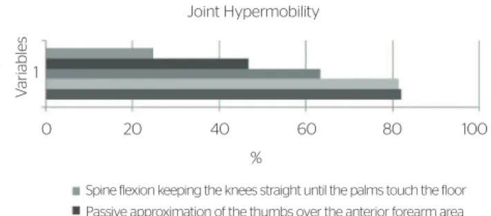
Among the children in JK and SK age assessed in this study, 300 (81.9%) presented elbow hyperexten-sion beyond 10°. Out of these children, 278 (92.7%) present joint mobility bilaterally, while 22 (7.3%) presented elbow mobility beyond 10° unilaterally (Table 3).
Knee hyperextension exceeding 10° occurred in 298 (81.4%) children, and in 244 (81.9%) the increased joint mobility was bilateral, and in 54 (18.1%) it was unilateral; with regard to the passive extension of the ifth inger (>90°), hypermobility was identiied in 243 (66.3%) children; 206 (84.8%) presented it bilaterally, and 37 (15.2%) unilater-ally. he passive approximation of the thumbs over the forearm anterior area occurred in 171 (46.7%) of the children assessed, 128 (74.9%) bilateral, and 43 (25.1%) unilateral. Lastly, lexing the spine while keeping the knees straight until the palms touched the loor occurred in 91 (24.8%) children (Table 3).
During the tests, we found that the highest occur-rence of hypermobility was elbow hyperextension (81.9%), followed by knee hyperextension (81.4%), pinky inger extension (66.3%), approximation of the thumb over the forearm anterior area (46.7%), and spine lexing while keeping the knees straight until the palms touched the loor (24.8%) (Figure 1). Bilateral mobility was more frequent than unilateral mobility, with a higher prevalence in female children.
Association analyses were performed among the ive hypermobility areas in relation to pain, to sex, and to pain and sex, but these were not statistically signiicant and they all presented Cramer’s V, which indicates a weak association among them.
DISCUSSION
Joint hypermobility was identiied in 54.1% of the chil-dren assessed, which difers from the indings in litera-ture. his fact might be related to the narrow and young age range. he presence of pain was identiied in 2.2% of the children evaluated, located in the knees, elbows, thumb, and lumbar spine, and the intensity varied from light to severe. herefore, the majority presents joint hypermobility, but not joint hypermobility syndrome. In literature, most indings on joint mobility in chil-dren are results of studies conducted with older ages24,25.
Table 2. Prevalence of unilateral and bilateral hypermobility
Elbow hyperextension
Knee hyperextension
Passive extension of
the fifth finger
Passive approx. of the thumbs over the forearm
Spine flexion with knee extended
n % n % n % n % n %
Bilateral 278 92.6 244 81.8 20 84.7 128 74.8 – –
Unilateral 22 7.4 54 18.2 37 15.3 43 25.2 – –
Total 300 81.9 298 81.4 243 66.3 171 46.7 91 24.8
Table 3. Joint hypermobility and musculoskeletal pain
Criteria Musculoskeletal pain Pain intensity Boys Girls
n % n % n %
Passive approximation of the thumbs over the
anterior forearm area 1 0.3 1 1 12.5 0 0
Passive extension of the fifth finger (>90°) 0 0 0 0 0 0 0
Elbow hyperextension beyond 10° 1 0.3 3 1 12.5 0 0
Knee hyperextension beyond 10° 5 1.3 2–3 2 25 3 37.5
Spine flexion keeping the knees straight until
the palms touch the floor 1 0.3 2 0 0 1 12.5
Roberto et al.23, detected the manifestation of joint hypermobility in 51 (56.9%) children, aged between 5 and 10 years, with musculoskeletal pain.
Oster and Nielsen24 showed the prevalence of joint hypermobility in 15.4% of school children. Gedalia et al.25, reported a 12% rate of hypermobility in a population of 260 school children. he results found in JK and SK children are probably related to the fact that, during childhood, the bones have more colla-gen, and are therefore more lexible. Concerning the prevalence of joint hypermobility among female chil-dren, our study is in accordance with Cavenaghi8 and Lamari9. Although there are not many explanations for the diferences among the sexes, Lamari, Chue-ire and Cordeiro2, suggest the inluence of bodily makeup, given that girls have higher fat and water percentage, favoring mobility, while boys have more muscles, which results in less joint mobility.
Increased joint mobility might cause postural alter-ation. Silva26 points that knee hyperextension can be related to a stretching in the length of ischiotibial muscles, which might result in lumbar hyperlordosis. Some illnesses or postural alterations, such as intoed knees or obesity, might be associated with knee hyper-extensions that can cause patellar micro traumas, orig-inating, in turn, patellar chondromalacia, characterized by the softening and deterioration of the patellofemo-ral joint cartilage27,28.
he low index observed in the score of spine lex-ing with straight knees until touchlex-ing the loor with the palms possibly relects a decrease in the posterior mobility of the torso, which can occur because of long periods of time spent in the sitting position or because of sedentariness29.
Joint hypermobility was a common inding in this study, and it is probably related to the fact that JK and SK children do not have completely mature
musculo-skeletal systems. For Nóbrega et al.30, the neuromuscu-lar system reaches maturity when the person is between 20 and 30 years old, and, as time goes by, the struc-ture and function of the muscles change. herefore, it seems to be a normal and physiological condition, considering that the JK and SK children in this study were undergoing a stage of maturation, with struc-tural alterations in the collagen ibers. It is expected that hypermobility declines as time goes by, with a loss of 20–30% between 30 and 70 years of age, regardless of sex. For Dantas31, biological changes emerge, such as modiication of joint capsules or muscles, respon-sible for mobility decline as age advances, due to an enlargement of muscle ibers and reduction in the quantity of water. Misner et al.32, report that, as time goes by, the calciication of cartilage and surround-ing tissues occurs, thus reducsurround-ing elasticity. However, 45.9% of the children assessed did not present joint hypermobility, in contrast to what is proposed in the literature in relation to age and maturation process, probably due to the inluence of intrinsic and extrin-sic factors.
We highlight that the increase of unilateral mobility in children in growth stages, especially during growth spurts, might lead to asymmetry, and, consequently, to inadequate postural alignment. For instance, the per-sistence of unilateral knee hyperextension might lead to the evolution of scoliosis. Kendall33 and Teixeira34 point that, to have an adequate posture, a balance between body segments used in positions of less efort and max-imal endurance is necessary.
On the course of human development, joint hyper-mobility might promote physiological alterations in the child’s posture during growth stages, and an inad-equate posture brings higher energy spending in daily activities as a consequence, predisposing the child to early fatigue28. According to Teixeira34, isolated hypermobility does not cause bad posture, since other factors are involved, such as excessive body weight, insuicient or inadequate physical activity, bad pos-tural habits, bone anomalies, protein deiciency char-acteristics. Barden et al.35, add that joint instability caused by mobility excess might compromise the pro-prioceptive systems.
It is necessary to follow the evolution of joint hypermobility during childhood and adolescence through longitudinal studies, because it is essential to establish its resolution period. If hypermobility is pro-longed or excessive, it may cause postural alteration, pain, and lesions. In addition, the occurrence of unilat-eral joint hypermobility as a probable cause of postural alterations must be investigated with the purpose of promoting its prevention.
Figure 1. Percentage of children who presented hypermobility in the diferent joints evaluated. n=366
Joint Hypermobility
0 20 40 60 80 100
%
V
ariables1
Spine flexion keeping the knees straight until the palms touch the floor Passive approximation of the thumbs over the anterior forearm area Passive extension of the fifth finger > 90°
Neves et al. Hypermobility in JK and SK children
CONCLUSION
he prevalence of joint mobility was high among JK and SK children, but it did not present statistically signiicant diference between the sexes, and was more frequent in the elbow and knee joints, where hyperextension occurred. he presence of pain was not prevalent in the study, and it might be a result of the narrow and young age range. Moreover, the dii-culty of assessing children between 5 and 6 years old must also be considered.
ACKNOWLEDGEMENTS
Financing of the Research Project “Detecção precoce de alterações posturais em crianças na idade pré-escolar: diagnóstico e orientações” (“Early detection of postural alterations in JK and SK children: diagnosis and guide-lines”) for the acquisition of equipment and materials: “Programa de Pesquisa para o SUS: gestão compartil-hada em saúde – PPSUS” (“Research Program for the Uniied Health System: shared health management – PPSUS”), edict 8-1 2008/2009 – Fundação Araucária (Araucária Foundation). Scientiic Initiation Scholar-ship: Fundação Araucária.
REFERENCES
1. Egri D, Yoshinari NH. Hipermobilidade articular generalizada. Rev Bras Reumatol. 1999;39(4):231-6.
2. Lamari NM, Chueire AG, Cordeiro JA. Analysis of joint mobility patterns among preschool children. São Paulo Med J. 2005;123(3):119-23.
3. Malfait F, Hakim AJ, Paepe A, Grahame R. The genetic basis of the joint hypermobility syndromes. Rheumatology. 2006;45(5):502-7.
4. Sztajnbok FR, Serra CRB, Rodrigues MCF, Mendoza E. Doenças reumáticas na adolescência. J Pediatr. 2001;77Suppl 2:S234-44.
5. Bird HA. Joint hypermobility-Reports from Special Interest Group of Annual General Meeting of the British Society for Rheumatology. Br J Rheumatol. 1992;31:205-8.
6. Vallespir SV, Salva JM, Gonzalez LA. Hipermobilidad articular en escolares de Palma. An Esp Pediatr.1991;35:17-20.
7. Lewkonia RM. The biology and clinical consequences of articular hypermobility. Editorial. J Rheumatol. 1993;20:220-2.
8. Cavenaghi S, Folchine ERA, Marino LHC, Lamari NM. Prevalência de hipermobilidade articular e sintomas álgicos em trabalhadores industriais. Arq. Ciên. saúde. 2006;13(2)66-70.
9. LamariN, MarinoLC, CordeiroJA, Pellegrini AM. Flexibilidade anterior do tronco adolescente após o pico da velocidade de crescimento em estatura. Acta Ortop.Bras. 2007;15(1):25-9.
10. Grahame R. Joint hipermobility and genetic collagen disordes: are they related? Arch. Dis. Child. 1999;80(2):188-91.
12. Marchand EAA. Condicionamento de flexibilidade. Revista Digital – Buenos Aires. 2002;8(53). Disponível em: http://www. efdeportes.com/.
13. Sutro CJ. Hypermobility of bones due to “overlengthened” capsular and ligamentous tissues-a case for recurrent intra-articular effusions. Surgery (Oxf.). 1947;21:67-76.
14. Cooper B, Oberdorfer M, Rumpf D, Malakhova O, Rudman R, Mariotti A. Trauma modifies strength and composition of retrodiscal tissues of the goat temporomandibular joint. Oral Dis. 1999;5(4):329-36.
15. Carter C, Wilkinson LE. Persistent joint laxity and congenital dislocation of the hypermobility. J Bone Joint Surg Br. 1964;46B(1):40-5.
16. Araújo LHP. Hipermobilidade articular em escolares da cidade de São Paulo. São Paulo. Tese de mestrado - Escola Paulista de Medicina;1991.
17. Beighton P, Solomon L, Soskolne CL. Articular mobility in an African population. Ann. Rheum. Dis.1973;32(5):413-18.
19. Vallespir SV, Salva JM, Gonzalez LA. Hipermobilidad articular en escolares de Palma. An. Esp. Pediatr.1991;35:17-20.
20. Forléo LH, Hilário MO, Peixoto AL, Naspitz C, Goldenberg J. Articular hypermobility in school children in São Paulo, Brazil. J Rheumatol. 1994;20(5):916-7.
21. Claro MT. Escala de faces para avaliação da dor em crianças: etapa preliminar. Ribeirão Preto, 1993. p. 1-50. Dissertação (Mestrado) – Escola de Enfermagem de Ribeirão Preto, Universidade de São Paulo (USP).
22. Torritesi P, Vendrúsculo DMS. A dor na criança com câncer: modelos de avaliação. Rev.latinoam.enferm. 1998;6(4):49-55.
23. Roberto AM, Terreri TRA, Szejnfeld V, Hilário MOE. Densidade mineral óssea em crianças. J Pediatr. 2002;78(6):523-28.
24. Oster J, Nielsen A. Growing Pains. A clinical investigation of a school population. Acta Paediatr. Scand. 1972;61(3):329-34.
25. Gedalia A, Person DA, Brewer EJ Jr, Giannini EH. Hypermobility of the joints in juvenile episodic arthritis/arthralgia. J Pediatr. 1985;107(6):873-6.
26. Silva, LRV. Avaliação da flexibilidade e análise postural em atletas de ginástica rítmica desportiva flexibilidade e postura na ginástica rítmica. Revista Mackenzie de Educação Física e Esporte. 2008;7(1):59-68.
27. Arnheim DD, Prentice WE. Princípios de Treinamento Atlético. Rio de Janeiro: Guanabara Koogan, 2002.
28. Bruschini S, Nery CAS. Aspectos ortopédicos da obesidade na infância e adolescência. In: Fisberg M. Obesidade na infância e adolescência. São Paulo: Fundação Editorial BYK; 1995:105-125.
29. Reis PF, Moro ARP, Contijo LA. A importância da manutenção de bons níveis de flexibilidade nos trabalhadores que executam suas atividades laborais sentados. Rev Prod On Line. 2003;3(3). Disponível em: http://producaoonline.org.br/rpo/article/view/563.
33. Kendall FP, Provance PG, McCreary EK. Musculos provas e funções. 4. ed. São Paulo: Manole, 1995.
34. Teixeira L. Educação Física Escolar Adaptada: Postura, Asma, Obesidade e Diabetes na Infância e Adolescência. São Paulo: EEFUSP/ EFP. 1993.
35. Barden JM, Balyk R, Raso VJ, Moreau M, Bagnall K. Atypical shoulder muscle activation in multidirectional instability. Clin Neurophysiol. 2005;116(8):1846-57.
Gerontologia: Atividade Física e Saúde do Idoso. Rev.Bras. Med. Esporte. Rio de Janeiro. 1999;5(6):207-211.
31. Dantas EHM, Pereira SAM, Aragão JCB, Ota AH. Perda da flexibilidade no idoso. Journal Fitness & Performance. 2002;1(3):12-20.