http://dx.doi.org/10.1590/1516-635x1704427-432
Macrophages
Author(s)
Lee HYI
Biswas DII
Ahn JIII
I School of Convergence Bioscience and
Technology, Seowon University, Cheongju, Chungbuk 361-742, Republic of Korea
II Department of Animal and Avian Sciences
and Center for Food Safety and Security System, University of Maryland, College Park, Maryland 20742, USA
III Department of Medical Biomaterials
Engineering, Kangwon National University, Chuncheon, Gangwon 200-701, Republic of Korea
Mail Address
Corresponding author e-mail address Juhee Ahn
Department of Medical Biomaterials Engineering, Kangwon National University, Chuncheon, Gangwon 200-701, Republic of Korea.
E-mail: juheeahn@kangwon.ac.kr
Keywords
Adhesion, Bacteriophage, Invasion, Salmonella, Toll-like receptor.
Submitted: October/2014 Approved: April/2015
ABSTRACT
This study was designed to assess the role of bacteriophage P22 in the adhesion, invasion, intracellular survival of, and cellular immune response to Salmonella Typhimurium in intestinal epithelial INT-407 and chicken macrophage-like HD11 cells. The ability of S. Typhimurium to adhere, invade, and survive to INT-407 and HD11cells was evaluated under Salmonella infection alone (control), phage treatment followed by Salmonella infection (PS), Salmonella infection followed by phage treatment (SP), and a combination treatment with Salmonella and phage (S+P). The number of S. Typhimurium associated on INT-407 cells was reduced from 4.2 to 2.7 log cfu/cm2 by phage treatment
(SP). The number of intracellular S. Typhimurium within INT-407 cells was significantly reduced to below the detection limit (0.7 log cfu/cm2)
compared with the control (3.4 log cfu/cm2). S. Typhimurium remained
inside HD11 cells at 49% and 17% levels in the absence and presence of phages, respectively, at 24 h post-infection (hpi). The expression levels of IFN-g, IL-10, IL-1b, IL-6, IL-8, iNOS, and IL-12 increased in HD11 cells regardless the absence and presence of phages, while those of IL-16, TLR2-1, TLR3, and TLR7 were decreased at 0 and 24 hpi. This study sheds new light on our understanding of the role of phages in
Salmonella adhesion, invasion, survival, and cellular immune responses.
INTRODUCTION
Salmonella enterica serovars are major causes of the human
infectious disease known as salmonellosis, resulting from the ingestion of contaminated food (Vikram et al., 2012).The ingested Salmonella
undergoes the infection process, including bacterial adherence, colonization, invasion, and propagation in the intestinal epithelium and macrophage cells (Antunes et al., 2010). Although innate immune responses are the first line of host defense against bacterial infection,
Salmonella can effectively induce cytoskeletal rearrangement and membrane ruffling of host cells by several effector proteins that are responsible for Salmonella internalization into the epithelial cells (Kubori & Galan, 2002, Yano & Kurata, 2011).The intestinal adhesion and invasion of the epithelium is an important stage to initiate the
Salmonella pathogenesis, depending on the type III secretion system (TTSS) encoded by Salmonella pathogenicity island (SPI) (Baxter et al., 2003). Therefore, the effective control of Salmonella infection is a high priority worldwide.
and invasion of epithelium cells and evade the cellular immune responses in the host cells. Therefore, a systematic approach for studying the adherence and invasion properties of extracellular S. Typhimurium competing with phages, the intracellular survival of
S. Typhimurium,and macrophage cellular immune responses to phages is essential to elucidate the pathogen-phage-macrophage interactions at cellular immune response levels and to design an effective phage therapeutic strategy.
Toll-like receptors (TLRs) recognize pathogen-associated molecular patterns (PAMPs), including lipoteichoic acid (TLR2), double-stranded RNA (TLR3), lipopolysaccharide (LPS; TLR4), flagell in (TLR5), single-stranded RNA (TLR7), and CpG oligodeoxynucleotides (ODN; TLR9) (Ishii et al., 2005). TLRs, as pattern recognition receptors (PRRs), can trigger inflammatory immune responses in macrophages, inducing the production of cytokines and chemokines (Iqbal et al., 2005; Kawai & Akira, 2006). However, the potential role of phages in macrophage-mediated immune response to intracellular pathogens still remains unclear. Therefore, the aims of this study were to examine the effect of phage P22 on the adhesion and invasion of
S. Typhimurium into the intestinal epithelium cell line INT-407, and assess the cellular immune response of Salmonella-infected macrophage cell line HD11 against phage P22.
MATERIALS AND METHODS
Bacterial strain and bacteriophage
The strain of Salmonella enteric subsp. enteric
serovar Typhimurium LT2 (ATCC 19585) was purchased from American Type Culture Collection (ATCC, Manassas, VA, USA) and cultivated in Trypticase Soy broth (TSB) (Difco, Becton, Dickinson and Co., Sparks, MD, USA) at 37oC for 20 h. After cultivation, cultures
were centrifuged at 3,000 × g for 20 min at 4oC
and resuspended in 0.1% sterile buffered peptone water (BPW) at approximately 108 cfu/mL. Salmonella
bacteriophage P22 (ATCC 97540) was purchased from ATCC and propagated with S. Typhimurium at 37oC
for 24 h. The culture was centrifuged at 13,000 × g
for 2 min, and the supernatant was filtered by using a 0.2-mm filter to eliminate bacterial lysates. The phages was purified according to the polyethylene glycol (PEG) precipitation method (Yamamoto et al., 1970).
Phage plaque assay
Phagetiter was determined by using a soft-agar overlay method (Bielke et al., 2007). The phages were
serially (1:10) diluted with phosphate buffered saline (PBS, pH 7.2) buffer. Each dilution was mixed with
S. Typhimurium cells in TSB (0.5% agar), pour-plated onto the surface of pre-warmed TSB (1.5% agar), and incubated 37oC for 24 h to enumerate lytic phages
expressed as plaque-forming unit (pfu).
Cell lines and culture conditions
Human intestinal epithelium cell line, INT-407 CCL-6, and chicken macrophage-like cell line, HD11, were purchased from American Type Culture Collection (ATCC). INT-407 cells were cultured in Dulbecco’s Modified Eagle Medium (DMEM; HyClone Laboratories Inc., Logan, UT, USA) supplemented with 10% fetal bovine serum (FBS) and 100 µg/mL gentamicin. HD11 cells were cultured in DMEM supplemented with 2 mM L-glutamine, 10% heat-inactivated chicken serum (Sigma-Aldrich, St. Louis, MO, USA), 100 U/mL penicillin, and 100 µg/mL streptomycin. The cells were incubated at 37oC with 5% CO
2.
Cell invasion assay
For the invasion assay, INT-407 and HD11 cells were seeded at 2 × 10⁶ cells/mL into 24-well plates
and 25 cm2 T-flask, respectively, and incubated to
approximately 90% confluence for 24-48 h at 37oC
under 5% CO2. The post confluent INT-407 or HD11 cultures were rinsed twice with PBS buffer and then stabilized in serum- and antibiotic-free DMEM for 1 h prior to bacterial infection. On the INT-407 cell monolayers, S. Typhimurium and phage P22 were infected simultaneously or sequentially at approximately 106 cfu/cm2 and 107 pfu/cm2, respectively: i) 1 h of
pre-treatment of INT-407 cell monolayers with phage P22 followed by 1 h of Salmonella infection (PS), ii) 1 h of Salmonella infection of INT-407 cell monolayers followed by 1 h of phage treatment (SP), and iii) 1 h of combined treatment of phage and S. Typhimurium (S+P). INT-407 cells infected with S. Typhimurium or phage P22 alone were used as control. On the HD11 cell monolayers, S. Typhimurium was infected for 1 h at 37oC. The infected HD11 cells were treated with
100 µg/mL of gentamicin at 37oC for 1 h to eliminate
adherent S. Typhimurium cells and then incubated with or without phage P22 at 37oC for 24 h.
Quantification of bacterial adhesion and invasion
The infected INT-407 and HD11 cell monolayers were rinsed 3 times with PBS to eliminate non-adherent
1% Triton X-100 for 15 min at 37oC. For intracellular
bacteria count, the INT-407 and HD11 cell monolayers were treated with 100 µg/mL of gentamicin at 37oC for
1 h and then lysed with 1% Triton X-100 for 15 min at 37oC to release intracellular bacteria. The collected
lysates were serially diluted with PBS to plate on TSA, incubated for 24 to 48 h at 37oC, and enumerate the
adherent and intracellular.
RNA extraction and cDNA synthesis
HD11 cell monolayers at 0 and 24 h post-infection (hpi) were rinse with PBS, lysed with 1 mLof TRIzol reagent (Life Technologies Co. Carlsbad, CA, USA), and then collected using a cell scrapper. The cell lysates were mixed with 200 µL of chloroform and incubated for 3 min at 25oC. The mixtures were centrifuged at
12,000 × g for 15 min at 4oC to collect the upper
aqueous phase (500 µL), which were mixed with 500 µL of isopropanol for 10 min at 25oC and centrifuged
at 12,000 × g for 10 min at 4oC. The pellet was rinsed
with 1 mL of 75% ethanol, air-dried to remove ethanol, and dissolved in 20 µL of RNase-free water at 40oC.
According to the protocol of qScript cDNA SuperMix (Quanta Biosciences, Gaithersburg, MD, USA), the RNA extract (1 µg) was mixed with 4 µL of 5X qScript cDNA SuperMix (MgCl2, dNTPs, RNase inhibitor protein, qScript reverse transcriptase, and oligo(dT) primer). The mixture was incubated subsequently at 42oC for
30 min and 95oC for 3 min.
Quantitative RT-PCR assay
The reaction mixture containing 10 µL of 2× QuantiTect SYBR Green PCR Master, 2 µL of each
primer, and 2 µL of cDNA, and 4 µL of RNase-free water was amplified using an iCycler iQ™ system (Bio-Rad Laboratories, Hemel Hempstead, UK). The PCR mixture was denatured at95 °C for 30 sec, followed by 40 cycles of 95 °C for 5 sec, 55 °C for 15 sec, and 72 °C for 10 sec. The relative expression levels of genes were estimated by the comparative method (Livak & Schmittgen, 2001). The CT values of target genes in Salmonella-infected HD11 cells with and without phage P22 were compared to those in non-infected HD11 cells. The reference gene (GAPDH) was used for normalization of inflammatory mediator gene expression.
Statistical analysis
All experiments were conducted with three replicates. Data were analyzed using the Statistical Analysis System software. The general linear model and least significant difference (LSD) procedures were used to evaluate the treatment as a fixed effect. Significant mean differences were calculated by Fisher’s LSD at p< 0.05.
RESULTS
Adhesion and invasion abilities of S. Typhimurium to INT-407 and HD11 cells
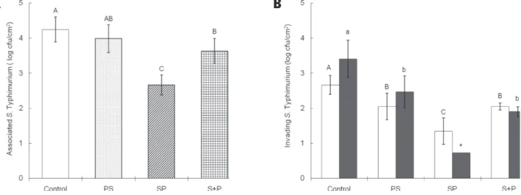
The adhesion ability of S. Typhimurium to INT-407 cells were evaluated under different treatment conditions: i) INT-407 infected with Salmonella or phage alone (control), ii) phage treatment followed by Salmonella infection (PS), iii) Salmonella infection followed by phage treatment (SP), and iv) combination treatment with Salmonella and phage (S+P) (Fig.
Figure 1 – Adhesion ability of S. Typhimurium (A) intracellular survival at 0 hpi (£) and 24 (■) hpi (B) to INT-407 cells infected with Salmonella or phage alone (control), phage treatment followed by Salmonella infection (PS), Salmonella infection followed by phage treatment (SP), and combination treatment with Salmonella and phage (S+P). Means with different letters (A-C and a-b) are significantly different at p < 0.05. The asterisk (*) indicates the bacterial count below the detection limit (0.73 log cfu/cm2).
1A).Compared with the control, the ability of S. Typhimurium to adhere to INT-407 cells significantly decreased in SP treatment, followed by S+P treatment. The associated numbers of S. Typhimurium were 4.24, 3.99, 2.65, and 3.63 cfu/cm2, respectively, for the
control, PS, SP, and S+P.
The invasion and survival properties of S. Typhimurium were evaluated in INT-407 cells treated with PS, SP, and S+P at 0 and 24 hpi (Fig. 1B). S. Typhimurium cells were more invasive in the control than in the phage treatments, including PS, SP, and S+P. The invasive ability was significantly decreased in SP, corresponding to the adhesion ability. The numbers of intracellular S. Typhimurium were 2.66, 2.05, 1.34, and 2.05 log cfu/cm2, respectively, at 0 hpi. The
phage treatment after Salmonella-infection effectively blocked the internalization of S. Typhimurium in theINT-407 cells. Compared with the control, the intracellular survival of S. Typhimurium in INT-407 cells was slightly increased in the control and PS at 24 hpi, while that was marginally decreased in SP and S+P. The intracellular survival of S. Typhimurium was evaluated in HD11 treated with and without phage (Fig. 2). The number of intracellular S. Typhimurium in HD11 was 3.76 log cfu/cm2 at 0 hpi, which was reduced to 3.44
log cfu/cm2 (51% reduction) in the absence of phage
and 2.93 log cfu/cm2 (83% reduction) in the presence
of phage at 24 hpi.
Figure2 – Survival of intracellular S. Typhimurium infected in HD11 cells treated without (–) and with (+) phage P22 for 24 h at 37oC. Means with different letters (a-b) are significantly different at p < 0.05.
Inflammatory-mediated gene expression in Salmonella-infected HD11 cells
The relative expression profiles of pro-inflammatory and TLR genes were determined in HD11 cells cultured with and without phage at 24 hpi (Fig. 3). The relative expression levels IFN-g, IL-10, IL-1b, IL-6, IL-8, iNOS,
and IL-12 mRNAs increased, while those of IL-16, TLR2-1, TLR3, and TLR7decreased in HD11 cells at 24 hpi. The levels of IL-13, IL-15, IL-17, IL-18, IL-3, TLR2-2, TLR4, and TLR5 mRNAs remained constant in HD11 cells at 24 hpi. The mRNA levels increased between 2 to 7-fold for IFN-g, 10, 1b, 6, 8, iNOS, and IL-12. Although no significant differences were observed in the expression levels of pro-inflammatory and TLR mRNAs between absence and presence of phages, the expression levels of pro-inflammatory cytokines were slightly higher in the presence of phages than the absence of phages.
DISCUSSION
Enteric bacteria can pass through the epithelial barrier, infect macrophage, and then develop survival strategies against the phagocytosis-associated cellular defense system. Phage can be a potential approach for controlling intracellular pathogens. This study describes the potential role of phage P22 in the adhesion and invasion of S. Typhimurium in the intestinal epithelial cells and the macrophage-mediated immune response to intracellular S. Typhimurium.
The phage treatment after Salmonella infection effectively excluded the attached S. Typhimurium from the INT-407 cells (Fig. 1A). Phage can be used as a possible approach to treat rather than prevent bacterial infections. This observation indicates that there were no significant changes in the lytic activity or binding affinity of phages against the adherent S. Typhimurium, resulting in the bacterial invasion of host cells.
There are two bacterial invasion mechanisms, including zippering (affinity binding of bacteria adhesins and host receptors) and triggering (injection of effector proteins into the host cells via TTSS) events (Ó Cróinín & Backert, 2012). Bacterial cells can internalize and even reproduce within the nonphagocytic epithelial cells (Kim et al., 2011).
The numbers of intracellular S. Typhimurium were reduced to below detection limit in the SP treatment (Fig. 1B). The phage treatment after Salmonella -infection most successfully inhibited the growth of intracellular S. Typhimurium in INT-407 cells. This implies that the phage-infected S. Typhimurium entered and was then lysed within the INT-407 cells. The number of intracellular S. Typhimurium in HD11 was significantly reduced to 2.93 log cfu/cm2 (83%
of survival processes, SPI-1 and SPI-2 (Flannagan et al., 2009). The significant reduction in the number of internalized S. Typhimurium in HD11 cells might be due to the inflammatory immune responses to the phage P22.
The phage P22 marginally induced immune responses in HD11 cells at 24 hpi (Fig. 3). The expression of IL-1β, IL-6, and IL-8 was up regulated in Salmonella -infected macrophages (Wigley, 2004, Withanage et al., 2004). IL-1β is a pro-inflammatory cytokine and
IL-6 is a pro- and anti-inflammatory cytokine (Lee et al., 2010). The inflammatory cytokines were inhibited by IL-10 and IL-13, which were increased during the initial stage of bacterial infection (Rothwell et al., 2004, Setta et al., 2012). The expression level of iNOS mRNA in bacteria-infected HD11 cells was enhanced by IFN-g but not IL-18 (He et al., 2011). IL-18 acts as an inducer of cellular immune responses against intracellular pathogens when combined with IL-12 (Biet et al., 2002). Lipopolysaccharide-induced TNF-afactor (LITAF) is involved in the initiation of cytokine cascade in response to Salmonella infection (Ma et al., 2010). TLR2, TLR4, and TLR5 are activated on the host cell surface, whereas TLR3 and TLR7 are activated within the host cells in response to bacterial infections (Ishii et al., 2005). TLR2 was primarily involved in the induction of anti-inflammatory cytokine, IL-10 (Netea
et al., 2004). TLR2 was stimulated by TLR-4 which was
not up-regulated after Salmonella
infection (Higgs et al., 2006). The TLR activation is mainly responsible for the cytokine induction in macrophages. However, the levels of TLR transcripts were decreased in this study. This result implies that the pro-inflammatory gene expression may be induced by a non-TLR-mediated recognition pathway (Bliss et al., 2005).
In conclusion, the most significant findings of this in-vitrostudy were:1) the phage treatment after Salmonella
infection effectively inhibited the invasion ability and intracellular survival of S. Typhimurium in epithelial INT-407 cells, and 2) although no significant macrophage-mediated inflammatory immune responses were observed in HD11 cells exposed to phage, the number of intracellular
S. Typhimurium was significantly reduced in HD11 cells at 24 hpi. This in-vitro study provides useful information for understanding the role of phage in inhibiting extracellular and intracellular pathogens and also improving phage therapy at the cellular level. However, further in-vivo studies are needed to elucidate the complex gene regulatory networks of the inflammatory immune system, which is the ongoing investigation of our laboratory.
ACKNOWLEDGEMENT
This study was supported by Research Grant from Kangwon National University.
REFERENCES
Antunes LC, Buckner MM, Auweter SD, Ferreira RB, Lolic PFinlay BB. Inhibition of Salmonella host cell invasion by dimethyl sulfide. Applied and Environmental Microbiology 2010;76:5300-5304.
Baxter MA, Fahlen TF, Wilson RLJones BD. HilE interacts with HilD and negatively regulates hilA transcription and expression of the Salmonella enterica serovar Typhimurium invasion phenotype. Infection and Immunity 2003;71:1295-1305.
Bielke L, Higgins S, Donoghue A, Donoghue DHargis BM. Salmonella host range of bacteriophages that infect multiple genera. Poultry Science 2007;86:2536-2540.
Biet F, Locht CKremer L. Immunoregulatory functions of interleukin 18 and its role in defense against bacterial pathogens. Journal of Molecular Medicine 2002;80:147-162.
Bliss TW, Dohms JE, Emara MGKeeler CL. Gene expression profiling of avian macrophage activation. Veterinary Immunology and Immunopathology 2005;105:289-299.
Flannagan RS, Cosio GGrinstein S. Antimicrobial mechanisms of phagocytes and bacterial evasion strategies. Nature Reviews Microbiology 2009;7:355-366.
Golkar Z, Bagasra OPace DG. Bacteriophage therapy: a potential solution for the antibiotic resistance crisis. Journal of Infection in Developing Countries 2014;8:129-236.
Han Y, Niu M, An LLi W. Involvement of TLR21 in baculovirus-induced interleukin-12 gene expression in avian macrophage-like cell line HD11. Veterinary Microbiology 2010;144:75-81.
He H, Genovese KJKogut MH. Modulation of chicken macrophage effector function by TH1/TH2 cytokines. Cytokine 2011;53:363-369.
Higgs R, Cormican P, Cahalane S, Allan B, Lloyd AT, Meade K, James T, Lynn DJ, Babiuk LAO’Farrelly C. Induction of a novel chicken Toll-like receptor following Salmonella enterica serovar Typhimurium infection. Infection and Immunity 2006;74:1692-1698.
Hong YH, Lillehoj HS, Lee SH, Dalloul RALillehoj EP. Analysis of chicken cytokine and chemokine gene expression following Eimeria acervulina and Eimeria tenella infections. Veterinary Immunology and Immunopathology 2006;114:209-223.
Iqbal M, Philbin VJ, Withanage GSK, Wigley P, Beal RK, Goodchild MJ, Barrow P, McConnell I, Maskell DJ, Young J, Bumstead N, Boyd YSmith AL. Identification and functional characterization of chicken Toll-like receptor 5 reveals a fundamental role in the biology of infection with
Salmonella enterica serovar Typhimurium. Infection and Immunity 2005;73:2344-2350.
Ishii K, Coban CAkira S. Manifold mechanisms of Toll-like receptor-ligand recognition. Journal of Clinical Immunology 2005;25:511-521.
Kawai T Akira S. Innate immune recognition of viral infection. Nature Immunology 2006;7:131-137.
Kim JS, Eom JS, Jang JI, Kim HG, Seo DW, Bang I-S, Bang SH, Lee ISPark YK. Role of Salmonella pathogenicity island 1 protein iacP in Salmonella enterica serovar Typhimurium pathogenesis. Infection and Immunity 2011;79:1440-1450.
Kubori T Galan JE. Salmonella type III secretion-associated protein InvE controls translation of effector proteins into host cells. Journal of Bacteriology 2002;184:4699-4708.
Lee SH, Lillehoj HS, Hong YH, Jang SI, Lillehoj EP, Ionescu C, Mazuranok LBravo D. In vitro effects of plant and mushroom extracts on immunological function of chicken lymphocytes and macrophages. British Poultry Science 2010;51:213-221.
Livak KJ Schmittgen TD. Analysis of relative gene expression data using real-time quantitative PCR and the 2-∆∆CT method. Methods
2001;25:402-408.
Lu TK Koeris MS. The next generation of bacteriophage therapy. Current Opinion in Microbiology 2011;14:524-531.
Ma J, Zhang Y-g, Xia YSun J. The inflammatory cytokine tumor necrosis factor modulates the expression of Salmonella typhimurium effector proteins. Journal of Inflammation 2010;7:42.
Netea MG, van der Graaf C, Van der Meer JWMKullberg BJ. Toll-like receptors and the host defense against microbial pathogens: bringing specificity to the innate-immune system. Journal of Leukocyte Biology 2004;75:749-755.
Ó Cróinín T Backert S. Host epithelial cell invasion by Campylobacter jejuni: trigger or zipper mechanism? Frontiers in Cellular and Infection Microbiology 2012;2:1-13.
Rothwell L, Young JR, Zoorob R, Whittaker CA, Hesketh P, Archer A, Smith ALKaiser P. Cloning and characterization of chicken IL-10 and its role in the immune response to Eimeria maxima. Journal of Immunology 2004;173:2675-2682.
Setta AM, Barrow PA, Kaiser PJones MA. Early immune dynamics following infection with Salmonella enterica serovars Enteritidis, Infantis, Pullorum and Gallinarum: Cytokine and chemokine gene expression profile and cellular changes of chicken cecal tonsils. Comparative Immunology, Microbiology and Infectious Diseases 2012;35:397-410.
Smith CK, Kaiser P, Rothwell L, Humphrey T, Barrow PAJones MA.
Campylobacter jejuni-induced cytokine responses in avian cells. Infection and Immunity 2005;73:2094-2100.
Vikram A, Jayaprakasha GK, Jesudhasan PR, Pillai SDPatil BS. Obacunone represses Salmonella pathogenicity islands 1 and 2 in an envZ-dependent fashion. Applied and Environmental Microbiology 2012;78:7012-7022.
Wigley P. Genetic resistance to Salmonella infection in domestic animals. Research in Veterinary Science 2004;76:165-169.
Withanage GSK, Kaiser P, Wigley P, Powers C, Mastroeni P, Brooks H, Barrow P, Smith A, Maskell DMcConnell I. Rapid expression of chemokines and proinflammatory cytokines in newly hatched chickens infected with
Salmonella enterica Serovar Typhimurium. Infection and Immunity 2004;72:2152-2159.
Yamamoto KR, Alberts BM, Benzinger R, Lawhorne LTreiber G. Rapid bacteriophage sedimentation in the presence of polyethylene glycol and its application to large-scale virus purification. Virology 1970;40:734-744.