THE
JOURNAL • RESEARCH • www.fasebj.org
Pth4, an ancient parathyroid hormone lost in eutherian
mammals, reveals a new brain-to-bone
signaling pathway
Paula Suarez-Bregua,* Eva Torres-Nuñez,* Ankur Saxena,†,‡Pedro Guerreiro,§ Ingo Braasch,{ David A. Prober,†Paloma Moran,kJose Miguel Cerda-Reverter,#Shao Jun Du,** Fatima Adrio,†† Deborah M. Power,§Adelino V. M. Canario,§John H. Postlethwait,{Marianne E. Bronner,† Cristian Cañestro,‡‡ and Josep Rotllant*,1
*Institute of Marine Research, Spanish National Research Council (IIM–CSIC), Vigo, Spain;†California Institute of Technology, Pasadena, California, USA;‡Department of Biological Sciences, University of Illinois, Chicago, Illinois, USA;§Center of Marine Sciences (CCMAR), University of Algarve, Faro, Portugal;{Institute of Neuroscience, University of Oregon, Eugene, Oregon, USA;kDepartment of Biochemistry, Genetics, and Immunology, University of Vigo, Vigo, Spain;#Institute of Aquaculture Torre de La Sal (IATS–CSIC), Castell´on, Spain; **Department of Molecular and Cellular Biology, University of Maryland, Baltimore, Maryland, USA;††Department of Cell Biology, University of Santiago de Compostela, Santiago de Compostela Spain; and‡‡Department de Gen`etica, Microbiologia i Estad´ıstica, Institut de Recerca de la Biodiversitat (IRBio), Universitat de Barcelona, Barcelona, Spain
ABSTRACT:Regulation of bone development, growth, and remodeling traditionally has been thought to depend on endocrine and autocrine/paracrine modulators. Recently, however, brain-derived signals have emerged as key regulators of bone metabolism, although their mechanisms of action have been poorly understood. We reveal the existence of an ancient parathyroid hormone (Pth)4 in zebrafish that was secondarily lost in the eutherian mammals’ lineage, including humans, and that is specifically expressed in neurons of the hypothalamus and appears to be a central neural regulator of bone development and mineral homeostasis. Transgenic fish lines enabled mapping of axonal projections leading from the hypothalamus to the brainstem and spinal cord. Targeted laser ablation dem-onstrated an essential role for ofpth4-expressing neurons in larval bone mineralization. Moreover, we show that Runx2 is a direct regulator ofpth4 expression and that Pth4 can activate cAMP signaling mediated by Pth receptors. Finally, gain-of-function experiments show that Pth4 can alter calcium/phosphorus levels and affect expression of genes involved in phosphate homeostasis. Based on our discovery and characterization of Pth4, we propose a model for evolution of bone homeostasis in the context of the vertebrate transition from an aquatic to a terrestrial lifestyle.—Suarez-Bregua, P., Torres-Nuñez, E., Saxena, A., Guerreiro, P., Braasch, I., Prober, D. A., Moran, P., Cerda-Reverter, J. M., Du, S. J., Adrio, F., Power, D. M., Canario, A. V. M., Postlethwait, J. H., Bronner, M E., Cañestro, C., Rotllant, J. Pth4, an ancient parathyroid hormone lost in eutherian mammals, reveals a new brain-to-bone signaling pathway. FASEB J. 31, 569–583 (2017). www.fasebj.org
KEY WORDS:runx • phosphate • calcium • hypothalamus • fgf23
Bone is a highly specialized vertebrate-specific tissue that functions as a supporting structure characterized by its resistance, stiffness, and high capacity for regeneration and repair. To achieve these characteristics, bones are first
shaped during early life and then constantly remodeled throughout adulthood. Bone remodeling is a lifelong process that relies on complex regulatory mechanisms to achieve proper rates of growth and differentiation.
ABBREVIATIONS: Actb1, actin-b1; BLAST, Basic Local Alignment Search Tool; BMD, bone mineral density; dpf, days post fertilization; eef1a1/1, elongation factor 1-a1 like 1; eGFP, enhanced green fluorescent protein; entpd5, ectonucleoside triphosphate diphosphohydrolase 5; fgf23, fibroblast growth factor 23; FISH, fluorescence ISH; HEK, human embryonic kidney; hpf, hours post fertilization; hrct, hypocretin gene; IBMX, 3-isobutyl-1-methylxantine; ISH, in situ hybridization; npt2a, sodium-phosphate cotransporter type IIa; MO, morpholino oligonucleotide; OGM, ohnologs gone missing; PFA, paraformaldehyde; phex, endopeptidase on the X chromosome; pl, primary lamella; pth1r, parathyroid hormone 1 receptor; PTH, parathyroid hormone; PTHLH, parathyroid hormone like-hormone; qPCR, quantitative PCR; slc34a1a, solute carrier family 34 sodium/phosphate cotransporter 1a; RACE, rapid amplification of cDNA ends; stc1l, stanniocalcin-1-like; TFBS, transcription factor binding site; Tg, transgenic; TPp, periventricular nucleus of posterior tuberculum; TSS, translation start site; TU, T ¨ubingen; vBMD, volumetric bone mineral density; VGD, vertebrate genome duplication; VOI, volume of interest; WGD, whole-genome duplication; WISH, whole-mount ISH; WT, wild-type
1Correspondence: Institute of Marine Research, Spanish National Research Council (IIM-CSIC), Vigo, Spain. E-mail: [email protected] doi: 10.1096/fj.201600815R
Vertebrates regulate bone mineralization and remodeling at 3 main levels: delivery of systemic hormones [e.g., parathyroid hormone (PTH) peptides and calcitonin], turnover of bone cells (e.g., osteoblasts, osteoclasts, and osteocytes), and auto-regulatory feedback loops mediated by cell-signaling products [e.g., fibroblast growth factor (FGF)-23, and ectonucleoside triphosphate diphosphohy-drolase (ENTPD)-5)] (1, 2). In addition to the complex network of endocrine, autocrine, and paracrine hormone signals that tightly regulate bone mineral metabolism, re-cent studies suggest that the brain is a pivotal regulator of bone homeostasis through 3 distinct pathways. The first pathway includes hormonal signals that arise from neuroendocrine neurons of the hypothalamus and are subsequently processed in the pituitary [e.g., leptin, pro-opiomelacortin-derived peptides, gonadotrophin hormone releasing hormone (GnRH), and thyrotropin releasing hormone] (3–6). The second pathway consists of neuronal factors released by the hypothalamus and distributed through the bloodstream [e.g., cocaine-and amphetamine-regulated transcript, neuropeptide Y, agouti-related pep-tide, and brain-derived neurotrophic factor (6)]. The third pathway consists of efferent neuronal signaling that regu-lates the sympathetic and parasympathetic nervous sys-tems (7). The high interconnectivity of the hypothalamus in the brain makes this structure one of the most powerful regulatory regions of the body, integrating signals not only from peripheral tissues but also from other areas within the brain itself. Understanding the novel regulatory axes be-tween brain and bone cells is therefore critical for a better understanding of bone biology.
Using a reverse genetic approach, we identified a new brain-to-bone pathway involving efferent neural signals from the hypothalamus to receptors on bone cells con-trolling skeletal mineralization and phosphate homeosta-sis in teleosts. The key factor of this new brain-to-bone pathway appears to be an ancestral PTH family member that has been conserved in ray-finned fish, lobe-finned fish, and noneutherian mammals, but surprisingly has been lost in eutherian vertebrates.
The PTH family of peptides plays a central role in phosphocalcic homeostasis, bone remodeling, and em-bryonic skeleton development in vertebrates (8–11). In tetrapods, PTH is synthesized by the parathyroid glands (PTGs) and plays a pivotal role in bone turnover by reg-ulating the metabolism of calcium and phosphate. Fish have homologs of PTH and PTH-like peptides (i.e., PTH2 and PTHLH). In contrast to tetrapods, however, none of these fish PTH peptides seems to regulate calcium and phosphate metabolism during bone turnover (10, 12). These taxon-specific differences are likely related to the transition from aquatic to terrestrial environments that occurred during the evolution of tetrapods, in which new mecha-nisms for controlling bone homeostasis became associated with the evolution of the PTH family and possibly facilitated the transition from an aquatic to terrestrial lifestyle, which was accompanied by greater stress on skeletal elements.
We describe experiments that demonstrate that the novel PTH family member Pth4 is unique to noneutherian vertebrates and acts as a neuropeptide produced by a specific subset of neurons located in the dorsal part of the
periventricular hypothalamus, with axonal projections leading from the hypothalamus to the brainstem and spinal cord. Loss- and gain-of-function experiments pro-vided functional evidence that Pth4 is a powerful regu-lator of bone mineral accrual acting through phosphate homeostasis. We also provide evidence that Runx2 di-rectly regulates Pth4 upstream and downstream, all Pth receptors facilitate activating cAMP signaling. Finally, in light of our new data and a reexamination of the evolution of the PTH family in fish and tetrapods, we propose a model describing the evolution of Pth signaling related to bone remodeling in the context of vertebrate evolution.
MATERIALS AND METHODS
Fish
Fish (zebrafish, Danio rerio) were cultured as previously described (13) and staged by hours post fertilization (hpf) or days post fer-tilization (dpf), according to standard criteria (14). Experiments were performed with a T ¨ubingen (TU)-wild-type (WT) strain [(TU), N ¨usslein-Volhard Laboratory, Max Planck Institute, T ¨ubingen, Germany]. For some experiments, embryo medium was supplemented with 0.003% (w/v) 2-phenylthiourea to inhibit embryo pigmentation (13). For histology, dechorio-nated embryos were fixed overnight at 4°C in 4% para-formaldehyde (PFA) in 13 PBS, washed in PBS, and stored at 4°C in 13 PBS for confocal imaging or dehydrated through a
methanol series and stored at220°C in 100% methanol for in situ
hybridization (ISH). Ethics approval (ref: AGL2014-52473R) for all studies was obtained from the Institutional Animal Care and Use Committee of the IIM-CSIC Institute in accordance with the Na-tional Advisory Committee for Laboratory Animal Research Guidelines licensed by the Spanish Authority (RD53/2013) and conformed to European animal directive (2010/63/UE) for the protection of experimental animals.
DNA cloning
The putative zebrafish pth4 gene was identified by searching the zebrafish genome database, Zv9, with translated Basic Local Alignment Search Tool [tBLASTn; National Institutes of Health, National Center for Biotechnology Information (NCBI), Bethesda, MD, USA; http://blast.ncbi.nlm.nih.gov/Blast.cgi] using the previously identified sequence of zebrafish Pth1a, Pth1b, Pth2 (Tip39), Pthlha, and Pthlhb paralogs (accession nos.: NM_212950, NM_212949, NP_991140, NP_001019798.2, and NP_001036789.1, respectively). Full-length cDNA was isolated by using sequence-specific primers (Supplemental Table S2) in 59 and 39 Smart RACE (rapid amplifi-cation of cDNA ends) cDNA amplifiamplifi-cation kit (TaKaRa, Mountain View, CA, USA), and sequenced by Sanger method. The zebrafish pth4 gene sequence has been submitted to the European Molecular Biology Laboratory (EMBL)/NCBI GenBank database (accession no. KT182088; https://www.ncbi.nlm.nih.gov/genbank).
Phylogenetic analysis and comparative genomic of conserved synteny
Multiple sequence alignment was made by the Muscle pctid-log algorithm, corrected manually for inconsistencies and trimmed to decrease the total number of gaps (Supplemental Fig. S3). Phylogenetic analysis was inferred by maximum likelihood us-ing PhyML 3.0 subjected to LG substitution model, a bionj starting tree improved by NNI, and computing aLRT SH-like
branch support (15). Analyses of conserved synteny were per-formed using automated pipeline tools of the Synteny Database variant Ens61 (16). Dot-plots represent the chromosomal distri-bution of human ohnologs [a special type of paralogs resulting by whole-genome duplication (WGD) (17)] of gene neighbors of the PTH family. The first 20 Mb of human chromosome 11 (Hsa11), which includes the human PTH gene, were analyzed taking the genomes of the urochordate Ciona intestinalis and the cepha-lochordate Branchiostoma floridae as outgroups for the identification of ohnologs resulting from the first and second rounds of verte-brate genome duplication [VGD1 and -2; for details see Cañestro et al. (18)]. Clusters in the Synteny Database were obtained by coupling results from the reciprocal best-hit BLAST pipeline with the use of a 50- or 25-gene sliding window analysis that links orthologous chromosome segments of 2 species by conserved synteny (16). The Synteny Database is especially useful to provide evidence of ohnologs gone missing (OGM) by uncovering the putative chromosomal region that still preserves paralogous syn-tenic conservation, but has lost a certain ohnolog of interest (18, 19). Real-time quantitative PCR
Temporal expression of pth4 was determined by real-time PCR. Total RNA was isolated at 0, 3, 6, 9, 12, and 14 hpf and 1, 2, 3, 4, 5, and 6 dpf, using Trizol reagent (Thermo Fisher Scientific) and first-strand cDNA was synthesized according to the Maxima First Strand cDNA Synthesis Kit (Thermo Fisher Scientific) pro-tocol starting with 500 ng of total RNA. The partial sequence of pth4 was amplified by using DreamTaq DNA Polymerase (Thermo Fisher Scientific) and specific primers (Supplemental Table S1). 18S ribosomal cDNA amplification was used as a positive control. Gene markers of phosphocalcic metabolism were analyzed by real-time quantitative PCR (qPCR). Total RNA from TU-WT and Tg(Xla.Eef1a1:pth4)iim010 adult male zebrafish (n = 6 per group) was extracted and reverse-transcribed as de-scribed above with specific primers (Supplemental Table S1). Each
sample was amplified in triplicate containing 12.5ml of Maxima
SYBR Green/ROX qPCR Master Mix (23) (Thermo Fisher Sci-entific), 0.5ml 0.2 mM of each primer, 10.5 ml nuclease-free water,
and 1mlcDNAtemplate.Real-time qPCR reactionswereanalyzed
on a 7500 Fast Real-Time PCR System (Thermo Fisher Scientific) with the following cycling conditions: initial denaturation at 95°C for 10 min, followed by 40 cycles at 95°C for 15 s and 60°C for 1 min. Gene expression was assessed in 2 independent experiments using the efficiency calibrated method (20), and relative mRNA expression levels were normalized to 18S, eukaryotic translation elongation factor 1-a1-like 1 (eef1a1/1) and actin b1(actb1). Whole-mount ISH and immunohistochemistry Whole-mount ISH (WISH) was performed using digoxigenin-labeled antisense riboprobes (21). Antisense and sense ribo-probes were made from linearized full-length zebrafish pth4, pth2, and pth1ra cDNA. For plastic sections, embryos with ISH
staining were fixed, dehydrated, Epon embedded, cut into 4mm
rostrocaudal transverse sections, mounted with Eukitt (EMS, Hatfield, PA, USA) as previously described (13) and imaged with a DM2500 microscope (Leica, Wetzlar, Germany). ISH of adult zebrafish sections (360 dpf) followed published methods (22). Samples were fixed in 4% PFA and 0.1 M phosphate buffer, for 2 d at 4°C, dehydrated and embedded in Paraplast (Sherwood, St. Louis, MO, USA). Serial 10-mm rostrocaudal transverse sec-tions were cut on a rotary microtome. Secsec-tions were mounted on 3-aminopropyltriethoxylane-treated slides, air-dried at room temperature overnight, and stored at 4°C in dry conditions and used for hybridization within 1 month. Double labeling of en-hanced green fluorescent protein (eGFP) immunofluorescence and fluorescence ISH (FISH) was performed in 3-dpf Tg(pth4:
eGFP)iim07 zebrafish embryos. A sheep anti-DIG-POD antibody (Roche Diagnostics, Indianapolis, IN, USA) was used and pth4 probe labeling was detected by tyramide signal amplification (TSA Plus Cyanine 3 and Fluorescein system; PerkinElmer, Waltham, MA, USA). Expression of eGFP was visualized with a mouse anti-eGFP primary antibody (Roche Diagnostics) fol-lowed by a horse anti-mouse fluorescein-conjugated secondary antibody (Vector Laboratories, Burlingame, CA, USA). For cryostat sectioning, Tg(pth4:eGFP)iim07 adult fish were fixed in 4% paraformaldehyde overnight at 4°C, washed with PBS, transferred to 15% sucrose, followed by 30% sucrose, and then embedded and frozen in optimal cutting temperature medium.
Cross-cryosections of 25 mm thickness were collected on
polylysine slides (Thermo Fisher Scientific) and allowed to dry. Slides were washed in PBS, mounted in Vectashield, and im-aged on a TCS SP5 (Leica) confocal microscope. Cartilage–bone staining was performed with a 2-color acid-free stain (21).
cAMP basedb- galactosidase reporter gene assay
To demonstrate whether Pth4 was able to bind and signal
through PTH receptors, we performed cAMP-based
b-galacto-sidase reporter gene assays. We amplified the predicted region for the pth4 mature peptide and full coding regions of the pth1ra, pth2r [splice variant pth2r (23) and pth1rb; previous names: pth1r, pth2r, and pth3r, respectively] from cDNA template by PCR using Pfu Ultra II HS DNA polymerase (Agilent Technologies, Santa Clara, CA, USA) and specific primers (Supplemental Table S2), and cloned the fragments into pSC-B-amp/kan (Agilent Tech-nologies). Inserts were subcloned into pcDNA3 (Thermo Fisher Scientific). All constructs were verified by sequencing both DNA strands. Signaling via cAMP was assessed using a human em-bryonic (HEK)-293 cell clone (Clon-Q) that stably expresses b-galactosidase under the control of human vasoactive intestinal peptide promoter placed downstream of tandem repetitions of
cAMP response elements (24). Cells were transfected with 1.5mg
for pcDNA3-pth4 or 1mg of pcDNA3-pth1ra, pcDNA3-pthr2, or
pcDNA3-pth1rb, using Lipofectamine LIX and Plus Reagent according to the manufacturer’s protocol (Thermo Fisher Scien-tific). A construct carrying the eGFP gene under the control of a cytomegalovirus constitutive promoter was also transfected to standardize transfection levels. After 24 h, transfection was assessed by analyzing the eGFP fluorescence and transfected cells with receptors were plated in 96-well plates (50,000 cells/well). Cells expressing Pth4 mature peptides were rinsed with PBS and lysed by thermal shock, spin dried at 1100 relative centrifugal force (rcf) for 5 min at 22°C, and the supernatant collected. Recombinant cells expressing each receptor were stimulated for 6 h at 37°C with 100ml of decreasing concentrations (1:2, 1:4, 1:8, 1: 16, 1:32, 1:64, and 1:128 dilutions) of peptide solution from cel-lular lysates containing 0.1 mM of 3-isobutyl-1-methylxantine (IBMX; Sigma-Aldrich, St. Louis, MO, USA), a phosphodiesterase inhibitor that prevent enzymatic degradation of cAMP. As
pos-itive controls, we used 0.001 mM forskolin and 1026M human (h)
PTH (Sigma-Aldrich) (25). Measurements were normalized for protein content, which was determined using the BCA protein assay kit (Thermo Fisher Scientific). Negative controls were per-formed using cells transfected with empty pcDNA3 vector (Thermo Fisher Scientific). All experiments were performed in triplicate. Data are presented as a percentage of negative control
values. cAMP basedb-galactosidase reporter gene assays were
performed, as previously described (26, 27). Transgene construction and microinjection
To ectopically express pth4, 3 independent transgenic (Tg) zebrafish lines were generated with the Tol2 transposon system: Tg (Xla.Eef1a1:pth4)iim010, Tg(Xla.Eef1a1:pth4)iim011, and Tg(Xla.
Eef1a1:pth4)iim012. The Tol2 vector was kindly provided by Koichi Kawakami, National Institute of Genetics, Mishimi, Japan (28). The pth4 PCR product was subcloned into pGEM-T easy vector (Promega, Madison, WI, USA) with BamHI and NotI re-striction sites. pGEM-T-pth4 was digested with BamHI and NotI and subcloned into pT2AL200R150G cut with the same restriction enzymes. The Tol2-transposon-based vector pT2AL200R150G contained an eGFP gene under the control of constitutive promoter of the elongation factor 1-a (Eef1a1). We replaced the eGFP gene with the pth4 gene. To generate pth4 reporter lines, zebrafish pth4 promoter sequences containing 2 and 1 kb upstream to the translation starting site (TSS) of the pth4 gene (GenBank accession no. CU856139.10) for the Tol2 transposon constructs used in this study were amplified from 6 dpf zebrafish genomic DNA with KOD Xtrem Hot Start DNA Polymerase Platinum (71975; Nova-gen, Madison, WI, USA) and specific primers (Supplemental Table S3). The PCR products (2394 and 890 bp) were diluted 1:10, and
1 ml was ligated into 1 ml of P-ENTR/D-TOPO Cloning Kit
(Thermo Fisher Scientific) and transformed according to the manufacturer’s instructions. Both promoter sequences were then ligated by LR Clonase into the destination vector pToleGFPDest (274; Lawson Lab, Malvern, PA, USA) containing the eGFP se-quence downstream, and sese-quenced. Construct P1 (1kb-pth4-Tol2-eGFP) contained 873 nucleotides upstream to the TSS of the pth4 gene. Construct P2 (2kb-pth4-Tol2-eGFP) contained 2377 nucleo-tides upstream to the TSS of the pth4 gene. Three P2 independent Tg zebrafish lines Tg(pth4:eGFP)iim07, Tg(pth4:eGFP)iim08, and Tg(pth4:eGFP)iim09 were generated. To study the regulatory re-gion of pth4, potentially cis-acting transcription factor binding sites (TFBSs) located in the zebrafish pth4 promoter sequence were identified with MatInspector software (29). Deletion and substi-tution mutations were created as has been described (30) or by using the QuickChange Site-Directed Mutagenesis Kit (Stratagene, La Jolla, CA, USA). The resulting clones were tested though re-striction analysis and confirmed by sequencing. All Tol2 constructs (Supplemental Tables S3, S4) were dissolved in distilled RNAase-free water to a final concentration of 50mg/ml. A total of 250 pg of construct and synthetic 59-capped mRNA (150 pg) encoding a transposase were coinjected into TU-WT embryos at the 1- or 2-cell stage, with 1% of phenol red as a tracer. Microinjection was per-formed under a dissection microscope (MZ8; Leica) fitted with a MPPI-2 pressure injector (ASI Systems, Omaha, NE, USA). The expression of eGFP was analyzed by direct observation of eGFP fluorescence under a fluorescence stereoscope and a confocal mi-croscope. The number of embryos positive for eGFP fluorescence was determined, and the fluorescence distribution was compared between different Tg lines to score activity and tissue specificity. Morpholino knockdown
Antisense morpholino oligonucleotides (MOs; GeneTools, Phil-omath, OR, USA) were designed against runx2a, runx2b, and runx3, as previously described, including a splice-target MO against both runx2a isoforms and translational start blocking MOs targeting of each isoform of runx2b (runx2bT1 and runx2bT2) and runx3 (31, 32). A scrambled MO with no known target in zebrafish, cMO, 59–CCTCTTACCTCAGTTACAATTTATA–39 was used as control. In addition, runx-MOs and/or p53 (p53-MO) (33) were also used to target runx and p53 genes, respectively. The MOs were resuspended in water to a final concentration of 0.5 mM. Approximately 2 nl was injected into 1- or 2-cell stage Tg reporter embryos. Knockdown embryos were analyzed for eGFP fluorescence at 2 dpf, and pth4 transcription was assessed by ISH and real-time qPCR.
Two-photon laser ablation
Embryos were mounted, imaged, and cells ablated as previously described (34). pth4:eGFP and bactin2:mCherry double-positive
Tg zebrafish were visualized throughout development. The hy-pothalamic area was imaged in vivo at 1, 2, 3, and 7 dpf. The 2 clusters of eGFP-expressing cells and the number of cells in each
stage of development were evaluated by analyzing each 2
mm-thick z-plane slice from a full confocal z-stack projection and scored. Two rounds of ablations were performed per experi-mental embryo at different time points (1 and 2 dpf). This pro-tocol was chosen as the most efficient of several attempted iterations for a complete ablation process at each time point and to achieve low neuronal recovery during development. After-ward, ablated and control larvae were imaged as full z-stack images to monitor recovery of the Pth4-expressing neurons. Micro-CT scanning and whole-body
mineral contents
Volumetric bone mineral density (vBMD) and whole-body cal-cium, phosphorus, and magnesium were determined in TU-WT and Tg(Xla.Eef1a1:pth4)iim010 adult male zebrafish (n = 20). Fish were euthanized with 2-phenoxyethanol (1:500), fixed in 2% PFA, and scanned with a high-resolution micro-CT SkyScan 1172 (micro-CT NV; Bruker, Kontich, Belgium) at 60 KV X-ray source
voltage and 167mA source current with an image pixel size of
5mm. Internal structures were reconstructed according to the
Feldkamp convolution back-projection algorithm (35). In the scanned area, including a portion of the head and the rostral portion of the spine, 4 different volumes of interest (VOIs) were selected to determine the vBMD, using standard software provided with the equipment (SkyScan CT-Analyzer 1.10.0.2; Bruker). vBMDs were calculated by direct calibration against the attenuation coefficients of 2 hydroxyapatite phantoms with
known densities (250 and 750 mg/cm3).
For whole-body calcium, phosphorus, and magnesium de-termination, fish were freeze dried until dry (dry weight) and dissolved in concentrated nitric acid (70%; 3 ml/g dry weight; Sigma-Aldrich). Nitric acid sample digestion were diluted 500-fold with demineralized water and analyzed for calcium, phosphorus, and magnesium using inductively coupled plasma optical emis-sion spectrophotometry (4300 DV, Optima; Perkin Elmer). Mineral content was expressed in milligrams per gram dry weight, based on the fish dry weight and the total sample digestion volume. Statistics
Data are expressed as means6SEM. Differences between groups
were assessed by Student’s t test. A value of P , 0.05 indicated statistical significance. Comparisons between numerical data were evaluated by 1-way ANOVA with the Tukey’s post hoc test. Data were deemed to be statistically similar if they shared at least 1 letter. Statistical analysis and figures were performed with PASW Statistics 18.0 (SPSS; IBM, Armonk, NY, USA) and Sig-maPlot 12.0 (San Jose, CA, USA), respectively.
RESULTS
Identification and genomic characterization
of the pth4 gene
Using BLAST analysis, we identified a previously un-annotated new zebrafish paralog of the pth gene family on zebrafish chromosome Dre22: 597996-600181 (GRCz10) that we call pth4. Results from 59- and 39-RACE revealed that the pth4 full-length transcript was 809 bp long and consists of an open reading frame of 366 bp, a 94-bp 59-UTR and a 349-bp 39-UTR (Supplemental Fig. S1). Aligning the
cDNA to the genome sequence predicted that the pth4 gene structure consists of 2 exons and 1 intron. The pth4 cDNA predicted translation was a 121-aa sequence that contained a secretion signal sequence of 24 aa in the N terminus (http://cbs.dtu.dk/services/SignalP/), as expected for PTH peptides. Predicted cleavage sites (http://www.cbs. dtu.dk/services/ProP/) should yield preprosequences of 31 AAs and a mature peptide of 90 aa. Protein sequence analysis revealed conserved regions including a dibasic cleavage site (RR) and an MHD motif.
Phylogenetic analysis and evolutionary origin
of the pth4 gene
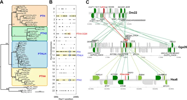
Phylogenetic analyses inferred by maximum likelihood and rooted with Pth protein sequences from the cepha-lochordate B. floridae (36) showed that zebrafish Pth4 be-longs to a well-supported clade that includes Pth4 from other teleosts and from spotted gars, coelacanths, chickens, turtles, and 2 marsupial mammals: opossums and Tas-manian devils (Fig. 1A). However, we could identify no Pth4 genes in human, mouse, or any eutherian mammalian genomes in Ensembl-v79. To investigate the evolutionary relationships of the pth4 of zebrafish and other teleosts in the context of the entire PTH-related gene family and to
better understand the apparent absence of an ortholog of PTH4 in humans, mice, and other eutherian mammals, we performed a comparative analysis of conserved syn-teny. Dot-plot analysis of conserved syntenies for 20 Mb encompassing the genomic neighborhood of the human PTH gene located on chromosome 11 revealed that paralogs of most PTH neighbor genes landed on Hsa12 and -19, where PTHLH and PTH2, respectively, were also located. This finding was consistent with a recent analysis of a 6-Mb window around PTH2 in Hsa19 (37). The dot plot also revealed other chromosomes, including Hsa1 and -6, with paralogs of numerous PTH neighbor genes, but in which no PTH-related gene has been identified (Fig. 1B). This situation would be expected if these regions formed part of the 4 paralogons that would have been generated by the 2 rounds of WGD that took place during early vertebrate evolution (38, 39) [reviewed in Cañestro (40)]. Analysis of gene clusters using the Synteny Data-base (16) revealed that paralogs of 21 of the 28 closest neighbor genes to the zebrafish pth4 gene in Dre22 were located in Hsa1 or -6 (Supplemental Fig. S2). Comparison of the genomic neighborhood of Pth4 in chromosome 26 of chickens (Fig. 1C; Gga26), which is more closely related to humans than to zebrafish, revealed that paralogs of 9 of the 10 nearest neighbor paralogs in a 150-kb window are
Figure 1. Phylogenetic and conserved synteny analysis of PTH related proteins. A) A phylogenetic tree inferred by maximum likelihood, showing the relationship between PTH4 proteins and other proteins of the PTH, PTH2, and PTHLH families. B)
Dot-plot analysis shows the distribution of paralogs (black dots) of the PTH neighbor genes in thefirst 20 Mb of human chromosome
11 (Hsa11 on the x axis) throughout all chromosomes of the human genome (y axis) using both the amphioxus B.floridae and
the ascidian C. intestinalis genomes as outgroups. Yellow shadow areas highlight conserved syntenic regions that probably originated in 2 rounds of WGD that took place during early vertebrate evolution (38, 39) [reviewed in Cañestro (40)], including Hsa12 and -19, where the ohnologs PTH2 and PTHLH are located (blue circles), and other regions including Hsa1 and -6, where PTH ohnologs are missing (PTH-OGM, red circle). C) Analysis of the Synteny Database revealed that the genomic neighborhoods of the pth4 gene in zebrafish and chickens show extensive conserved synteny with Hsa6, suggesting that the putative paralogon region in which the ancestral PTH4-OGM should have been located before it was lost (transparent red) in the lineage leading to humans after the split of the lineage leading to current armadillos, whereas it has been preserved (red) in chicken Gga26 and teleosts such as in zebrafish Dre22. Sequence references may be found in Supplemental Fig. S3.
specifically in Hsa6. This finding strongly suggested that an ancestral PTH4 ohnolog was present in the last com-mon ancestor of ray-finned fish (Actinopterygii, including zebrafish) and lobe-finned fish (Sarcopterygii, including coelacanths, chickens, and humans), but was lost in the lineage leading to humans after the eutherian–metatherian split; furthermore, our analysis revealed that the PTH4-OGM (ohnolog gone missing) should have been in Hsa6 before it was lost, whereas it was preserved in other noneutherian vertebrate lineages, including chickens and teleosts, such as zebrafish.
Temporal and spatial expression ofpth4
during embryonic and larval development We examined the spatial expression of pth4 by ISH in zebrafish. Starting at 24 hpf, embryos displayed pth4 expression in 2 bilateral spots in the lateral ventral di-encephalon corresponding to the region of the developing hypothalamus (Fig. 2A, B). We observed a gradual num-ber and movement of cells within the 2 bilateral clusters during development (Fig. 2A–F). At 3 dpf, pth4 appeared to be specifically expressed by 2 subsets of neurons in the lateral hypothalamus, distant from pth2-expressing cells (Fig. 2G, H). In adult fish (360 dpf), bilateral cell clusters were detected in the dorsal zone of the periventricular hypothalamus (Fig. 2I). Expression of endogenous pth4 was never detected by eGFP expression and ISH outside this hypothalamic area in embryonic and later stages. Analysis of temporal expression of pth4 by real-time PCR (Fig. 2J) revealed that pth4 was maternally transcribed, being detected in embryos as early as 0 hpf and showing a progressive decrease until 9 hpf. Zygotic expression of pth4 appeared to increase from 12 hpf through subsequent development and then to increase at 4, 5, and 6 dpf.
A neural network for pth4-expressing cells
Having shown that pth4 was specifically expressed in the hypothalamus, we generated independent zebrafish Tg lines containing eGFP reporter constructs including 1 kb (P1 = 1kb-pth4-Tol2-eGFP) or 2 kb (P2 = 2kb-pth4-Tol2-eGFP) fragments upstream of the pth4 TSS. A search for eGFP signal in all P1 and P2 Tg zebrafish lines and in situs to the pth4 transcript was performed carefully throughout the organism and showed that the eGFP expression patterns replicated the spatiotemporal pattern of endogenous pth4 transcript (Fig. 3). At 1 dpf, the P2 line labeled 2 bilateral clusters of 4–6 neural bodies, each as well as their axonal projections, which extended both to the anterior and pos-terior part of the brain from the lateral hypothalamus (Fig. 3A, B). Double labeling experiments showed a robust colocalization of endogenous pth4 transcripts and eGFP signal (Fig. 3C–C0). At 3 dpf, the number of labeled neu-ronal bodies had increased up to 14 per cluster, and long neural projections labeled by eGFP extended ventrally from the caudal part of the diencephalon through the midbrain and hindbrain and along the spinal cord (Fig. 3D, E). Posterior projections were arranged in 2 parallel axes showing long-range neuronal fibers running through
the spinal cord (Fig. 3F). At 5 dpf, the number of neural cell bodies did not appear to have changed, but projections formed a complex and branched neural network (Fig. 3G). One-year-old adult fish showed a high density of branched fibers around the cluster of neurons in the dorsal
Figure 2. Spatial and temporal expression of PTH 4 (pth4) transcripts in zebrafish embryos. A–F) Gradual movement of the expression domain during development from caudoventral to a more rostrodorsal situation with reference to the eye position. Lateral (A, C, E) and ventral (B, D, F) views of whole-mount ISH of pth4 expression of 1 dpf (A, B), 2 dpf (C, D), and 3 dpf (E, F) wild-type zebrafish embryos. Staining with the pth4 antisense probe showed expression in bilaterally symmetric regions of the developing
hypothalamus. G, H) Rostrocaudal transverse sections (4mm thick)
of Epon-embedded, whole-mount double ISHs of pth2 (G, red arrowheads) and pth4 (G, black arrowheads) in 3 dpf zebrafish identified pth4 expression domains in the lateral hypothalamus (H).
I) Rostrocaudal transverse section (10 mm thick) of
paraffin-embedded ISH of pth4 expression in adult zebrafish displayed pth4 transcripts exclusively in bilateral cell clusters located in the dorsal zone of the periventricular hypothalamus. J) Real-time PCR analyses of the temporal expression pattern of pth4 mRNA. e, eye; Hd, dorsal zone of the periventricular hypothalamus; hy, hypothalamus; Hv, ventral zone of the periventricular hypothalamus; m, midbrain; t, telencephalon; TPp, periventricular nucleus of posterior
area of the periventricular hypothalamus (Fig. 3I) and throughout the spinal cord (Fig. 3H).
To further characterize the cluster of neural bodies expressing pth4 in the hypothalamus, we crossed the P2 line to a hcrt:tdTomato line [hypocretin gene (hrct), a marker of the lateral hypothalamus in zebrafish embryos] (Fig. 3J). Although both genes were expressed in the hy-pothalamic region, they were expressed in different neu-ral cell types, revealing the presence of different neuneu-ral subpopulations in this part of the hypothalamus.
The discovery of this complex pth4-expressing neural network sending widespread projections from the hypo-thalamus throughout the brain and spinal cord suggests that Pth4 is a neuropeptide playing a systemic role throughout the entire animal.
Functional characterization of pth4 promoter
and upstreamrunx regulation
The minimal regulatory region required for pth4 promoter activity was identified by generating serial deletions of the 2-kb promoter sequence upstream of the pth4 TSS and
directed mutagenesis of the 1-kb promoter sequence up-stream of the TSS (Fig. 4A). Injection of the 2kb-pth4-Tol2-eGFP (n = 174) or 1kb-pth4-Tol2-2kb-pth4-Tol2-eGFP (n = 208) reporter constructs showed the same efficient eGFP expression, suggesting that essential regulatory elements of pth4 were located in the first 1-kb sequence. Promoter efficiency in all reporter constructs injected was normalized to the 2kb-pth4-Tol2-eGFP construct activity. Deletion of the re-gion 941–554 bp did not alter the pth4 pattern replicated by eGFP expression, but decreased the efficiency of expres-sion to 43%. Deletion of the region 544–262 bp, however, resulted in a complete loss of eGFP expression in all of 144 injected embryos, demonstrating that this critical 292-bp fragment contained regulatory elements essential for pth4 expression. TRANSFAC sequence analysis revealed the presence of at least 6 potential transcription factor-binding boxes within this critical 292 bp (Fig. 4A and Supplemental Fig. S4). Mutations designed to disrupt consensus-binding sequences of the predicted transcription factors (Supple-mental Fig. S4, red) significantly affected eGFP expression in all injected embryos. Mutations in boxes 1 and 4 de-creased the number of embryos expressing eGFP by.50%, similar to the 941–554 deletion (Fig. 4A). Mu-tations in boxes 2, 3, and 5 decreased the efficiency of expression by 77% or more of the injected embryos. Mutation in box 6 resulted in a complete loss of eGFP expression in all injected embryos. This mutated con-served region had homology to the core binding site for RUNX transcription factors. Thus, these results sug-gested that RUNX transcription factors most likely act as key regulators of pth4 promoter activity.
The expression pattern of zebrafish runx paralogs (i.e., runx1, runx2a, runx2b, and runx3) is highly dynamic, and all of them have been described as playing a role in carti-lage and bone development (31). Among these paralogs, the runx2a expression pattern includes a bilateral domain compatible with the 2 clusters of pth4-positive cells in the hypothalamus (41, 42). To test whether zebrafish Runx2a regulates the pth4 promoter in vivo, we used runx2a-specific antisense morpholino-knockdown approach and tested for 2kb-pth4-Tol2-eGFP activity, as well as for pth4 expression. Knockdown by injecting runx2a-MO into the P2 reporter line revealed that by 2 dpf, the formation of the 2 clusters of neuronal eGFP-positive cell bodies was sig-nificantly altered: 6.3% of injected embryos did not show any pth4 positive cells, 41.4% showed absence of one of the neural clusters and the other was severely reduced (in most cases only 1 cell), and 52.3% showed 2 clusters but with a reduced number of positive cells (Fig. 4B, C). To provide further evidence that the runx2a morphant phe-notypes are specific and not related to nonspecific off-target effects [e.g., p53 mediated apoptosis (33)], we also analyzed eGFP expression in vivo in runx2a morphants that were coinjected with a p53 morpholino. Similar to previous experiments, the formation of the 2 eGFP+ clus-ters of neurons was severely affected suggesting that our phenotype was not caused by nonspecific apoptosis (Fig. 4D). Animals injected with p53-MO alone did not exhibit a phenotype in our region of interest (data not shown). Moreover, WISH (Fig. 4E–G) as well as real-time qPCR (Fig. 4H) showed significant reduction of pth4 transcript
Figure 3. Whole-mount confocal imaging of the stable Tg(pth4: eGFP)iim07 zebrafish line. A–G) Pth4 cells and projections at 1 (A, B), 2 (C–C0), 3 (D–F), and 5 dpf (G) monitored as eGFP fluorescence. Double labeling of eGFP immunofluorescence
and fluorescence pth4 ISH (FISH) on 3-dpf Tg(pth4: eGFP)
iim07 zebrafish line (C–C0) demonstrates identical expression of eGFP (green color) and endogenous pth4 transcripts (red). Confocal imaging of 25-mm cross-sections from adult fish, showing the Pth4 anterior–posterior caudal projections in the spinal cord (H ) and Pth4 bilateral clusters in the periventricular hypothalamic area (I ). J ) Double pth4:eGFP and hcrt:tdTomato zebrafish line showing the specific spatial colocalization of Pth4 and Hcrt neural clusters in the lateral hypothalamus at 5 dpf. e, eye; hd, dorsal hypothalamus; hv, ventral zone of periventricular hypothalamus; hy, hypothalamus; m, midbrain; SC, spinal cord; TPp, periventricular nucleus of posterior tuberculum; VC,
expression in runx2a morphants, similar to the decrease of expression observed for eGFP.
Results from the promoter deletions and direct muta-genesis, together with knockdown experiments, show that Runx2a is a direct upstream activator of pth4 expression. That the expression of pth4 was not completely abolished in 100% of the animals and that MOs against runx2bT1, runx2bT2, and runx3 also affect pth4 expression (unpublished results),
suggest the possibility of a certain degree of redundancy or cooperative action among runx paralogs.
Pth4 interacts with all 3 zebrafish Pth receptors
Analysis of the zebrafish genome revealed the presence of 3 Pth receptors (i.e., Pth1ra ENSDARG00000020957, Pth1rb
Figure 4. Functional analysis of the pth4 promoter through eGFP expression. A) DNA fragments containing 2- and 1-kb sequences upstream from the ATG of pth4 are in gray. Vertical lines mark increments of the functional 2kb-pth4 promoter deleted. Filled circles: conserved regions selected for mutations. Box 1, positions 507–513 (TFBSs): CREB/HRE/ATF6); box 2, 433–439 (TFBS: HMX2/LXRE); box 3, 397–402 (TFBS: HOXA5/BRN5/LHX6/VAX2/ISL1); box 4, 329–338 (TFBS: AP1/NFE2/MEIS1/ PBX1_MEIS1/VDR_RXR); Box 5, 321–323 (TFBS: VMAF/CREB/MARE); and box 6, 518–522(TFBS: RUNX2/RUNX3). Arrow and hatched section: the start of transcription and 59UTR, with eGFP coding sequences substituting at the native TSS. Promoter
activity was assessed as the proportion of embryos displaying 1 or more fluorescent Pth4 cells at 3 dpf and normalized to the
wild-type 2kb-pth4 promoter results. B–H) Knockdown of runx2a affects pth4 expression. B–D) Morphology of the control (B, ventral view), runx2a-MO-injected (C, ventral view), and runx2a-MO- and p53-MO-injected (D, ventral view) Tg(pth4:eGFP)iim07 embryos
at 48 hpf. Note the reduced eGFPfluorescence in the runx2a-MO-injected embryos. Scale bars, 100 mm. E–G) WISH using pth4
riboprobe on control (E, ventral view), runx2a-MO-injected (F, ventral view), and runx2a-MO- and p53-MO-injected (G, ventral view) Tg(pth4:eGFP)iim07 embryos at 48 hpf. Note the reduced pth4 expression in the runx2a-MO-injected embryos. H ) Real-time qPCR analysis of pth4 expression in control (white bars), runx2a-MO-injected (black bars), and runx2a-MO- and p53-MO-injected (gray
bars) Tg(pth4:eGFP)iim07 embryos at 48 hpf. Results normalized to eef1a1/1 expressed as means 6 SEM of 2 independent
ENSDARG00000018418, and Pth2r ENSDARG00000006678) that can act downstream of Pth4. To determine whether a specific receptor mediates Pth4 signaling, we expressed zebrafish Pth receptors in HEK cells and performed a cAMP-basedb-galactosidase reporter gene assay. All re-ceptors showed similar basal induced cAMP production. Pth4 stimulation induced a dose–response increase in cAMP production and no significant differences were detected with any of the 3 receptors transiently transfected (Fig. 5A, B). These results suggest that Pth4 signaling could be mediated promiscuously by all 3 Pth receptors. We also transfected cells with human Pth and found that all 3 zebrafish receptors increased cAMP production, with Pth1ra being activated the most efficiently and Pth2r the lowest, which is consistent with in vitro analysis published using human PTH and zebrafish receptors (25) (Fig. 5B). It is interesting to note that zebrafish Pth4 activated Pth1ra with efficiency similar to that of human PTH, suggesting that human PTH (i.e., PTH1) and zebrafish Pth4 exert equivalent effects at the ligand–receptor level.
In humans, PTH peptides play crucial roles in bone homeostasis via PTH1R on osteoblasts (43). Therefore, in addition to Pth4-Pthr signaling, we also analyzed the spatial expression of zebrafish pth1ra in adult zebrafish. Expression of pth1ra was specifically found in gills (Fig. 5C, D); craniofacial bones, such as ceratobranchial and oper-culum (Fig. 5C); intestinal epithelium (Fig. 5E), and spinal cord (Fig. 5F). These results are consistent with a Pth4-Pth1ra signaling pathway controlling bone mineral metabolism through the main target organs (i.e., gills, in-testinal epithelium, and bone) that are known to promote ionic fluxes via absorption/reabsorption or mobilization.
Targeted pth4-expressing neuron ablation
results in reduced bone mineralization during embryo development
Using the zebrafish Tg(pth4:eGFP)iim07 Tg line, in which Pth4-expressing neurons were labeled by eGFP (Fig. 3), 2-photon laser ablation was used to eliminate the mCherry-positive nuclei (bactin2:H2A-mCherry) of individual pth4: eGFP-positive neural bodies in the hypothalamus at 2 se-quential time points (1 and 2 dpf). Successful ablation was demonstrated by the instant loss of mCherry and sub-sequent loss of eGFP (Fig. 6A–D). Observations of the morphology after ablation did not uncover damaging ef-fects to neighboring tissue. When examining ablated lar-vae at 7 dpf, 5 d after conducting the double (1 and 2 dpf) laser ablation protocol, the lack of pth4:eGFP-positive neural bodies was evident in comparison to control larvae (Fig. 6B, D). Absence of pth4:eGFP-positive neurons resulted in a significant decrease in Pth4 expression levels, as assayed by whole-embryo real-time qPCR (Fig. 6I). A 2-color acid-free cartilage and bone stain (Fig. 6E–H) showed that ablated larvae showed a marked decrease in bone mineralization of craniofacial structures, such as the notochord tip, operculum, otolith, cleithrum, and cerato-branchial arch 5 at 7 dpf, whereas the formation of head cartilage was unaffected. These experiments, therefore, sug-gest that the pth4:eGFP-positive neurons in the hypothala-mus are essential brain regulators of bone mineralization.
Likewise, it is important to remark that ablating pth4-expressing neurons should affect all factors downstream of Pth4, so the effect we detect in bone mineralization could be secondary to the release of other factors from the brain that act downstream of Pth4.
Overexpression of pth4 induces loss of BMD
and changes in whole-body mineral contents in Tg zebrafish
To investigate the biological role of Pth4 in adult fish, we created a zebrafish Tg line ubiquitously expressing pth4 driven by the eef1a1 promoter Tg(Xla.Eef1a1:pth4)iim010. The strong, widespread, and stable Tg Pth4 expression was assessed by real-time qPCR (Supplemental Fig. S5).
Figure 5. Pth4 interaction with zebrafish Pth receptors. A, B) Effects of recombinant Pth4 mature peptide dilutions (A) and synthetic hPTH (B) on galactosidase activity in HEK293
cells stably expressing cAMP-responsive b-galactosidase
re-porter gene and transiently transfected with zebrafish pth1ra, pth1rb, and pth2r genes in the presence of a phosphodiesterase
inhibitor, IBMX. Results represent the means 6 SEM of 2
independent biological replicate experiments, each performed with 3 technical replicates. Measurements were normalized for protein content and expressed as a percentage of basal levels (HEK293 cells transfected with empty pcDNA3 vector).
Values of P , 0.05 (indicated by letters on bars) were
considered statistically significant. Data are statistically similar if bars share at least 1 letter. C–F) ISH of adult zebrafish sections shows specific pth1ra expression in gills (C, D), craniofacial bones (C ), intestine (E ), and spinal cord (F ). g, gills; op, opercular bone; cb, ceratobranchial bone; sc, spinal cord; pl, primary lamella; sl, secondary lamella; ie, intestinal epithelium.
Characterization of whole-body mineral contents (i.e., calcium, phosphorus, and magnesium) by spectropho-tometry in adult male zebrafish (n = 20) showed a signif-icant decrease in minerals in pth4 Tg fish compared with control TU-WT (Fig. 7A). We wondered whether the de-crease in whole-body mineral contents could be related to ion mobilization from bone. To test this hypothesis, we analyzed volumetric bone mineral density (vBMD) and found a significantly lower vBMD in all volumes of in-terest analyzed in pth4 Tg zebrafish (P, 0.01; Fig. 7B). We concluded, therefore, that Pth4 can mobilize minerals from bone to extracellular fluids and then the increase of mineral concentration at the extracellular level leads to its removal from the body, which affects calcium/phosphorus com-position in the whole-body and skeletal structure.
Phosphate homeostasis is disrupted inpth4
Tg zebrafish
To investigate whether pth4-dependent changes in body calcium/phosphorus contents and in vBMD could be re-lated to alterations in expression levels of genes involved in mineral homeostasis, we performed real-time qPCR to determine the levels of expression of several phosphocalcic metabolism regulatory genes (Supplemental Table S1) in adult fish of both TU-WT and pth4-Tg lines (Fig. 8). Results revealed that pth4-Tg fish had a significant (P, 0.05) up-regulation of the solute carrier family 34 type II sodium/ phosphate cotransporter member 2b gene (slc34a2b, npt2b) but a significant (P , 0.01) down-regulation of a set of phosphate-regulating genes, including the endopeptidase on the X chromosome (phex), ectonucleoside triphosphate diphosphohydrolase 5 (entpd5), and solute carrier family 34 type II sodium/phosphate cotransporter member 1a
(slc34a1a, npt2a). Notably, the down-regulation of npt2a was accompanied by a significant (P , 0.025) up-regulation of fgf23, which has been described as a key regulator of phosphate homeostasis (44). Finally, no sig-nificant differences were found on stanniocalcin-1-like (stc1l) gene expression levels. Overall, results of pth4-overexpression experiments revealed that pth4 not only had an important role affecting the expression of effector proteins responsible for bone metabolism through regula-tion of phosphate homeostasis (i.e., npt2a, npt2b, phex, and entpd5) but also affected a regulatory pathway (fgf23). DISCUSSION
The ancestral Pth4, which was lost in eutherian vertebrates, is a regulator of bone homeostasis
The PTH gene family regulates bone homeostasis in ver-tebrates. To date, 3 ohnologs have been described—PTH, PTH2, and PTHLH (45)—that appear to have originated via 2 rounds of 1R/2R WGDs during early vertebrate evolution (38) (Fig. 9A). In teleosts, moreover, pth and pthlh each have 2 paralogs—ptha/pthb and pthlha/pthlhb—that originated in the extra round of WGD that occurred during early teleost evolution (3R or teleost genome duplication) (37). We identified and characterized a novel Pth family member, Pth4. Our phylogenetic inferences and analysis of conserved syntenies shared by the pth4 genomic neighborhood in zebrafish and its ohnologons in chickens and humans provide evidence consistent with the hy-pothesis that Pth4 is the fourth missing PTH ohnolog resulting from 1R/2R-WGDs. Our genome survey shows that Pth4 is present in ray-finned fish and in noneutherian
Figure 6. Two-photon laser ablation of Pth4 neurons. A–D) Whole-mount confocal imaging of the pth4:eGFP+
/bactin2:H2A-mCherry+double Tg embryo, showing the Pth4 neural hypothalamic bodies at 1 (A, C) and 7 dpf (B, D), monitored as eGFP
fluorescence in control (A, B) and laser-ablated (C, D) embryos. E–H) Two-color acid-free cartilage and bone staining of control (E, ventral view, and F, lateral view) and Pth4 neuron-ablated (G, ventral view, and H, lateral view) 7 dpf embryos. Note the lack of mineralized craniofacial bones with an incipient mineral deposit on teeth in Pth4 neuron ablated embryos. I ) Real-time qPCR analysis of pth4 expression in control (white bars) and Pth4 neuron-ablated (black bars) embryos at 7 dpf. Results are normalized
to actb1 and expressed as means 6 SEM of 2 independent experiments. nt, notochord tip; op, operculum; ot, otolith;
vertebrates but absent in eutherians because of a gene loss that likely occurred at the base of the eutherian mamma-lian clade (i.e., a eutherian OGM; Fig. 9A). For the sake of clarity and to better reflect the evolutionary origin of the Pth family, we propose a change in the nomenclature of this vertebrate family, in which PTH and PTHLH should be renamed to PTH1 and -3, respectively. Accordingly, after the nomenclature of teleost genes duplicated during the 3R, ptha and pthb should be thought of as pth1a and pth1b; pth2 remains as pth2; pthlha and pthlhb should be thought of as pth3a; and pth3b and the newly identified gene should be called pth4.
From brain to bone: Pth4, a hypothalamic neural signaler downstream of Runx that regulates bone remodeling in zebrafish
Expression analysis during zebrafish development revealed that pth4 is maternally transcribed. By 24 hpf, however, pth4 is expressed exclusively in a bilateral subset of neurons located in the hypothalamus. Transgenic re-porter lines expressing eGFP under the pth4 and hrct pro-moters identified 2 distinct subpopulations of neuronal cell bodies in the lateral hypothalamus. The pth4:eGFP re-porter line also mapped a pth4-positive complex network of long axonal projections from the dorsal zone of the periventricular hypothalamus to the brainstem and spinal cord. Promoter dissection identified a region of 544–262 bp that is critical for pth4 expression. Results of TRANSFAC sequence prediction, promoter region deletions and di-rected mutagenesis uncovered 5 potential regulatory ele-ments driving pth4 expression. In particular, Runx-factor binding sites appeared to be critical for pth4 expression.
Loss of Runx2a activity abrogated both pth4 expression and the activity of the pth4:eGFP reporter line, dem-onstrating that pth4 is downstream of runx2a. In mam-mals, RUNX2 is a multifunctional transcription factor implicated in bone development and mineralization by
Figure 7. A) Whole-body calcium, phosphorus and magnesium analysis. B) Representative images for VOI1: end lapilli otoliths to
end of sagittal otoliths; VOI2: end sagittal otoliths to 5 vertebrae (included pectoralfins and scales); VOI3: end sagittal otoliths to
5 vertebrae but includes only a cylinder (diameter of 1 mm) with the center in the middle of the vertebral bodies; and VOI4:
VOI1+VOI2; and vBDM analysis. a, analfin; c, caudal fin; d, dorsal fin; pec, pectoral fin; pel, pelvic fin. Results are expressed as
means6SEM. *P, 0.05, **P , 0.025, ***P , 0.01.
Figure 8. Phosphate homeostasis is disrupted in pth4 Tg fish. Real-time qPCR analysis of mineral homeostasis regulatory factors in TU-WT (white bars) and Tg(Xla.Eef1a1:pth4)iim010 adult male zebrafish (black bars). mRNA expression levels of fibroblast growth factor 23 (fgf23), phosphatase orphan 1 (phospho1), phosphate-regulating gene with homologies to endopeptidase on the X chromosome (phex), (entpd5), solute carrier family 34 (type II sodium/phosphate cotransporter), member 1a (slc34a1a, npt2a), member 2a (slc34a2a, npt2b), and stanniocalcin-1-like (stc1l) were determined. Results normalized
to 18S are expressed as means6SEMof 2 independent experiments.
regulating numerous genes of chondrogenic/osteogenic signaling pathways (46). RUNX2 binding sites have been identified on the COL10A1 and SP7 promoter controlling their transcriptional regulation (47, 48). Using the pth4 eGFP reporter lines, we demonstrated that the ablation of pth4-positive neurons altered bone mineralization during larvae development, affecting craniofacial structures, such as the notochord tip, operculum, otolith, cleithrum, and ceratobranchial arch. However, no difference in cartilage development was found. Taken together, these findings suggest that a population of hypothalamic neurons that express pth4 under the regulation of the conserved Runx2a bone transcription factor form a complex neural network throughout the body that plays a major role in the mech-anisms regulating embryonic bone mineralization in zebrafish.
Pth4 can activate cAMP/PKA signalingvia Pth
receptors and regulates expression of genes involved in calcium/phosphorus homeostasis Zebrafish pth4 is predicted to encode a putative protein of 121 aa that includes PTH-specific motifs and dibasic cleavage sites for preprosequences conserved in other se-creted members of the PTH family (11, 49). We demon-strated that Pth4 can recognize the 3 zebrafish Pth receptors (i.e., Pth1ra, Pth1rb, and Pth2r) to activate cAMP/PKA signaling. In fish, previous analyses by real-time PCR found that pth2r was expressed in the central nervous system, kidney and scales (23, 45, 50), and pth1rb in the kidney, intestine, and vertebral bone (51–53). In the current
study, WISH revealed that pth1ra is expressed in the gills, intestinal epithelium, craniofacial bones, and spinal cord (Fig. 5). The fact that Pth4 shows the same affinity for all 3 receptors in cell culture suggests that this neuropeptide not only acts on the skeleton, either directly or indirectly through the spinal cord, but also, in ectopic/overexpression experiments, has the potential to function in gills, intestine, kidney, and scales, all of which are involved in mineral homeostasis.
Gain-of-function experiments using a Tg line that overexpresses pth4 demonstrate that an excess of this neuropeptide induces a significant reduction of whole-body calcium/phosphorus levels and results in a signifi-cant loss of vBMD, acting through the regulation of phosphate homeostasis. Our analysis of factors involved in the regulation of phosphate levels revealed that pth4 overexpression induced a significant increase in fgf23 and npt2b levels and a decrease in npt2a, phospho1, phex, and entpd5 expression levels. FGF23 is a key phosphaturic factor that acts in the bone–kidney axis in mammals and provides a signal from bone to adjust phosphate fluxes through NPT2a in kidney by promoting phosphate ex-cretion or reabsorption (44, 54). In addition, systemic phosphate levels are managed by NPT2b through active transport in the small intestine (55). In zebrafish, fgf23 is expressed in the corpuscles of Stannius (CS), on the teleost kidney, to regulate ion homeostasis. Expression of npt2b in the intestine plays an important role in phosphate ab-sorption (56). Thus, the significant decrease in whole-body phosphorus content in Tg fish could be related to up-regulation of fgf23 and down-up-regulation of npt2a, which in
Figure 9. Evolutionary history of the PTH family in vertebrates. A) Analysis reveals 4 PTH ohnologs (PTH1–4) that arose because of 2 rounds of 1R/2R WGD in early vertebrates and third round (3R) in teleosts (a/b paralogs). The absence of Pth4 in eutherian mammals is related to an ancestral gene loss that likely occurred when this clade diverged from metatherian mammals. B) This loss is concomitant with the recruitment of PTH1 from parathyroid glands in eutherians to provide an equivalent function to the Pth4 neuropeptide in bone mineral homeostasis, as it is represented by zebrafish and human diagrams, in which both PTH peptides provoke similar responses in phosphate and calcium homeostasis, acting in bone and regulating the expression of the same regulatory genes in homologous organs.
turn would produce phosphate excretion through the kidney. Such an effect may be counteracted by phosphate absorption via the intestine, consistent with high levels of npt2b. In accordance with our results, other studies have demonstrated that loss of function of phospho1, phex, or entpd5 in hypophosphatemic disorders led to a significant decrease in vBMD and abnormalities in mineralization (1, 57, 58). One of our most striking results was that despite hypocalcemic conditions in Tg fish, the expression of stc1l did not appear to occur, as would be expected from its function in zebrafish (59). This result suggests that anti-hypercalcemic hormones other than stc1l (e.g., calcitonin) may act in teleosts to reduce calcium transport across ep-ithelia as its mammalian counterpart (60).
Evolutionary model for the role of the PTH family in bone homeostasis in aquatic and terrestrial vertebrates
Despite the lack of parathyroid glands, fish can respond to changes in serum mineral levels and consequently regu-late mineral concentrations within a narrow range. During the past 2 decades, several studies focused on identifying PTH paralogs and explored their involvement in bone mineral balance in vertebrates. Six peptides and 3 recep-tors had been identified previously, revealing a higher complexity of this gene family in noneutherian vertebrates than in eutherians. Pthlh (i.e., Pth3) has been reported to promote calcium uptake from surrounding water via branchial and intestinal epithelia (61), stimulating calcium reabsorption and phosphate secretion through Npt2a in the kidney (53) and promoting osteoclast activity in scales via Pthr (62). In addition to calcium uptake, Pth3 seems to affect Piconcentration in the serum of fish species (10). In
contrast to placental mammals, no previous evidence supported the hypothesis that Pth1 or Pth2 played a de-cisive role in mineral balance in fish. In eutherians, PTH1 is considered to be the principal regulator of calcium/ phosphate metabolism and bone turnover. This hormone is produced by the parathyroid glands in response to low calcium or high phosphate serum levels and exerts its endocrine function on kidney and bone by interacting with other systemic and local factors, such as vitamin D, FGF23 and calcitonin to restore normocalcemia in a negative feedback loop (63, 64). Our studies suggest that zebrafish Pth4 acts on bone to mobilize calcium and phosphate through Pth1r signaling. Moreover, a PTH-like peptide previously identified in pufferfish and designated PTH-L (here renamed as Pth4) shares a high similarity in the 1- to 34-aa sequence with zebrafish Pth4 and showed calcio-tropic activity in an in vitro scale assay (12). This finding probably reflects the ancestral bony vertebrate condition that has been conserved in noneutherians but lost in eu-therian mammals. Concomitantly Pth was recruited to perform an equivalent function in the latter (Fig. 9).
In terrestrial vertebrates, bone provides an internal storage of minerals that have an important metabolic function by mobilizing calcium and phosphate for mineral homeostasis. In fish, calcium-driven mineral homeostasis has been a matter of debate because aquatic vertebrates
have an abundant supply of calcium in freshwater and marine environments. As a consequence, it is likely that the skeleton would act only as an internal calcium source under conditions of mineral shortage. Instead, the post-cranial dermal skeleton (scales) would be more susceptible to resorption than the endoskeleton (62). Conversely, phosphate levels in water are generally low and unlikely to account for all body functions. Thus, the circulating concentration of phosphate depends on dietary Piintake,
intestinal absorption, renal filtration/secretion and reab-sorption, and exchange from intracellular and bone res-ervoirs. For this reason, fish face mineral challenges different from those of terrestrial vertebrates with respect to the availability of these 2 elements (65, 66). In conditions of severe phosphate shortage (e.g., low Pidietary
avail-ability or systemic Pi homeostasis disorder), fish would
need expeditious mobilization of phosphate ions to ensure the survival. Under these circumstances, the scales and endoskeleton would be the faster and more accessible sources for phosphate mobilization (Fig. 9B). In this con-text, ancient Pth4 would act in bone to promote phosphate mobilization and consequently mobilization of calcium. Our findings suggest a prominent role for Pth4 in main-tenance of systemic phosphate homeostasis via control of bone mineral content, the major reservoir for calcium and phosphate. We cannot, however, exclude potential actions of Pth4 in gills and in intestinal reabsorption of phosphate through epithelia. During the vertebrate transition from the aquatic to terrestrial life, the parathyroid glands arose via transformation of the gills (67), with PTH1 emerging as the endogenous master regulator of bone mineral ho-meostasis. We hypothesize that during evolution of tet-rapods, PTH1 gradually became the principal regulator of the calcium/phosphate metabolism and Pth4 was then lost in the eutherian vertebrates caused by redundancy. Our findings illustrate, therefore, the relevance of the identification of gene loss events to better understand the evolution and diversification of animal lineages (68).
ACKNOWLEDGMENTS
The authors thank Yi-lin Yan (University of Oregon) for providing the tip39 (PTH2) expression vector; Prof. N. Lawson (University of Massachusetts Medical School, Worcester, MA, USA) and Prof. K. Kawakami (National Institute of Genetics, Mishima, Japan) for providing the iTol2 constructs; D.A.P. for the hcrt:tdTomato line and Prof. K. M. Kwan (University Of Utah, Salt Lake City, UT, USA) for the bactin2:H2A-mCherry line; In´es Pazos and Jes ´us M´endez [Scientific and Technolog-ical Research Assistance Centre (CACTI), University of Vigo] for their advice and assistance with the confocal microscope; Rub´en Chamorro and Rosa Ceinos (IIM–CSIC) for their help in
handling and care of the fish and David Guede and Jos´e R.
Caeiro (University of Santiago de Compostela, Santiago, Spain) for their advice and assistance with the micro-CT SkyScan. This work was funded by the Spanish Economy and Competitiveness Ministry Project ALG2011-23581 and AGL2014-52473R (to J.R.); the Portuguese Foundation for Science and Technology (Project PTDC/BIA-ANM/4225/2012-phos-fate; to P.G.); U. S. National Institutes of Health/Office of the Director Grant R01OD011116 (alias R01 RR020833; to J.H.P.); Generalitat de Catalunya (Grant SGR2014-290) and the Spanish Economy and Competitiveness Ministry (Project BFU2010-14875; to C.C.). Partial funding was
obtained from Science and Innovation Ministry (AGL2010-22247-C03-01; to J.M.C.-R.), a Campus do Mar Ph.D. grant, and Xunta de Galicia (Santiago, Spain; Project AGL2014-52473R) (to P.S.B.). C.C. and J.R. are co-senior authors.
AUTHOR CONTRIBUTIONS
J. Rotllant and P. Suarez-Bregua conceived, designed, and coordinated the study; C. Cañestro, I. Braasch, and J. H. Postlethwait performed comparative genomics analyses; P. Suarez-Bregua and E. Torres-Nuñez performed pro-moter and mutation analysis; J. Rotllant performed transgenic lines creation and screening with support from P. Suarez-Bregua and E. Torres-Nuñez; J. Cerda-Reverter and P. Suarez-Bregua performed receptor binding studies; P. Suarez-Bregua, A. Saxena, and M. E. Bronner performed laser ablation studies; P. Moran, D. A. Prober, P. Guerreiro, D. M. Power, S. J. Du, F. Adrio, D. M. Power, and A. V. M. Canario provided technical support and contributed to methodology; and P. Suarez-Bregua, C. Cañestro, and J. Rotllant wrote and revised the manuscript with input from A. Saxena, J. H. Postlethwait, and M. E. Bronner.
REFERENCES
1. Huitema, L. F., Apschner, A., Logister, I., Spoorendonk, K. M., Bussmann, J., Hammond, C. L., and Schulte-Merker, S. (2012) Entpd5 is essential for skeletal mineralization and regulates phosphate homeostasis in zebrafish. Proc. Natl. Acad. Sci. USA 109, 21372–21377
2. Lu, Y., and Feng, J. Q. (2011) FGF23 in skeletal modeling and remodeling. Curr. Osteoporos. Rep. 9, 103–108
3. Blair, H. C., Zaidi, M., Huang, C. L.-H., and Sun, L. (2008) The developmental basis of skeletal cell differentiation and the molecular basis of major skeletal defects. Biol. Rev. Camb. Philos. Soc. 83, 401–415
4. Driessler, F., and Baldock, P. A. (2010) Hypothalamic regulation of bone. J. Mol. Endocrinol. 45, 175–181
5. Elefteriou, F. (2008) Regulation of bone remodeling by the central and peripheral nervous system. Arch. Biochem. Biophys. 473, 231–236 6. Quiros-Gonzalez, I., and Yadav, V. K. (2014) Central genes, pathways
and modules that regulate bone mass. Arch. Biochem. Biophys. 561, 130–136
7. Bajayo, A., Bar, A., Denes, A., Bachar, M., Kram, V., Attar-Namdar, M., Zallone, A., Kov´acs, K. J., Yirmiya, R., and Bab, I. (2012) Skeletal parasympathetic innervation communicates central IL-1 signals regulating bone mass accrual. Proc. Natl. Acad. Sci. USA 109, 15455–15460
8. Kronenberg, H. M., Lanske, B., Kovacs, C. S., Chung, U. I., Lee, K., Segre, G. V., Schipani, E., and J¨uppner, H. (1998) Functional analysis of the PTH/PTHrP network of ligands and receptors. Recent Prog. Horm. Res. 53, 283–301, discussion 301–303
9. Potts, J. T. (2005) Parathyroid hormone: past and present. J. Endocrinol. 187, 311–325
10. Guerreiro, P. M., Renfro, J. L., Power, D. M., and Canario, A. V. M. (2007) The parathyroid hormone family of peptides: structure, tissue distribution, regulation, and potential functional roles in calcium and phosphate balance infish. Am. J. Physiol. Regul. Integr. Comp. Physiol. 292, R679–R696
11. Pinheiro, P. L. C., Cardoso, J. C. R., Gomes, A. S., Fuentes, J., Power, D. M., and Can´ario, A. V. M. (2010) Gene structure, transcripts and calciotropic effects of the PTH family of peptides in Xenopus and chicken. BMC Evol. Biol. 10, 373–387
12. Canario, A. V. M., Rotllant, J., Fuentes, J., Guerreiro, P. M., Rita Teod´osio, H., Power, D. M., and Clark, M. S. (2006) Novel bioactive parathyroid hormone and related peptides in teleostfish. FEBS Lett. 580, 291–299
13. Westerfield, M. (2007) The Zebrafish Book. A Guide for the Laboratory Use of Zebrafish (Danio rerio), University of Oregon Press, Eugene, OR
14. Kimmel, C. B., Ballard, W. W., Kimmel, S. R., Ullmann, B., and Schilling, T. F. (1995) Stages of embryonic development of the zebrafish. Dev. Dyn. 203, 253–310
15. Guindon, S., Dufayard, J.-F., Lefort, V., Anisimova, M., Hordijk, W., and Gascuel, O. (2010) New algorithms and methods to estimate maximum-likelihood phylogenies: assessing the performance of PhyML 3.0. Syst. Biol. 59, 307–321
16. Catchen, J. M., Conery, J. S., and Postlethwait, J. H. (2009) Automated identification of conserved synteny after whole-genome duplication. Genome Res. 19, 1497–1505
17. Wolfe, K. (2000) Robustness: it’s not where you think it is. Nat. Genet. 25, 3–4
18. Cañestro, C., Catchen, J. M., Rodr´ıguez-Mar´ı, A., Yokoi, H., and Postlethwait, J. H. (2009) Consequences of lineage-specific gene loss on functional evolution of surviving paralogs: ALDH1A and retinoic acid signaling in vertebrate genomes. PLoS Genet. 5, e1000496 19. Catchen, J. M., Braasch, I., and Postlethwait, J. H. (2011) Conserved
synteny and the zebrafish genome. Methods Cell Biol. 104, 259–285 20. Pfaffl, M. W. (2001) A new mathematical model for relative
quantification in real-time RT-PCR. Nucleic Acids Res. 29, e45 21. Rotllant, J., Liu, D., Yan, Y. L., Postlethwait, J. H., Westerfield, M., and
Du, S. J. (2008) Sparc (Osteonectin) functions in morphogenesis of the pharyngeal skeleton and inner ear. Matrix Biol. 27, 561–572 22. Cerd´a-Reverter, J. M., Mart´ınez-Rodr´ıguez, G., Anglade, I., Kah, O.,
and Zanuy, S. (2000) Peptide YY (PYY) andfish pancreatic peptide Y (PY) expression in the brain of the sea bass (Dicentrarchus labrax) as revealed by ISH. J. Comp. Neurol. 426, 197–208
23. Rubin, D. A., Hellman, P., Zon, L. I., Lobb, C. J., Bergwitz, C., and J¨uppner, H. (1999) A G protein-coupled receptor from zebrafish is activated by human parathyroid hormone and not by human or tel-eost parathyroid hormone-related peptide: implications for the evo-lutionary conservation of calcium-regulating peptide hormones. J. Biol. Chem. 274, 23035–23042
24. S´anchez, E., Rubio, V. C., and Cerd´a-Reverter, J. M. (2009) Characterization of the sea bass melanocortin 5 receptor: a putative role in hepatic lipid metabolism. J. Exp. Biol. 212, 3901–3910 25. Hoare, S. R., Rubin, D. A., J¨uppner, H., and Usdin, T. B. (2000)
Evaluating the ligand specificity of zebrafish parathyroid hormone (PTH) receptors: comparison of PTH, PTH-related protein, and tuberoinfundibular peptide of 39 residues. Endocrinology 141, 3080–3086 26. S´anchez, E., Rubio, V. C., Thompson, D., Metz, J., Flik, G., Millhauser, G. L., and Cerd´a-Reverter, J. M. (2009) Phosphodiesterase inhibitor-dependent inverse agonism of agouti-related protein on melano-cortin 4 receptor in sea bass (Dicentrarchus labrax). Am. J. Physiol. Regul. Integr. Comp. Physiol. 296, R1293–R1306
27. Chen, W., Shields, T. S., Stork, P. J. S., and Cone, R. D. (1995) A colorimetric assay for measuring activation of Gs- and Gq-coupled signaling pathways. Anal. Biochem. 226, 349–354
28. Kawakami, K. (2007) Tol2: a versatile gene transfer vector in vertebrates. Genome Biol. 8, S7.1–S7.10
29. Cartharius, K., Frech, K., Grote, K., Klocke, B., Haltmeier, M., Klingenhoff, A., Frisch, M., Bayerlein, M., and Werner, T. (2005) MatInspector and beyond: promoter analysis based on transcription factor binding sites. Bioinformatics 21, 2933–2942
30. Wang, J., and Wilkinson, M. F. (2001) Deletion mutagenesis of large (12-kb) plasmids by a one-step PCR protocol. Biotechniques 31, 722–724
31. Flores, M. V., Lam, E. Y. N., Crosier, P., and Crosier, K. (2006) A hierarchy of Runx transcription factors modulate the onset of chondrogenesis in craniofacial endochondral bones in zebrafish. Dev. Dyn. 235, 3166–3176
32. Kalev-Zylinska, M. L., Horsfield, J. A., Flores, M. V., Postlethwait, J. H., Chau, J. Y., Cattin, P. M., Vitas, M. R., Crosier, P. S., and Crosier, K. E. (2003) Runx3 is required for hematopoietic development in zebrafish. Dev. Dyn. 228, 323–336
33. Robu, M. E., Larson, J. D., Nasevicius, A., Beiraghi, S., Brenner, C., Farber, S. A., and Ekker, S. C. (2007) p53 activation by knockdown technologies. PLoS Genet. 3, e78
34. Saxena, A., Peng, B. N., and Bronner, M. E. (2013) Sox10-dependent neural crest origin of olfactory microvillous neurons in zebrafish. eLife 2, e00336
35. Feldkamp, L. A., Davis, L. C., and Kress, J. W. (1984) Practical cone-beam algorithm. J. Opt. Soc. Am. A 1, 612–619
36. On, J. S. W., Duan, C., Chow, B. K. C., and Lee, L. T. O. (2015) Functional pairing of class B1 ligand-GPCR in cephalochordate pro-vides evidence of the origin of PTH and PACAP/glucagon receptor family. Mol. Biol. Evol. 32, 2048–2059