Product Design Engineering
Optimization of a Perfusion Bioreactor for Tissue
Engineering
Dino Miguel Fernandes Freitas
Product Design Engineering
Optimization of a Perfusion Bioreactor for Tissue
Engineering
Dino Miguel Fernandes Freitas
Master thesis realized under the supervision of Doctor Henrique Amorim Almeida Professor at the School of Technology and Management of the Polytechnic Institute of Leiria.
To my Family
and Ritinha
“Que me pode o Destino conceder Melhor que o lapso sensual da vida Entre ignorâncias destas? Sábio deveras o que não procura, Que, procurando, achara o abismo em tudo E a dúvida em si mesmo. Pomos a dúvida onde há rosas. Damos Quase tudo do sentido a entendê-lo E ignoramos, pensantes. … Terei razão, se a alguém razão é dada”
Agradecimentos
Ao Professor Doutor Henrique Almeida, meu orientador e grande amigo, a quem agradeço a sua generosa paciência, exigência e ambição. Os contínuos estímulos para prosseguir este trabalho e o contínuo apoio moral e cientifico foram sempre determinantes para que continuasse a desenvolver esta dissertação. Agradeço profundamente a amizade com que me prendou durante todos estes anos do meu percurso académico e profissional, que revelam uma pessoa dedicada, competente e honesta.
Ao Professor Doutor Paulo Bártolo, meu co-orientador, meu amigo, meu chefe e meu exemplo pessoal e profissional. Palavras são parcas para descrever o meu profundo agradecimento não só pelo constante apoio profissional e académico bem como pessoal. Agradeço todas as orientações científicas que me deu na realização deste trabalho bem como todo o rigor e contributo crítico ao longo de toda a minha vida profissional e académica. A sua constante disponibilidade para me auxiliar tornam todas as minhas etapas sempre mais fáceis de superar. Agradeço a todos os meus familiares e amigos por todo o apoio, contributo e incentivo prestado e por estarem sempre presentes quando mais preciso e principalmente por compreenderem os meus momentos de constante ausência. Agradeço especialmente ao André Vieira, Joana Maia, João Pascoal, Carolina Luís, Paulo Carvalho, Tânia Luís, Sérgio Santos, Vítor Hugo, Jair Cruz e Ruben Santos por estarem sempre presentes, mesmo sem pedir nada em troca (às vezes). À Ana Rita por todo o apoio e motivação em todo o meu percurso académico e profissional. Foi graças à sua extrema paciência e compreensão, especialmente quando mais tive ausente, e também do seu contínuo apoio ao longo de todos estes anos que foi possível chegar até aqui. Acreditou sempre no meu valor e deu-me sempre a força necessária para ir superando os meus obstáculos. Obrigado.
Ao Alexandre, meu irmão e meu melhor amigo. Por me apoiar incondicionalmente e perdoar todos os meus momentos de ausência. Por me apoiar mesmo sem palavras. Agradeço a boa disposição e acima de tudo o amor de “maninho” com que me presenteia diariamente.
Por último, aos meus Pais, principalmente por estarem presentes. Por me apoiarem incondicionalmente e darem-me a força necessária para continuar a lutar pelo que quero. Por me alegrarem e animarem nos momentos mais difíceis e por nunca me deixarem baixar os braços acreditando sempre em mim e principalmente por me apoiarem no percurso que escolhi. Agradeço há minha mãe pela amizade de uma amiga e o amor de uma mãe. Agradeço ao meu pai pelos sacrifícios e determinação nas vitórias por quem ama.
Resumo
A regeneração de tecidos e/ou de órgãos é a solução para colmatar a falta de órgãos e tecidos, tanto actualmente como num futuro próximo. Este domínio da medicina tem crescido bastante e tem substituído algumas terapias convencionais. Os seus principais objectivos são restaurar, manter ou melhorar as funções dos tecidos. A Engenharia de Tecidos utiliza um dador para a recolha de tecido que é depois desassociado em células individuais. Estas células podem ser directamente implantadas, ou influenciadas a proliferar para um tecido organizado. Esta última situação pode ocorrer dentro de uma estrutura 3D vascularizada, conhecida como scaffold. Depois da implementação das células, esta estrutura pode ser inserida num bioreactor de perfusão permitindo a proliferação e diferenciação celular antes da implementação. Por forma a optimizar o processo de cultura in vitro, está em curso o desenvolvimento de um bioreactor de perfusão, sendo que uns dos pontos críticos assentam nas entradas e saídas do fluido na câmara de cultura.
O presente trabalho tem por objectivo avaliar por meio de métodos numéricos o fluxo do fluido obtido no interior da câmara de cultura por forma a optimizar o processo de proliferação celular.
Palavras-chave: Scaffolds, Bioreactor de Perfusão, Simulação Numérica, Optimização Computacional, Comportamento Vascular, Cultura Celular, Engenharia de Tecidos.
Abstract
Tissue engineering aims to repair and regenerate damaged tissues by developing biological substitutes mimicking the natural extracellular matrix. It is evident that scaffolds, being a tri-dimensional matrix, are of extreme importance providing the necessary support for the new tissue. This new tissue is cultivated in vivo or in vitro in a bioreactor in which is placed the scaffold with cells. In order to control the cell culture process inside of a bioreactor it is essential to know the fluid flow inside and around the scaffold in order to know witch parameters must be controlled in order to obtain optimum conditions to cell culture. The wall shear stress must be adequate to the tissue to be cultivated, i.e., bone, muscle, cartilage and it is known that a proper stimulus is necessary to improve the cell proliferation inside the scaffold.
This work considers a novel perfusion bioreactor and it is intended to optimize the fluid flow within the chamber and the scaffold by assessing the turbulence kinetic energy, the velocity and the wall shear stress.
Keywords: Scaffolds, Perfusion Bioreactor, Numerical Simulation, Computational
Index of Figures
Figure 1 Multidisciplinary nature of the tissue engineering field. ... 1
Figure 2 Scaffold-based strategies for engineering personalized bone grafts (adapted from Bhumiratana and Vunjak-Novakovic, 2012). ... 3
Figure 3 Tissue engineering process involving the cell seeding on scaffolds, in vitro culturing and patient implantation, adapted from Bartolo et al., (2012) and Liu and Czernuszka (2006). ... 5
Figure 4 Bioreactors classification. ... 7
Figure 5 Flowchart of the topics and objectives that are addressed in the thesis. ... 9
Figure 6 Mesenchymal stem cells differentiation (Caplan and Bruder, 2001). ... 15
Figure 7 Properties of a bioreactor to use in TE. ... 21
Figure 8 Example of static culture systems a) T-flasks, b) Well plates and c) Petri dishes. ... 23
Figure 9 Examples of Spinner flasks from Chemglass®(left) and Capitol Scientific® (right). ... 25
Figure 10 Wavy-walled bioreactor (Chen and Hu, 2006)... 25
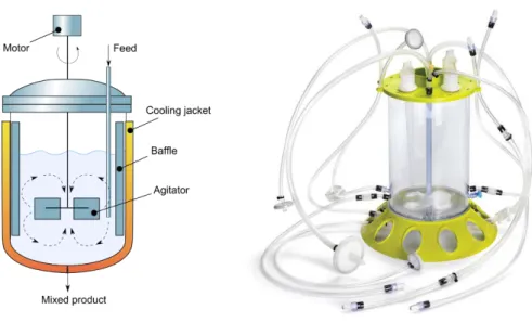
Figure 11 Schematic of a stirred vessel (left) (Redondo, 2014) and a stirred bioreactor from Merckmillipore® (right). ... 26
Figure 12 Rotating wall vessel (RWV) bioreactor from Synthecon® ... 28
Figure 13 Illustration of a STLV and HARV bioreactor (Ghosh and Kaplan, 2008) ... 29
Figure 14 Schematic of a Rotating Shaft Bioreactor (Chen et al., 2004) ... 30
Figure 15 Biaxial Rotating Bioreactor (Singh et al., 2005) ... 30
Figure 16 Schematic of a bioreactor that apply controlled mechanical forces (Martin et al., 2004) .... 32
Figure 17 Schematic illustration of a bioreactor assembly that demonstrates (a) perfusion flow and (b) hydrostatic compression (Orr and Burg, 2008) ... 33
Figure 18 Illustration of a (a) perfusion bioreactor and (b) an example of a perfusion bioreactor (Chen and Hu, 2006; C.I.T., 2014). ... 34
Figure 19 Bidirectional perfusion bioreactor in a U tube design (Wendt et al., 2003). ... 35
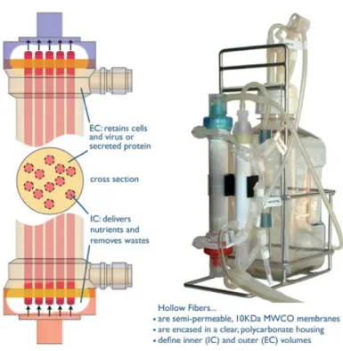
Figure 20 Basic hollow fibre bioreactor design and the HF Primer™ small-scale bioreactor (Hirschel et al., 2011). ... 36
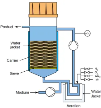
Figure 21 Schematic of the fluidized bed or packed bed bioreactor (Cabrita et al., 2003). ... 37
Figure 22 Schematic diagram of the pulsatile flow bioreactor system (Cooper et al., 2007). ... 37
Figure 23 Classification of the rheological behavior of fluids (adapted from Nguyen and Choi, 2012). ... 40
Figure 24 Model and measures of the perfusion bioreactor. ... 46
Figure 25 Model of the perfusion bioreactor demonstrating the four pistons configurations, a) Open-Open, b) Open-Close, c) Close-Open and d) Close-Close positions. ... 47
Figure 26 Membrane configuration to redirect the fluid flow where a) is the Parallel Flow configuration, b) the Inwards Flow configuration and c) the Outward Flow configuration. ... 48
Figure 27 Design of the scaffold used in this work, a) detail of the filament pattern; b) lateral view of
the scaffold... 49
Figure 28 Computational simulation schematic. ... 49
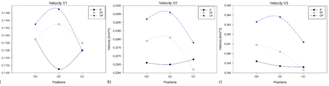
Figure 29 Velocity results for the three input velocities without scaffold. ... 52
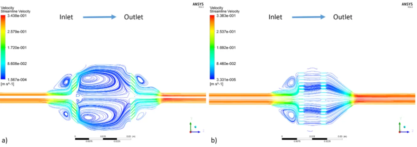
Figure 30 V1 velocity streamlines without scaffold for a) the OF-OC and b) the IF-OC. ... 52
Figure 31 V2 velocity streamlines without scaffold for a) the OF-OC and b) the PF-CC. ... 53
Figure 32 V3 velocity streamlines without scaffold for a) the OF-OC and b) the PF-CC. ... 53
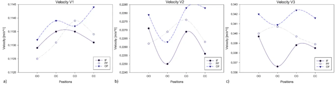
Figure 33 Velocity results for the three input velocities with scaffold. ... 54
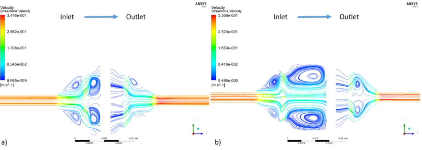
Figure 34 V1 velocity streamlines with scaffold for a) the OF-CC and b) the PF-OO. ... 54
Figure 35 V2 velocity streamlines with scaffold for a) the OF-CC and b) the IF-OC. ... 55
Figure 36 V3 velocity streamlines with scaffold for a) the OF-CO and b) the IF-OC. ... 56
Figure 37 Turbulence results for the three input velocities without scaffold... 56
Figure 38 V1 turbulence results without scaffold for a) the IF-CC and b) the PF-CC. ... 57
Figure 39 V2 turbulence results without scaffold for a) the IF-OO and b) the PF-CC. ... 57
Figure 40 V3 turbulence results without scaffold for a) the IF-OC and b) the PF-CC. ... 58
Figure 41 Turbulence results for the three input velocities with scaffold. ... 58
Figure 42 V1 turbulence results with scaffold for a) the IF-CC and b) the PF-CC. ... 59
Figure 43 V2 turbulence results with scaffold for a) the IF-OO and b) the PF-CC. ... 59
Figure 44 V3 turbulence results with scaffold for a) the IF-CO and b) the PF-OO. ... 60
Figure 45 Pressure results for the three input velocities without scaffold. ... 60
Figure 46 V2 pressure results without scaffold for a) the OF-CO and b) the PF-OO. ... 61
Figure 47 Pressure results for the three input velocities with scaffold. ... 62
Figure 48 V2 pressure results with scaffold for a) the IF-CO and b) the PF-OC. ... 62
Figure 49 Scaffold velocity results for the three input velocities. ... 63
Figure 50 V2 scaffold velocity results for a) the IF-CO, b) the PF-OC and c) the OF-CO. ... 63
Figure 51 Scaffold velocity results for the three input velocities. ... 64
Figure 52 V2 wall shear stress results for a) the IF-CO, b) the PF-OC and c) the OF-CO. ... 65
Figure 53 Velocity comparison in percentage between the different combinations with scaffold. ... 67
Figure 54 Turbulence comparison in percentage between the different combinations with scaffold. 68 Figure 55 Pressure comparison in percentage between the different combinations with scaffold. .... 68
Figure 56 Scaffold velocity comparison in percentage between the different combinations. ... 69
Figure 57 Wall shear stress comparison in percentage between the different combinations. ... 69
Index of Tables
Table 1 Core areas within Tissue Engineering, adapted from Kuppan et al., (2012), Jeong et al., (2007)
and Tabata (2001). ... 2
Table 2 Most relevant growth factors for tissue engineering applications, adapted from Tessmar and
Gopferich (2007), Boontheekul and Mooney (2003) and Rose and Oreffo (2002). ... 4
Table 3 Relationship between scaffold characteristics and the corresponding biological effect
(Mahajan, 2005). ... 6
Table 4 Most common culture mediums (Butler, 2004) ... 15 Table 5 Example of limit and optimal stresses of several types of cells. ... 20 Table 6 Main requirements of a bioreactor for the use in TE (Chen and Hu, 2006; Lyons and Pandit,
2005; Pörtner et al., 2005; Korossis et al., 2005; Martin et al., 2004). ... 22
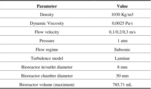
Table 7 Mesh conditions used in the CFD analysis. ... 50 Table 8 Fluid characteristics and chamber properties used in the CFD analysis. ... 50
List of acronyms
0-9
2D Two-dimensional
3D Three-dimensional
B
BME Basal Medium Eagle
BMP Bone morphogenetic protein
C
CAD Computer-aided Design
CFD Computational Fluid Dynamics
cm centimetres
D
DMEM Dulbecco’s Modified Eagle’s Medium
DNA Deoxyribonucleic acid
dyn Dyne (unit)
E
ECM Extracellular Matrix
EMEM Eagle’s Minimal Essential Medium
EGF Epidermal growth factor
F
FGF Fibroblast growth factor
G
GAG Glycosaminoglycans
GMEM Glasgow Minimum Essential Medium
H
HARV High Aspect Ratio Vessel
HEPES 4-(2-hydroxyethyl)-1-piperazineethanesulfonic acid
HFB Hollow Fibre Bioreactor
I
IGF Insulin-like growth factor
K
kPa kilopascalL
L-15 Leibovitz MediumM
min minute ml millilitre mm millimetreN
NASA National Aeronautics and Space Administration
P
Pa Pascal (unit)
PCL Poly(ε-caprolactone)
PDGF Platelet-derived growth factor
R
rpm Revolutions per minute
RPMI Roswell Park Memorial Institute Medium RSB Rotating Shaft Bioreactor
RWPV Rotating-wall Perfusion Vessel
RWV Rotating-wall Vessel
S
SMC Smooth Muscle Cells
STLV Slow Turning Lateral Vessel
T
TCP Tricalcium Phosphate
TE Tissue Engineering
TKE Turbulence Kinetic Turbulence
TGF-β Transforming growth factor-β
U
USA United States of America
W
Index
AGRADECIMENTOS... V RESUMO ... VII ABSTRACT ... IX INDEX OF FIGURES ... XI INDEX OF TABLES... XIII LIST OF ACRONYMS ... XV
INTRODUCTION ... 1
1.1 CELL CULTURE AND BIOREACTORS ... 6
1.2 RESEARCH AIMS ... 8
1.3 THESIS STRUCTURE ... 9
1.4 RESEARCH FUNDING AND PUBLICATIONS ... 10
STATE-OF-THE-ART ... 13
2.1 CELL CULTURE IN TISSUE ENGINEERING ... 13
2.1.1IMPORTANCE OF ENVIRONMENTAL AND OPERATIONAL VARIABLES ... 16
2.1.2CELL DAMAGE AND CRITICAL TENSIONS ... 18
2.2 BIOREACTORS FOR TISSUE ENGINEERING ... 20
2.2.1BIOREACTORS, CONCEPTS AND DEFINITIONS ... 20
i. Classification and design of bioreactors... 21
COMPUTATIONAL MODELLING OF FLUID DYNAMICS ... 39
3.1 FLUID CLASSIFICATION ... 39
3.2 COMPUTATIONAL MODELLING ... 40
3.2.1NAVIER-STOKES EQUATION ... 40
3.2.2TURBULENCE KINECT ENERGY ... 43
SIMULATION OF THE PERFUSION BIOREACTOR ... 45
4.1 METHODS ... 45
4.1.1PERFUSION BIOREACTOR DESIGN ... 45
4.1.2SCAFFOLD DESIGN ... 48
4.1.3SIMULATION CONDITIONS AND SETTINGS ... 49
4.2 RESULTS AND DISCUSSION ... 51
4.2.1CFDSIMULATION RESULTS ... 51
i. Velocity Results ... 51
ii. Turbulence Results ... 56
iii. Pressure Results ... 60
iv. Scaffold Velocity ... 62
v. Wall Shear Stress Results... 63
CONCLUSIONS AND FUTURE WORKS ... 66
5.1 CONCLUSIONS ... 66
5.2 FUTURE WORKS ... 71
Introduction
Tissue Engineering is an emerging field that complies different areas of science focusing essentially on the use of cells, biomaterials, computational methods and fabrication processes (Figure 1) merging principles of areas such as biology, engineering and medicine in order to create biological tissues that are functionally defective or damaged (Eshraghi and Das, 2010; Bártolo et al., 2009a; 2009b; 2008; Gibson, 2005; Tan et al., 2005; Vozzi et al., 2003; Risbud, 2001). Skalak and Fox (1988), defined Tissue Engineering as the “application of the principles and methods of engineering and life sciences toward the fundamental understanding of structure-function relationships in normal and pathological mammalian tissues and the development of biological substitutes to restore, maintain, or improve tissue and organ functions” (Bártolo et al., 2008).
The main reason for the appearance of this new research domain that rapidly expanded was to address the organ shortage problem that comprises not only full organs to be substituted by the damaged ones, as well as to regenerate tissues and gene therapy (Table 1). In the near future, diseases such as Parkinson, Alzheimer, osteoporosis, cancer and many others can be treated through the regeneration of the damaged or diseased tissues.
The National Science Foundation (2003) of the United States of America (USA) published an historical overview of this emerging field and also, more recently, Bártolo et al. (2012) presented the perspectives till 2030 for this specific domain of research.
Table 1 Core areas within Tissue Engineering, adapted from Kuppan et al., (2012), Jeong et al., (2007) and Tabata
(2001).
Purpose Techniques/Methodology
Tissue Regeneration
In vitro production of tissue constructs Cell scaffolding, bioreactor , microgravity
In vivo natural healing process Cell scaffolding, controlled release, physical
barrier
Ischemia therapy Angionesis
Organ Substitution
Immunoisolation Biological Barrier
Nutrition and oxygen supply Angionesis
Temporary assistance for organ function Extracorporeal system
Gene therapy
Inhibiting induction of a specific gene, or by editing undesirable genomic mutations
Intracellular transfer of nucleic acid drugs to modulate cellular functions and responses by expressing exogenous proteins
In order to create new tissues there are three general strategies: Cell-based strategies; Growth-factor-based strategies; and Scaffold-based strategies as is illustrated in Figure 2. The scaffold-based strategies is one of the most common and has been adopted by several authors, such as: Bhumiratana and Vunjak-Novakovic (2012), Norotte et al. (2009), Bártolo et al. (2008), Matsumoto and Mooney (2006), Mistry and Mikos, (2005), Fuchs et al. (2001), Langer (1997) and Langer and Vacanti (1993).
Figure 2 Scaffold-based strategies for engineering personalized bone grafts (adapted from Bhumiratana and
Vunjak-Novakovic, 2012).
i. Cell-based strategies, consists in the implantation, in vivo, of isolated cells or cells
substitutes so that they may synthesize their own Extracellular Matrix (ECM). Using this method is intended to limit surgery complications by only allowing the replacement of the damaged cells. The main limitations of this strategy is the immunological rejection and failure of the encapsulated cells.
ii. Growth-factor-based strategies uses growth factors and controlled-released systems to
create new tissues. Growth factors are basically proteins that are produced by cells that are functioning as signalling molecules. With these proteins, it is possible to promote cell adhesion, proliferation, migration and differentiation of the cultured cells (Tessmar and Gopferich, 2007; Boontheekul and Mooney, 2003; Rose and Oreffo, 2002). Table 2 indicates the most relevant growth factors for bone regeneration and wound healing applications.
Table 2 Most relevant growth factors for tissue engineering applications, adapted from Tessmar and Gopferich
(2007), Boontheekul and Mooney (2003) and Rose and Oreffo (2002).
Bone Regeneration
Growth factor Relevant activities
Transforming growth factor-β (TGF-β) Proliferation and differentiation of bone Bone morphogenetic protein (BMP) Differentiation of bone forming cells
Insulin-like growth factor (IGF) Stimulates proliferation of osteoblasts and the synthesis of bone matrix
Fibroblast growth factor (FGF) Proliferation of osteoblasts
Platelet-derived growth factor (PDGF) Proliferation of osteoblasts
Wound Healing
Growth factor Relevant activities
Platelet-derived growth factor (PDGF) Active in all stages of healing process
Epidermal growth factor (EGF) Mitogenic for keratinocytes
Transforming growth factor-β (TGF-β) Promotes keratinocyte migration, ECM synthesis and remodelling, and differentiation of epithelial cells
Fibroblast growth factor (FGF) General stimulant for wound healing
iii. Scaffold-based methods provides a substrate for the implanted cells, i. e., scaffolds
provide an initial biochemical and biophysical substrate in order to support the cells until they start producing their own ECM and stimulate new tissue formation (Bartolo et al., 2012). There are two main methods within this strategy, which can be used in an open or closed culture system. The first one starts with an in vitro culture of cells and then they are seeded inside the scaffold pores, whereas the second method consists on isolating the cells from the body by a permeable membrane allowing the exchange of nutrients and wastes, protecting cells from the immune response of the body. The last step of this culture strategy regards the implantation of the cell-matrix in the body.
Using scaffolds as a temporary support matrix for cells is the most commonly used strategy for tissue engineering (Fuchs et al., 2001). With this approach, the cells are harvested from living tissue, from the patient (autograph) or from a different individual (allograph) and then they will be cultured in vitro on a scaffold in order to obtain the tissue-scaffold construct for
transplantation (Liu and Czernuszka, 2006). Besides providing the initial biochemical and biophysical substrate to improve the cell growth, scaffolds also serve as a temporary template to accommodate and aid in the definition, formation and orientation of the new tissue throughout the 3D space (Bártolo et al., 2012). In Figure 3 is illustrated this strategy taking into account all the essential factors.
Figure 3 Tissue engineering process involving the cell seeding on scaffolds, in vitro culturing and patient
implantation, adapted from Bartolo et al., (2012) and Liu and Czernuszka (2006).
There are several main purposes that a scaffold must achieve. It must serve as an adhesion substrate for cells, allowing cell attachment, proliferation and differentiation; it must deliver and retain cells and growth factors; it has to enable diffusion of cell nutrients and oxygen; and must provide temporary mechanical and biological environment to the newly grown tissue enabling tissue regeneration in an organized way (Billiet et al., 2012; Truscello et al., 2012; Guillotin and Guillemot, 2011; Bártolo et al., 2009a; 2009b; 2008; Tan and Teoh, 2007; Liu
and Czernuszka, 2006; Kreke et al., 2005; Gomes and Reis, 2004; Gross and Rodriguez-Lorenzo, 2004; Leong et al., 2003; Kreeger and Shea, 2002; Hutmacher, 2001; Kim and Mooney, 2001; Langer and Vacanti, 1993). In order to accomplish all this goals, the scaffolds must fulfil several biological and physical requirements (Table 3) that will affect cell survival, signalling, growth, propagation and reorganization and also cell shape modelling and gene expression (Bártolo et al., 2012; Billiet et al., 2012; Reverchon and Cardea, 2012; Vasanthan et al., 2012; Bártolo et al., 2009a; 2009b; 2008; Sanz-Herrera et al., 2009; Leong et al., 2008; Chen et al., 1997; Mooney et al., 1992).
Table 3 Relationship between scaffold characteristics and the corresponding biological effect (Mahajan, 2005).
Scaffold Characteristics Biological Effects
Biocompatibility Cell viability and tissue response
Biodegradability Aids tissue remodelling
Porosity Cell migration inside the scaffold - Vascularisation
Chemical properties of the material Aids in cell attachment and signalling in cell environment. Allows release of bioactive substances
Mechanical properties Affects cell growth and proliferation response
1.1 Cell Culture and Bioreactors
In Tissue Engineering, cell culture plays a major role in the construction of tissue replacement. Normally cells are harvested from embryonic tissue being these type of cells used in primary cultures because they have an enormous potential to differentiate and grow into different types of tissues.
There is several types of cells ideal for processing new tissues ex situ like mesenchymal stem cells; permanent or established cell strains; and hybridomas. Considering the primary cells, there are a vast diversity of cell lines that are used in cell culture such as epithelial cells; connective tissue cells; muscle cells; nerve cells; and blood and lymphatic cells.
For a successful tissue construct, there are some requirements that most provide a stable and propitious environment for culturing the cells. In order to accomplish a successful cell culture, one has to consider the operational and environmental variables such as pH, temperature, medium control and oxygenation. All these conditions must be considered in order to have an
excellent cell growth within the scaffold and to control all these variables in tissue engineering, bioreactors are often used because one may control and visualize the progress of the undergoing cell culture.
A bioreactor is usually defined as an equipment in which occurs biological and/or biochemical processes in a controlled environment. There are several types of bioreactors and they are classified accordingly to the environment which unfolds the tissue culture depending in the type of stimulus involved and they can also be classified regarding the type of tissue that will be cultured.
Due to the specificity of each type of cell and the tissue that will be created, there was a need to develop various types of bioreactors with more focus on its operational and ability to apply stimulus. Essentially the bioreactors are divided in two groups, the static culture group and the dynamic culture bioreactors (Figure 4).
Figure 4 Bioreactors classification.
Static culture systems are the most simple to operate and manage because one just needs to considerer the environmental conditions in order to occur cell growth within the scaffold (e.g. petri dish, t-flask, culture bag) while dynamic systems apply stronger stimulus to the cells, either directly or indirectly.
In dynamic cultures, there are agitation systems that provide a homogeneous environment and such systems like spinnerflask, Wavywalled Bioreactor (WWB) or stirred vessel bioreactor have the main purpose of increasing mass transfer during the cell culture. Another type of
dynamic bioreactors are the rotational bioreactors that uses movement about one or more axes to create agitation in the culture medium and therefore increasing the mass transfer.
Mechanical stimulation systems have the advantage of direct stimulation of the cells, not only aiding the increase of cell proliferation as also will improve the differentiation and alignment of the cells within the scaffolds. Depending on the type of cells that will are to be cultured, and more intrinsically, the different tissues that will be constructed, there are different types of stimulus that can be applied using these systems.
One of the main purposes of the perfusion bioreactors is to solve the problems of the non-uniformity of the cell proliferation within the scaffold. This can be accomplished by controlling the medium flow velocity through the scaffold and cells while continuously renewing the culture medium and cell retention.
Optimised cell culturing can be accomplished in vitro with the aid of equipment’s such as bioreactors by taking into account all these factors since operational to the environmental variables.
1.2 Research Aims
The design of a perfusion bioreactor for tissue engineering applications is the key topic of this research, where problems as diffusion of the flow within the scaffolds, cell damage and critical tensions, homogenization of the cell distribution throughout the scaffold and optimization and prediction of fluid behaviour around and within the scaffold, allowing further improvement of scaffolds design, present a critical challenge in this research topic.
Taking into account the different problems that are inherent to cell culturing in tissue engineering, this research work must consider the following main objectives:
• The design optimization of a perfusion bioreactor;
• The evaluation of the inlet and outlet fluid flow within the chamber and scaffold; • Analysis of the critical tensions that are admissible in cell culturing.
1.3 Thesis Structure
This thesis is structured into 5 chapters as illustrated in Figure 5.
Figure 5 Flowchart of the topics and objectives that are addressed in the thesis.
The first chapter presents an overview of the tissue engineering field and more specifically the use of scaffolds. An in depth description of scaffolds, regarding its purpose, functionality, biological and physical requirements and characteristics.
The second chapter comprises the state-of-the-art related with Cell Culture and Bioreactors in Tissue Engineering. It is comprehensively described the process of tissue culture using several types of cells along with their differentiation process into a specific tissue. It is also described the several bioreactors and their properties according to the tissue needed to be cultured. The third chapter describes the properties of fluids and how to characterize it computationally using several mathematical models in the computational calculations in order to determine the fluid behaviour across the bioreactor.
The fourth chapter of this work describe the methods used in this work since the design of the perfusion bioreactor and the scaffold as also the simulation parameters. It will also be exposed and discussed the results obtained from the simulations.
The fifth and last chapter presents the conclusions as also the pros and cons of the work carried out under the perfusion bioreactor and future works in order to continue improving the tissue culture in bioreactors.
1.4 Research Funding and Publications
The research work undertaken in this thesis was supported by the following research projects:
1. Internacional Research Exchange for Biomedical Devices Design and Prototyping “IREBID”, funded by the People Marie Curie Actions, International Research Staff Exchange Scheme.
2. Development of Scaffolds with Controlled Microstructure for Bone Tissue Engineering (PTDC/EME-PME/104498/2008), funded by the Portuguese Foundation for Science and Technology.
3. Strategic Project UI 4044 (Pest-OE/EME/UI4044/2011), funded by the Portuguese Foundation for Science and Technology.
4. Rede IberoAmericana de Biofabricação: Materiais, Processos e Simulação, funded by the CYTED – Ciencia Y Tecnologia Para El Desarrollo.
5. BIOMAS – Biomanufacturing and Engineering Scaffolds, funded by the Portuguese Agency for Innovation.
6. BIOMAS II – Biomanufacturing and Engineering Scaffolds, funded by the Portuguese Agency for Innovation.
The following publications and patents also emerged from the research work carried out:
Articles in proceedings edited as book:
1. D. Freitas, H.A. Almeida & P.J. Bártolo (2014) “Perfusion bioreactor fluid flow optimization”, CENTERIS/ProjMAN/HCist 2014 Book of industry papers, poster papers and abstracts, J. Varajão et al. (Eds.), SciKA (ISBN: 978-989-97433-4-2), 159 (CENTERIS/ProjMAN/HCist 2014, 15/10/2014-17/10/2014).
2. D. Freitas, H. Almeida & P. Bártolo (2014) “Permeability evaluation of flow behaviors within perfusion bioreactors”, New Trends in Mechanism and Machine Science – From Fundamentals to Industrial Applications, P. Flores & F. Viadero (Eds.), Springer (ISBN: 978-3-319-09410-6), 761-768 (5th European Conference on Mechanism Science – EUCOMES 2014, Guimarães, Portugal, 16/09/2014-20/09/2014).
3. D. Freitas, H. Almeida & P. Bártolo (2014) “Optimization of a Perfusion Bioreactor for Tissue Engineering”, Biodental Engineering III, R.N. Jorge et al (Eds.), CRC Press (ISBN: 978-1-138-02671-1), 145-149 (Porto, Portugal, 22/06/2014-23/06/2014). 4. R.F.B. Pereira, D.M.F. Freitas, A.P.O. Tojeira, H.A. Almeida, N. Alves & P.J. Bártolo
(2012), Computational optimization of a novel bioreactor for tissue engineering applications, Proceedings of the 1st International Conference on Design and Processes for Medical Devices (PROMED), E. Cerretti et al (Eds.), Neos Edizioni (ISBN:978-88-6608-058-9):217-221 (Brescia, Italy, 02/05/2012-04/05/2012).
5. D.M. Freitas, R. Pereira, A. Tojeira, P.J. Bártolo, N.M. Alves, C. Capela, A. Mendes & H.A. Almeida (2011), Influência das Variáveis Dimensionais e de Rotação das Câmaras de Cultura de Bioreactores no Crescimento Celular, 4º Congresso Nacional de Biomecânica, L. Roseiro & A. Neto (Eds.), Sociedade Portuguesa de Biomecânica (ISBN:978-989-97161-0-0):407-412 (Coimbra, Portugal, 04/02/2011-05/02/2011).
Articles in international journals edited with review:
1. D. Freitas, H.A. Almeida & P.J. Bártolo (2014) “Perfusion bioreactor fluid flow optimization”, Procedia Technology (ISSN: 2212-0173), 16: 1238-1247.
2. R. Pereira, D. Freitas, A. Tojeira, H.A. Almeida, N. Alves & P.J. Bártolo (2014), Computer modelling and simulation of a bioreactor for tissue engineering, International Journal of Computer Integrated Manufacturing (ISSN: 0951-192X (Print) 1362-3052 (Online)), 27(10): 946-959.
Articles in proceedings:
1. D. Freitas, P.J. Bártolo, H.A. Almeida, "Análise Computacional De Diferentes Tipos De Entradas De Fluxo Em Bioreactores De Perfusão", 6º Congresso Nacional de Biomecânica (CNB2015), Monte Real, Portugal, 06/02/2015-07/02/2015.
2. D. Freitas, H.A. Almeida & P.J. Bártolo (2014) “Fluid Flow Optimization of a Perfusion Bioreactor”, International Conference on Biofabrication (ICB 2014), Pohang, Korea, 28/09/2014-01/10/2014.
3. A.P. Tojeira, D.M. Freitas, R.F. Pereira, P.J. Bártolo, N. Alves, C. Capela, A. Mendes & H.A. Almeida (2010), Computational analysis of bioreactor chamber for tissue engineering applications, Rapid Product Development (RPD 2010), Marinha Grande, Portugal, 20/09/2010-21/09/2010.
4. R. Pereira, A. Tojeira, D.M. Freitas, P.J. Bártolo, N. Alves, C. Capela, A. Mendes & H.A. Almeida (2009), Bioreactores em Engenharia de Tecidos, 1as Jornadas do Curso de Mestrado em Concepção e Desenvolvimento de Produto, Leiria, Portugal, 19/12/2009.
Intellectual Property – Patents:
1. Bioreactor Multifuncional para a Engenharia de Tecidos, D.M. Freitas, A.P. Tojeira, R.F. Pereira, P.J. Bártolo, N.M. Alves, A.L. Mendes, C.A. Capela & H.A. Almeida, Patente de Invenção Nacional Nº 105176, 21/03/2013.
State-of-the-art
In this chapter it is presented the state-of-the-art related with Cell Culture and Bioreactors in Tissue Engineering. It is thoroughly descript the several cells used for tissue construct along with the different differentiation of each kind of cell in the specific tissue. It is also described the several bioreactors and their properties according to the tissue needed to be cultured.
2.1 Cell Culture in Tissue Engineering
In the broad scope of Tissue Engineering, cell culture has been a carrier for the advances in the construction of tissue replacement for tissue/organ damage. The cell culture process is characterized by two phases, the acquisition of cells from a donor; and the cell growth in nutrient-rich media and specific growth factors for each cell type.
The stem cells primary culture, i.e., the first cell culture established from cells obtained directly from animal tissues, is generally established from embryonic tissue since in this type of tissue the cells are easily dispersed and have a high potential for growth and high differentiation. After the completion of the primary culture it is selected one type of cell (Butler, 2004).
Recent studies have demonstrated that there is some type of cells ideal for process future tissues ex situ being the following ones the most common:
• Mesenchymal stem cells: cells originating from the embryonic tissue and from multipotent cells that by differentiation processes origin specific cells type. Their use in tissue engineering is primarily explained by the versatility of becoming any kind of cell lineage.
• Permanent or established cells strains: cells that derived from a primary culture, but due to some changes are able to divide and proliferate indefinitely (primary culture);
• Hybridomas: Cells that are obtained by fusing lymphocytes with tumor cells and are capable of expressing monoclonal antibodies.
From the primary cells (primary culture) it can be selected the following types of cell lines (Figure 6):
• Epithelial cells: cells disposed in layered formation coating organs or cavities which easily proliferate in a cell culture;
• Fibroblasts: cells that make up the majority of human tissues and are characterized by having a fibrous matrix, as a good example of this type of cells is the fibroblasts. The growth in culture is fast and efficient (doubling time 18-24 hours), becoming the most studied cell type under Tissue Engineering.
• Muscle cells: muscle tubules are formed from precursor cells which form a complex and multinucleated containing structural proteins (actin and myosin). The precursor cells, i.e., myoblasts may undergo differentiation forming aligned myotubes.
• Nerve cells: this cell type is responsible for transmitting electrical impulses. Neurons are highly differentiated cells and therefore difficult to grow in culture media. However, the addition of growth factors can lead to the formation of neurites (cytoplasmatic growth external to the neuron);
• Blood and lymphatic cells: cells growing in culture medium in the form of suspension. An example of this type of cells are the lymphocytes (white blood cells). These cells have high importance for the culture of cells, since they secrete immunoregulatory compounds.
Figure 6 Mesenchymal stem cells differentiation (Caplan and Bruder, 2001).
In order to provide an environment conducive to the growth of cells in a three-dimensional structure or scaffold it is necessary to pay special attention to the culture medium, which can be defined chemically as a complex liquid medium capable of supporting cell growth over several generations. The composition of the culture medium varies depending on the cell type (Table 4) (Butler, 2004).
Table 4 Most common culture mediums (Butler, 2004)
Culture Medium Observations
BME Basal Medium Eagle, originally conceived for L mice and HeLa cell.
EMEM Eagle’s Minimal Essential Medium, is used in a variety of cells.
DMEM Dulbecco’s Modified Eagle’s Medium, possess four times more amino acids and vitamins concentration that the BME.
GMEM Glasgow Minimum Essential Medium, possess two times more amino acids and vitamins concentration that the BME.
RPMI 1640 Roswell Park Memorial Institute Medium is often used for the culture of lymphocytes and hybridomas.
L-15 Leibovitz Medium, used in environments with absence of CO2 for the culture of fibroblasts.
Ham’s F-12 It’s a medium with a complex composition used for the culture of several cell lines.
199 Medium 199 has broad species applicability, particularly for cultivation of non-transformed cells.
2.1.1 Importance of environmental and operational variables
Generically primary cells, which subsequently gives origin to a tissue, require a ribbed nutrient medium for the correct growth and proliferation. However, in certain environments human cells require external control of conditions such as temperature, pH, product concentration of cell growth, among other parameters.
Temperature
The cell growth and development take place in vivo at a temperature of 37°C. In vitro these conditions are ensured by incubator systems, however, the increase of an incubator throughout all the mechanical systems of the bioreactor limits the accessibility to the culture itself. More recently, have been suggested heated atmospheres systems using heating plates; another proposed solution is insertion of the heating units within the culture system, which can be monitored by computer using low-voltage circuits and circuit breakers which avoid the electric shock (Pörtner, 2009; Minuth et al., 2005).
pH
Human primary cells are extremely sensitive to variations of pH. Therefore, the culture medium is controlled at this level by a buffer solution which maintains the pH between 7,0 and 7,3, these values are considered in the literature as optimum values (Pörtner, 2009). In several experiments, the solution of carbon dioxide-bicarbonate at 5% (Equation 1) has been widely used since it resembles to the solution existent in the blood, at in vivo conditions (Pörtner, 2009):
𝐶𝐶𝐶𝐶2+ 𝐻𝐻2𝐶𝐶 ↔ 𝐻𝐻𝐶𝐶𝐶𝐶3−+ 𝐻𝐻+ (1)
Under different conditions of 5% concentration of CO2 the physiological pH becomes slightly alkaline (under normal atmospheric conditions – 0,3% CO2 concentration) which can be countered by reducing the concentration of NaHCO3 or by adding other buffer solutions such as sodium phosphate or HEPES, which can be monitored by the pH indicator phenol red (Pörtner, 2009; Minuth et al., 2005).
The cell activity in vivo, is usually associated with the activity of sodium-potassium pump, whose function is to transport nutrients and other ions from the extracellular medium to the intra-cellular (Minuth et al., 2005). In addition to the necessary activity to normal cells ions is necessary to ensure power supply in the form of carbohydrates, is essentially reported, the glucose concentrations of 446 mg/dl. In addition, amino acids are also added (which are only precursors activity of protein synthesis), vitamins, minerals and other compounds.
In addition to all these components, is also generally associated with a serum concentrations ranging between 5 and 10%, and whose function is to provide certain growth and protect cells from shear stress. An example of such a serum are the fetal of calf or horse. However, the addition of this component to the culture medium of cells entails some disadvantages, such as: (1) the composition of the solution is not well defined; (2) the high cost of serum; (3) the difficult purification of the solution; (4) variation of loads in the solution; (5) the risk of contamination and spread of viruses.
However, the absence of this component in the growth medium and cell differentiation greatly delays the culture process (Pörtner, 2009; Butler, 2004).
Oxigenation
To achieve the aerobic metabolic cycles of the cells is necessary to take into account the distribution of gas in the culture medium, the transfer of nutrients and also the wastes of cellular reactions. Transferring sufficient amount of oxygen to cells is difficult essentially due to the low oxygen solubility in the culture medium (about 0,2 mmol/L). However in order to have a viable cell culture is necessary to have an equal or approximated concentration of both O2 and Glucose (Martin and Vermette, 2005). In recent culture systems, the oxygen concentration is kept constant between 20% and 100% of air saturation that creates a balance between oxygen needs and tolerance to the formation of harmful free radicals that causes cytotoxic effects on cells.
Oxygenation can be performed by two methods: (1) gas sparging and (2) without oxygen bubbles. The gas sparging method is often used since it proves to be simple to provide the oxygen to the bioreactor. This method allows a high oxygen transfer due to a high interfacial area per bubble. However, the bubbles can lead to failure in cell viability, since the bubbles can damage the cells. The preferable process in bioreactor systems is the method without bubbles
of the used gas that can be divided in two methods, the superficial gas sparging and the permeable membrane (porous or diffusion) (Bliem et al., 1991).
On the superficial gas sparging process, the culture medium is directly exposed to a controlled atmosphere rich in O2 and 5% CO2 constantly occurring diffusion of these gases and keeping them in the culture medium.
The permeable membranes transfer the gas to the culture medium and they can be defined in two different types: porous or diffusion. In the first case, the culture medium is in continuous contact with the gas through pores arranged on the membrane. The interface created in the pores is controlled by pressure effects and hydrophobic forces. In the other end, the diffusion membranes diffuse the oxygen from the initially gas phase to a soluble membrane in oxygen and then into the culture medium. Although these two cases avoid the formation of bubbles they are influenced by factors such as the concentration of oxygen, the porosity of the membrane and the surface area of the membrane. However, these processes of oxygenation present difficulties in their implementation, namely, the complexity of the process, control of intrinsic variables of the process, the need for high membrane area exposed to the gas phase, difficulties in maintenance and cleaning of the membrane. In addition, the deposition of proteins (derived from the culture medium) at the base of the membrane, modify its hydrophobic property.
2.1.2 Cell damage and critical tensions
Human cells are very sensitive to changes in the culture environment, the hydrodynamic forces and the accumulation of toxic residues derived from the cellular respiration. Knowledge of the critical shear stress for cell development is of vital importance in TE because of this knowledge depends greatly on the success of the strategies adopted.
Human cells are highly sensitive to shear stresses due to their size (cell size ranging between 10 and 30 μm) and also because of the cell contents are only protected by the cell membrane. While, in some cultures, cells can grow freely in suspension, others require connection to a biocompatible structure to occur adhesion, proliferation and differentiation of cells. In these previous circumstances, if the connecting surface is fixed, the cell has an increased susceptibility to experience shear stress, since the cell cannot move freely and adapt to the forces imposed by the environment. In this case, it should be imposed laminar flow, to minimize shear forces (Minuth et al., 2005).
Although excessive shear stresses may be harmful to the cells, it is desirable that there are controlled stresses, since these have a positive effect on adhesion, proliferation and cell differentiation, such as: (1) increase the permeability of the cell; (2) increase the secretion of extra-cellular proteins; (3) influence the shape of the cell.
The application of the shear stress and the definition of its value depends on the following factors (Minuth et al, 2005): (1) cell type; (2) method of culture (in suspension /fixed); (3) time of exposure to shear stresses. Regarding the exposure time, most of the studies on cell viability associated with critical shear stress reports short-term experiments (less than 24 hours). However long time cultures with levels of shear stresses away from the critical values may be advantageous, this study also shows that, apparently, the cells cultured in suspension support higher levels of shear stresses that the fixed cells (Minuth et al., 2005).
The culture of mesenchymal stem cells with a goal for osteogenic cell differentiation has been widely studied and it is concluded that this differentiation process is very sensitive to shear stresses caused by fluids. Godara et al., (2008) mentioned that throughout the process of cell differentiation, the critical shear stresses varies and after clarified that shear stresses ranging between 10-5 Pa and 10-4 have some responsibility in the osteogenesis process (in addition to the chemical differentiation factors), and that the differentiation of osteocytes is enhanced by the action of shear stress ranging 0,5 and 1,5 Pa. According to Cartmell et al. (2003), the optimal shear stress value to maximize cell viability of the osteoblasts is about 5×10⁻⁵ Pa under the influence of a flow of 0,01 ml/min and that flow values above 0,2 ml/min are negative to cell viability of osteoblasts. Porter et al. (2005) reported that the shear stresses above 5,7×10⁻² Pa are associated with the death of cells within the scaffold. Similarly, Schinagl et al. (1999) demonstrated that the chondrocytes present in the hyaline cartilage suffer damage with values of shear stress of about 0,1 Pa. For the culture of cardiomyocytes Radisic et al. (2008) indicated that low values of shear stress leads to phenotypic changes of cardiac cells (primarily elongation) but values above 0,24 Pa (2,4 dyn/cm-2) can lead to cell death without apoptosis. Park et al. (2008) have shown that hepatocytes exposed to shear stresses exceeding 0,5 Pa (5 dyn/cm2) show a decreased production of albumin and urea, but shear stresses lower than 0,033 Pa (0,33 dyn/cm2) does not affect the normal function of the cell (Table 5).
Table 5 Example of limit and optimal stresses of several types of cells.
Cell Type Shear Stress Reference
Osteogenesis
Osteoblasts 5x10
-5 Pa (optimal stress) Cartmell et al., 2003
5,7x10⁻² Pa (cell death) Porter et al., 2005
Osteocytes 0,5 to 1,5 Pa (optimal stress) Godara et al., 2008
Chondrogenesis
Chondrocytes 0,1 Pa (normal stress) Schinagi et al., 1999
Myogenesis
Smooth Muscle Cells 0,5 to 2,5 (optimal stress) Martin and Vermette, 2005 Cardiomyocytes 0,24 Pa (cell death) Radisic et al., 2008
Others
Hepatocytes 0,033 Pa (optimal stress) Park et al., 2008 0,5 Pa (critical stress)
Heart Valves Cells 2,2 Pa (optimal stress) Martin and Vermette, 2005
2.2 Bioreactors for Tissue Engineering
2.2.1 Bioreactors, concepts and definitions
The bioreactors are generally defined as locations in which occurs the development of biological, biochemical and biophysical processes in controlled and monitored environmental conditions (e.g. pH, temperature, pressure, concentration of nutrients). The reproducibility, control and automation of the process are important aspects when culturing new tissues (Lyons and Pandit, 2005; Martin et al., 2004). These devices have been used for many years in the fermentation of food, water treatment, manufacture of pharmaceuticals, and also in the production of recombinant proteins (e.g. growth factors, antibiotics and vaccines). More recently, the concept has been adapted in order to be used in TE taking into account the requirements of this field (Figure 7) (Pörtner et al., 2005; Martin and Vermette, 2005; Korossis et al., 2005).
Figure 7 Properties of a bioreactor to use in TE.
The use of bioreactors in TE aims to provide an environment conducive to adhesion, proliferation and differentiation of living animal cells. Moreover, these devices provide the necessary conditions to perform in vitro studies about the effects of mechanical and biomechanical stimuli on cells. The most critical aspect in a bioreactor is related to the mass transfer (nutrients, gases and waste removal) (Martin and Vermette, 2005; Pörtner et al., 2005), since in vivo cells benefit from their proximity to blood capillaries of 100-200μm (Rouwkema et al., 2008), which is extremely difficult to replicate in vitro.
i. Classification and design of bioreactors
Bioreactors may be classified according to many aspects, including:
• The environment in which unfolds the tissue culture - cell culture may be conducted in static or dynamic conditions;
• The type of stimulus involved - the bioreactor can apply several kinds of stimuli during cell culture or act in various ways to provide the same type of stimulus (e.g. stirring action, infusion mechanical compression or rotation);
• The tissue in culture - each tissue requires different requirements from the bioreactor (the stimulus involved or the level design).
A bioreactor is designed according to the desired size, complexity and functionality. However, regardless of these considerations a bioreactor must possess a number of basic requirements as described in Table 6.
Table 6 Main requirements of a bioreactor for the use in TE (Chen and Hu, 2006; Lyons and Pandit, 2005;
Pörtner et al., 2005; Korossis et al., 2005; Martin et al., 2004).
Bioreactor Main Requirements
Adequacy of vascularity of the cells/tissue providing nutrients in the proper time and amount
Application of stimuli in order to increase the adhesion, proliferation and differentiation
Avoid flow turbulence and excessive pressure on the culture medium, which can damage cells and impair the formation of new tissue
Control of environmental, chemical and physical conditions of culture
Ensure aseptic and sterilized conditions
Ensure the biocompatibility of the building materials
Monitoring of cell growth and formation of new tissue
Allow removal of the waste
generated by cells Easily place the scaffold inside
Maintain a high degree of reproducibility
Allow the operation over long periods of time
Be simple and ease to sterilize, clean and perform the components maintenance
The general requirements listed above are determined by the needs of the mechanical, physical, biophysical and biomechanical level of the tissue in culture. For example, the use of pulsed mechanical/perfusion instead of continuous stimulation on tubular scaffolds with Smooth Muscle Cells (SMC) increases the structural organization of blood vessels and their mechanical properties; while applying dynamic stresses to chondrocytes in appropriate environments stimulates the synthesis of Glycosaminoglycans (GAG) and increases the mechanical properties of the formed cartilage (Chen and Hu, 2006). Studies on cell proliferation in vitro have shown that mechanical stimuli have a positive impact regarding the formation of new tissue, improving the acceleration of the processes of cell differentiation and proliferation (Chen and Hu, 2006;
Pörtner et al, 2005; Martin et al. 2004). This effect is particularly evident in the formation of cartilage, bone and cardiovascular tissue (Pörtner and Giese, 2007). The most important stimulus on cellular differentiation and proliferation are the shear forces resulting from the fluid passage from the surfaces and pores of the scaffold enabling signal transmission/stimulation of the cells (Chen and Hu, 2006).
As a result of the specific needs of each type of cell in the culture process, various types of bioreactors were studied in particular on the level of operation and ability to apply stimuli.
a. Systems of static culture
The static culture systems are the most simple ones and includes systems like T-flasks, Well plates and Petri dishes (Figure 8), which are designed to culture cells in static conditions (Kumar et al., 2004). The main characteristics of these devices include ease of use, low cost and the possibility of sterilization. In return, these systems have several limitations such as: (1) the weak agitation of the medium which translates into low nutrient concentrations in certain places; (2) poor reproducibility; (3) difficulty of changing the culture medium, and (4) low amount of cell numbers. Furthermore the application of stimuli and the monitoring and control of environmental conditions, such as pH, moisture and CO2 is practically impossible (Pörtner and Giese, 2007; Pörtner et al, 2005; Kumar et al, 2004).
These devices are widely used in cell culture for shorter periods of time being used mainly to increase the number of cells (Pörtner et al, 2005; Cabrita et al, 2003). After this period the cells are transferred to bioreactors in which will occur the development of the new tissue (Lyons and Pandit, 2005; Kumar et al, 2004).
Figure 8 Example of static culture systems a) T-flasks, b) Well plates and c) Petri dishes.
Static culture in 3D scaffolds generally produces tissues with small thickness and located at the periphery of the structure. The created tissue is characterized by its heterogeneity, a result of
inadequate mass transfer (Porter et al., 2005). In order to solve the mass transfer limitations of these simple culture systems other bioreactor systems were developed such as, spinner flasks, Rotating-Wall Vessel (RWV), perfusion bioreactors, mechanical compression bioreactors, among others (Zhang et al., 2009; Bueno et al., 2004).
b. Agitation Systems
The agitation bioreactors provides a homogeneous environment, presenting results that are superior to the static culture. The main purpose of these systems (e.g. Spinner flask, wavy-walled bioreactor (WWB) and Stirred vessel bioreactor), is the increase in mass transfer (Kumar et al., 2004).
Spinner Flask
Spinner flask is the simplest type of bioreactor (Figure 9), in which the porous matrices containing the fixed cells are suspended on needles in the cap. Stirring of the medium is performed by using a magnetic bar, depending on its rotation, helps to induce the mixing of oxygen and nutrients to increase the homogeneity of the medium and consequently tissue growth (Chen and Hu, 2006). These devices can allow monitoring of variables such as pH and CO2 (Kumar et al., 2004), the rotation speed of the medium between 50 and 80 rpm. The culture medium is renewed periodically (usually in periods of 2-3 days) to maintain or increase the concentration of nutrients. In this system the bottle allows aeration of the culture medium and the resulting gas exchange. The nutrient delivery and waste removal occurs by convection and by the fluid passage on the surface of the porous matrix. Their disadvantages are related to the agitation of the medium, which can generate high shear stress values and turbulent regimes harmful to the cell culture (Zhang et al., 2009; Chen and Hu, 2006; Lyons and Pandit, 2005; Korossis et al., 2005; Martin et al., 2004).
Figure 9 Examples of Spinner flasks from Chemglass®(left) and Capitol Scientific® (right).
Wavy-walled bioreactor
In order to overcome the limitations of the turbulence and shear stresses observed in Spinner flask systems, was developed a new bioreactor, the Wavy-Walled Bioreactor (WWB) (Figure 10). This machine has ribbed walls which allow reducing turbulence levels of the medium and its shear stresses (Bueno et al., 2004). Studies conducted allowed to verify that this new system has an increase in cell proliferation and deposition/formation of extra-cellular matrix in polymeric scaffolds containing chondrocytes (Chen and Hu, 2006; Bueno et al, 2004).
Bilgen et al., (2006) studied the hydrodynamic parameters involved in the bioreactor WWB, comparatively to the Spinner flask and its influence on the cartilage tissue engineering. This study concluded that the fluid flow is significantly different between them and it was also found that the position of the scaffold inside the bioreactor it is an important parameter because alters the hydrodynamic behaviour of the fluid and the uniformity tensions on the scaffold.
Stirred vessels bioreactors
This type of bioreactor (Figure 11) it’s characterized by obtaining a homogeneous hydrodynamic environment and also because it is easy to operate. Generally it comprises a container (where the cell culture takes place) pipes, sensors, valves, pumps and motor. This system is in motion, usually at a rotation speed of 50-80 rpm (Martin and Vermette, 2005), while the sensors provide continuous monitoring of parameters such as temperature, pH, glucose, among others (Wang et al., 2005; Kumar et al., 2004). This system is similar to spinner flask glass however allows the control over a large number of variables, therefore presenting greater reproducibility. These bioreactors have been used in the culture and differentiation of various cells types of stem cells from human to rats (Serra et al., 2009; Cabrita et al., 2003). It is important to refer that the agitation of the medium can originate high shear stresses harmful to the cells which is a disadvantage of this type of equipment. One possible solution involves changing the shape and/or the diameter of the rotor blade (Wang et al., 2005).
Figure 11 Schematic of a stirred vessel (left) (Redondo, 2014) and a stirred bioreactor from Merckmillipore®
(right).
Serra et al., (2009) used successfully this bioreactor to culture pancreatic stem cells from rats using a process of controlled expansion of the pancreatic stem cells in order to apply in the development of new cell therapies. In another study Martin and Vermette (2005) mention the success of culturing cartilage with a thickness of 0.3-0.5 mm using this bioreactor, a value still far from clinical implant thickness (2-5 mm).
c. Rotation Systems
The rotation bioreactors base their operation on the application of a rotation about one or more axes. This movement helps to increase the agitation of the culture medium and thus increase the mass transfer. Some bioreactor examples include Rotating-wall vessel bioreactor (RWV), Rotating Shaft bioreactor and biaxial rotating bioreactor.
Rotating-wall vessel
Despite the importance of shear forces in modelling the mechanical properties of new tissue, high shear stresses can lead to cell death (Chen and Hu, 2006). Because of this were developed bioreactors that during its operation can apply low shear stress values.
The RWV bioreactor (Figure 12) developed by the National Aeronautics and Space Administration (NASA) operates in a microgravity environment. This equipment was the first to combine the dynamic culture environment with low shear stress allowing a high mass transfer (Chen and Hu, 2006; Lyons and Pandit, 2005; Pörtner et al., 2005). In this bioreactor, the scaffold containing the cells is found floating in constant motion by the action of the forces involved (gravity, centrifugal and tangential force). The RWV bioreactor is presented as an alternative to overcome mass transfer limitations, typical of the bioreactors with low agitation (Martin et al., 2004). This equipment has been further modified to allow the fluid inlet at one end and its output through a filter located in the centre of the cylinder, originating the Rotating Wall Perfusion Vessel bioreactor (RWPV). The RWPV is used for cartilage engineering in a gravitic environment and was shown that the proliferation of cartilage tissue and heart tissue was superior at a structural and functional point of view to static culture and spinner flask (Chen and Hu, 2006).
Figure 12 Rotating wall vessel (RWV) bioreactor from Synthecon®
Klement et al., (2004) used the bioreactor RWV to determine its influence on the number of processes involved in the formation of bone tissue. To do this, bone tissues have used embryonic rat at four different stages of differentiation. In the study carried out it was found that tissues in the three advanced stages of differentiation, when placed in the bioreactor showed substantial growth, differentiation and mineralization.
Other examples include crop rotation, the Slow Turning Lateral Vessel (STLV) and High Aspect Ratio Vessel (HARV). The STLV is a horizontal rotating bioreactor (Figure 13) consists of two concentric cylinders. The inner cylinder is stationary and consists of a silicone membrane that permits the exchange of gases, while the outer cylinder is of the rotary type and comprises a waterproof material. The rotation speed is variable and adjustable (between 15-40 rpm) allowing the scaffold remains suspended in the bioreactor stationary point due to the dynamic equilibrium of forces (Chen and Hu, 2006; Lyons and Pandit, 2005; Martin and Vermette, 2005). The HARV bioreactor is very similar to STLV and comprises two concentric cylinders, which have a membrane for gas exchange at the base. This equipment is characterized by having a large diameter, a small length and have low rotation velocities, about 12-15 rpm (Martin and Vermette, 2005). In this system the cylinders are connected to two independent motors allowing different rotation speeds and thus different levels of stress and turbulence (Lyons and Pandit, 2005). The operation is carried out to a horizontal working volume of 100-110 ml (Chen and Hu, 2006).
Figure 13 Illustration of a STLV and HARV bioreactor (Ghosh and Kaplan, 2008)
The limitations associated with the above bioreactors include the possibility of a heterogeneous cell proliferation occur due to the low voltages involved and even possible damage due to collisions with the wall of the bioreactor (Zhang et al., 2009; Chen and Hu, 2006). These bioreactors have also some limitations in culturing tissues with high mass (Martin and Vermette, 2005).
Rotating Shaft Bioreactor
To solve problems related to non-uniformity of the growth of tissues Chen et al., (2004), developed a Rotating Shaft Bioreactor (RSB). This equipment consists of a central horizontal axis of stainless steel, in which is coupled 22 support needles, as shown in Figure 14. This device has two distinct phases: a gas phase and a liquid phase (medium). Through a rotation provided by a motor, the assembly cells/scaffold is continuously transiting between phases at a given rotational speed, which helps to increase the efficiency of mass transfer (Chen and Hu, 2006, Chen et al., 2004).
Figure 14 Schematic of a Rotating Shaft Bioreactor (Chen et al., 2004)
Biaxial rotation bioreactor
Hutmacher et al., (2006) developed a biaxial rotational bioreactor (Figure 15) which allows continuous movement of the culture medium and the control and monitoring in real time of key variables for the cell growth. This system allows the rotation of the culture chamber into two separate axes (x, z) and independent rotation speeds. The container and the reservoir are connected, allowing the culture medium to circulate between them, resulting in a perfusion system. Thus, it is possible to maximize mass transfer across the scaffold and achieve greater uniformity in the new tissue (Hutmacher et al., 2006; Singh et al., 2005).