UNIVERSIDADE DE LISBOA
FACULDADE DE MEDICINA
Effect of Memantine in Aging: the Impact
on Hippocampal Dependent Memory.
Júlia Sabrina Ferreira Pinho
Mestrado em Neurociências
UNIVERSIDADE DE LISBOA
FACULDADE DE MEDICINA
Effect of Memantine in Aging: the Impact
on Hippocampal Dependent Memory.
Júlia Sabrina Ferreira Pinho
Dissertação orientada por:
Professora Doutora Maria José Diógenes
Doutora Luísa V. Lopes
Mestrado em Neurociências
O trabalho apresentado foi realizado no Instituto de Farmacologia e Neurociências, Faculdade de Medicina de Lisboa e Unidade de Neurociências, Instituto de Medicina Molecular
IV
Resumo:
A potenciação de longo termo (Long-term potentiation- LTP) é considerada a base neurofisiológica de diferentes tipos de memória, tal como a memória espacial. No envelhecimento, está bem descrito um défice da memória dependente do hipocampo, em função do aumento da idade, que é classicamente correlacionado com a diminuição da LTP.
Estudos recentes no nosso laboratório, focados nos mecanismos da LTP na área CA1 de fatias de hipocampo de rato, demonstraram que a magnitude da LTP pode ser potenciada no envelhecimento. Contudo é crucial entender este paradoxo entre a LTP aumentada no envelhecimento e os défices de memória apresentados em animais velhos.
Os receptores de glutamato do tipo NMDA são essenciais para a potenciação de longo termo na área CA1 do hipocampo.
Curiosamente, apesar de ser uma antagonista parcial dos receptores NMDA, a memantina é um fármaco amplamente utilizado no tratamento da doença de Alzheimer com propriedades de melhoria cognitiva. Assim, utilizando este fármaco, estudámos de que forma a alteração da activação dos receptores NMDA poderia estar relacionada com o aumento da magnitude da LTP e os défices de memória observados em animais velhos.
A memantina (10, 5 e 1 mg/kg/dia) ou um controlo salino foram cegamente administrados intraperitonealmente durante 14 dias a ratos machos wistar com 10-15 ou 70-80 semanas de idade. Foram usados ensaios comportamentais para avaliar a memória dependente de hipocampo (Morris Water Maze), a actividade locomotora (Open Field) e a ansiedade (Elevated Plus Maze). Potenciais pós-sinapticos excitatórios (fiel excitatory postsynaptic potentials- fEPSP) foram registados na área CA1 do hipocampo para avaliar a LTP e a transmissão sináptica basal. A LTP foi induzida por um protocolo θ-burst (4 bursts, 100Hz, 4 estimulos, separados por 200 ms) e a transmissão sináptica basal foi analisada por curvas Input-Output. Os níveis dos receptores AMPA (subunidade GluR1) e NMDA (subunidade 2B) foram quantificados por immunoblot.
V
A memantina apresentou um efeito dependente da dose em animais velhos. Para doses altas (10 mg/kg/dia) a memantina diminuiu a magnitude da LTP em 40.2% conduzindo à perda de memória dependente do hipocampo. Para doses moderadas (5 mg/kg/dia) é observada uma diminuição da magnitude da LTP de 21.7% sem afectar a aprendizagem. Em animais tratados com doses baixas (1 mg/kg/dia) não são observadas alterações quer na LTP quer na aprendizagem. A memantina em animais jovens não apresenta, independentemente da dose, efeitos tanto na LTP como na memória dependente do hipocampo.
Não são observadas alterações na transmissão sináptica basal, em qualquer grupo de animais, bem como nos níveis de receptores NMDA (subunidade 2B) e AMPA (subunidade GluR1).
Em suma, estes resultados sugerem que a LTP aumentada em animais velhos é um fenómeno compensatório e não patológico. O crescente bloqueio da LTP, através de uma antagonista parcial dos receptores NMDA, conduz a prejuízo da aprendizagem ao invés de melhoria da aprendizagem, o que significa que a LTP aumentada é necessária para que os processos de aprendizagem ocorram.
VI
Abstract:
Hippocampal long-term potentiation (LTP) is considered the neurophysiological basis of different types of memory, such as spatial memory. In ageing, there is a well-documented age-dependent decay of hippocampal age-dependent memory, which is classically associated with LTP impairments. Recent studies in our laboratory, focused on LTP mechanisms in the CA1 area of rat hippocampal slices, have shown that hippocampal LTP magnitude can also be enhanced upon ageing. Therefore it is crucial to understand this apparent paradox correlating the higher LTP to the memory deficits displayed by these aged animals.
NMDA glutamate receptors are essential for CA1 hippocampal long-term potentiation. Interestingly, in spite of being a partial antagonist of NMDA receptors, memantine is a drug widely used in the treatment of Alzheimer’s disease, having cognitive enhancing properties. Thus, using this drug, we now studied whether changes on NMDA receptor activation could be related to the increased LTP magnitude and memory impairments observed in older animals.
Memantine (10, 5 and 1 mg/kg/day) or saline vehicle were blindly intraperitonealy (ip) administrated for 14 days to 10-15 (young) and 70-80 (old) weeks old male Wistar rats. Behaviour assays were used to evaluate hippocampal dependent memory (Morris Water Maze), locomotor activity (Open Field) and anxiety (Elevated Plus Maze) across the different age groups and pharmacological treatments. Field-excitatory post-synaptic potentials were recorded from the CA1 area of the hippocampus to evaluate LTP and basal synaptic transmission. LTP was induced by a θ-burst protocol (4 bursts, 100Hz, 4 stimuli, separated by 200 ms) and basal synatic transmission was analized through Input-Output Curves. The levels of GluR1 and NMDA(2B) subunits of glutamate receptors were quantified by immunoblot analysis.
Memantine induces a dose dependent effect in old animals: for higher doses (10 mg/kg/day) memantine decreases the LTP magnitude by 40.2%, and leads to hippocampal dependent memory impairments. For moderate doses (5 mg/kg/day) a decrease of 21.7 % in LTP is observed, whereas learning remains unchanged. The lower doses did not affect neither LTP nor the learning performance. In contrast, the administration of memantine to young animals, did not change LTP or hippocampal dependent memory, regardless of the dosage.
The basal synaptic transmission and the levels of AMPA (GluR1 subunit) and NMDA (subunit 2B) receptors were not affected in any of the age groups or dose.
VII
Overall, these results suggest that the higher LTP observed in old animals is a compensatory phenomenon, rather that a pathological one. The age-dependent blockade of LTP by a partial antagonist of NMDA receptors, leads to learning deficits in spite of learning improvements, implies that the higher LTP observed may be required for the learning process to occur.
1
Abstract: ... VI
Abreviations List: ... 3
1 - Introduction ... 5
1.1 – Aging ... 5
1.1.1-
LTP and aging ... 5
1.2 – Memantine ... 8
2 - Aim: ... 11
3 - Techniques and Methods ... 13
3.1 – Drugs ... 13
3.2 - Intraperitoneal injection ... 13
3.3- Behaviour Assays: ... 14
3.3.1 - Open Field Test ... 14
3.3.2 - Elevated Plus Maze ... 16
3.3.3- Morris Water Maze ... 18
3.4- Extracellular electrophysiology recordings ... 21
3.4.1 - Hippocampal slice preparation ... 21
3.4.2 - LTP induction ... 23
3
.4.3 - Basal Synaptic Transmission - Input / Output Curve ... 25
3.5 - Western Blotting ... 26
3.6 - Statistics ... 28
4 - Results ... 29
4.1 - Behaviour assays: ... 29
4.1.1 - Open Field ... 29
4.1.2 - Elevated Plus Maze ... 31
2
4.2 – Extracellular electrophysiology recordings ... 35
4.2.1 – Input/output curve ... 35
4.2.2 – Long term Potentiation (LTP) ... 38
4.3 – Western Blotting results ... 41
5 – Discussion ... 47
6 - Bibliography ... 51
3
Abreviations List:
AD – Alzheimer´s DiseaseAMPA – L-α-amino-3-hydroxy-5-methylisoxazole- 4- propionate
CaMKII - Calcium / Calmodulin-dependent Kinase II cAMP - Cyclic Adenosine Monophosphate
CNS – Central Nervous System
CREB - cAMP Response Element Binding protein EPM – Elevated Plus Maze
fEPSP – field Excitatory Post-Synaptic Potential GABA – γ-Aminobutyric Acid
GHS-R1A - Growth Hormone Secretagogue Receptor type 1A
GluR1 – Glutamate Receptor 1 subunit of AMPA Receptor
H3 - Histamine receptors subunit 3 IP - Intraperitoneal
LTP – Long-Term Potentiation
MAPK - Mitogen-activated Protein Kinases mRNA – Messenger Ribonucleic acid MWM – Morris Water Maze
NMDA – N-methyl-D-aspartic Acid Receptor OF- Open Field
PKA- Protein Kinase A PKC - Protein Kinase C
5
1 - Introduction
1.1 – Aging
Aging is a complex process with a multifactorial origin. Different processes and homeostatic systems, with central roles in cellular physiology, have been identified as playing important roles in healthy aging. Among them are the mechanisms underlying synaptic plasticity, that ultimately modify the dynamic of the neural network that support cognition (Backman et al., 2010; Bishop et al., 2010; Fenech, 2010; Lopez-Lluch et al., 2010). Changes in this processes and also in dentritic morphology can affect and promote many of the alterations observed in aging (Burke and Barnes, 2006; Luebke et al., 2010) such as decreased cellular connectivity (Bondareff and Geinisman, 1976; Geinisman et al., 1977; Wilson, 2005), calcium homeostasis dysfunction (Toescu and Vreugdenhil, 2010), changes in gene expression (Burke and Barnes, 2006) and also protein homeostasis (Douglas and Dillin, 2010).
1.1.1-
LTP and aging
The molecular and physiological alterations observed in aging are associated to impairments in learning and memory processes that are recognized as one of the major features of
the aging process (Diogenes et al., 2011; Zhang et al., 2006). Clearly, memory impairments in
aging are not solely related to a loss of synaptic plasticity (Lynch et al., 2006), but instead there are multiple processes altered. In old rats there is no reported loss in hippocampal CA1 pyramidal cells (Rapp and Gallagher, 1996; Rasmussen et al., 1996). Most biophysical properties of pyramidal cells remain unchanged, such as the spontaneous firing rates of single cells (Barnes, 2003). On the other hand, there is a decrease in the size of neuronal trees (which has been correlated with synaptic plasticity) (Burke and Barnes, 2006) and a decline in the expression of the protein and mRNA encoding for different NMDA receptor subunits (Bai et al., 2004; Magnusson, 2000) (Clayton et al., 2002; Sonntag et al., 2000).
Long-term potentiation (LTP) is considered a model for the basic mechanisms involved in memory formation (Bliss and Collingridge, 1993). LTP is characterized as a reinforcement of the
6
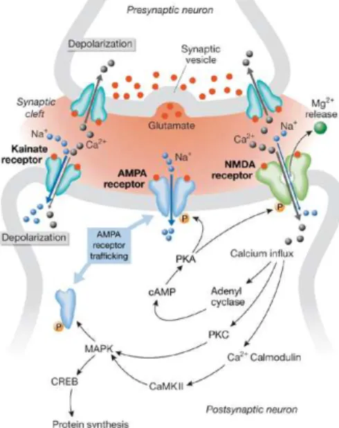
postsynaptic potential in response for a high frequency stimuli, where glutamate plays a central role (Lomo, 1966). Therefore, stimulation of a certain circuit will induce permanent changes that increase the efficiency of synaptic transmission in that same stimulated synapse. One of the main brain areas where synaptic plasticity has been best studied is the hippocampus, whose main circuit is glutamatergic. In basal conditions, glutamate activates several classes of metabotropic receptors and three major types of ionotropic receptors: α-amino-3-hydroxy-5-methyl-4-isoxazolepropionate (AMPA), N-methyl-D-aspartate (NMDA) and kainate receptors (Figure 1.1.1.1). Ionotropic
receptors are ligand gated ionic channels permeable to the monovalent cations Na+ and K+ and,
depending on the subtype, also to the divalent cation Ca2+ (Danysz and Parsons, 2003).
Figure 1.1.1.1 - Glutamate receptors and synaptic plasticity. The arrival of a series of impulses at the presynaptic terminal triggers the release of glutamate, which binds to glutamate receptors at the postsynaptic membrane. Once activated, AMPA and kainate receptors conduct sodium ions, which initiate postsynaptic depolarization. Membrane potential changes leading to the release of magnesium ions that block NMDA receptors. Calcium influx through NMDA channels sets off a chain of events that establish long-term potentiation. Kainate receptors at the presynaptic end also seem to facilitate synaptic transmission at specific synapses by augmenting neurotransmitter release. Abbreviations: AMPA, α-amino-3-hydroxy-5-methyl-4-isoxazolepropionate; CaMKII, calcium / calmodulin-dependent kinase II; CREB, cAMP response element binding protein; MAPK, mitogen-activated protein kinase; NMDA, N-methyl-D-aspartate; PKA, protein kinase A; PKC, protein kinase C.
7
AMPA receptors are largely impermeable to Ca2+ and participate in most forms of fast
synaptic transmission (Danysz and Parsons, 2003). The kainate receptors are also involved in synaptic transmission. Although the contribution of this receptors is controversial they produce bi-directional modulation of both excitatory and inhibitory transmission and in some cases may trigger also metabotropic cascades (Huettner, 2003). In contrast, NMDA receptors usually do not contribute to basal synaptic transmission and are only activated under certain conditions. These
receptors have three major features: they present high permeability to Ca2+ ions;
voltage-dependently blockage by Mg2+ ions and a slow gating kinetics (Danysz and Parsons, 2003).
When a LTP is induced by a high frequency signal (or convergence of several signals) that arrives to the glutamatergic synapse, this leads to a massive glutamate release which triggers a sequence of events described in Figure 1.1.1.1 (Danysz and Parsons, 2003). In these conditions,
AMPA / kainate receptors are activated, while NMDA receptors remained blocked by Mg2+. The
continuous activation of AMPA / kainate receptors leads to a significant influx of Na2+ ions into
the pos-synaptic cell, which in turn, decreases the membrane potential removing the Mg2+ ion from
the blocked NMDA receptor. At this stage, Ca2+ ions can freely enter the cell via the NMDA
receptor channel and initiate a number of enzymatic processes that lead to the permanent strengthening of synaptic transmission. This is manifested post synaptically as an enhancement of AMPA receptor sensitivity and number.
Classically it is stated that LTP magnitude is directly proportional to the learning ability, meaning that, a weak performance in the Morris Water Maze task is usually associated with a lower LTP magnitude (Lynch, 2004). However, there are data showing a lack of direct correlation between animals performance in Morris Water Maze tasks and LTP magnitude. Recently, data from the lab, demonstrate that hippocampal slices taken from old animals, which have an impaired hippocampal dependent memory when compared to younger animals, have an increased LTP magnitude induced by θ-burst stimulation (Diogenes et al., 2011).
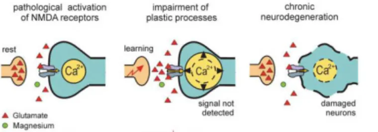
Hypothetically, the higher LTP together with the impairments in hippocampal dependent memory can be explained by an exacerbation of glutamate receptors activation. Excessive
activation of these receptors would lead to a continuous Ca2+ influx into the cells, thus would
ultimately result in neuronal damage, synaptic dysfunction or even cell death and consequent further decline of cognitive function in a mechanism similar to the already proposed for Alzheimer´s disease (AD) (Figure 1.1.1.2).
8
Figure 1.1.1.2 – It is hypothesized that in Alzheimer’s disease, due to the overactivation of the glutamatergic system, Mg2+ is no longer effective in its ability to control NMDA receptor activation (Danysz and Parsons, 2003). Abbreviators: Ca2+ - calcium ion.
Although, the changes in LTP observed upon aging are controversial. Most of the studies describe that old animals present impairments in LTP that are specific for lower-intensity stimulus (Rosenzweig and Barnes, 2003). For high-frequency stimulation protocols (e.g. 100 Hz for 1 s) no age-related deficits are observed either in CA1 or Dentate gyrus both in vivo and in vitro (Barnes, 1979; Diana et al., 1994a; Diana et al., 1994b; Landfield and Lynch, 1977; Landfield et al., 1978; Rosenzweig et al., 2003).
These differences might be related to the type of stimulation protocol and the areas of the
dendritic tree, particularly because age-related changes have been described in basal, but not apical, dendrites of CA1 pyramidal neurons (Lynch, 2004).
Importantly, most of the alterations observed in healthy aging, namely those that lead to excitotoxicity, are also present in acute insults and in neurodegenerative diseases such as Alzheimer’s disease.
1.2 – Memantine
Aging increases the vulnerability to suffer from diseases such as cancer or neurodegenerative disorders as AD. AD begins with mild memory impairments and evolves into a major loss of cognitive abilities (Alloul et al., 1998).
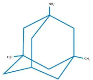
Currently the therapeutic approaches used in patients with AD are based on drugs that ameliorate cognitive symptoms such as the acetylcholinesterase inhibitors. Memantine (1-amino-3, 5-dimethyladmantane), a partial antagonist of the glutamate NMDA receptors (Parsons et al.,
9
1999), particularly of the 2B subtype is being used for the symptomatic treatment of AD since its safety and efficacy has been demonstrated (Chen and Lipton, 2006; McShane et al., 2006; Parsons
et al., 2007; Rogawski and Wenk, 2003). Memantine with a molecular formula of C12H21N.HCl
and a molecular weight of 215.77 g/mol is an uncompetitive NMDA receptor antagonist (IC50 of ~
3 µM) (Parsons et al., 1995). This drug presents good tolerability and is predicted to act selectively at NMDA receptors in vivo producing improvements in the therapy of dementia (Ditzler, 1991; Gortelmeyer and Erbler, 1992; Pantev, 1993) or on learning and memory (Lipton, 2006). However, memantine can produce psychotomimetic effects that appear only if the recommended titration of dosage from 5 to 20 mg over 3–4 weeks is skipped, or when memantine is combined with dopaminomimetic therapies (Parsons et al., 1997).
However, it is well recognized that the blockade of NMDA receptors could lead to impairments of neuronal plasticity and learning (Collingridge and Bliss, 1995). In 1972, Merz and collaborators demonstrated that memantine is effective in the central nervous system (CNS), revealing its potential for the treatment of Parkinson’s disease, spasticity and cerebral disorders like coma, cerebrovascular and geronto-psychiatric disturbances (Grossmann and Schutz, 1982; Miltner, 1982a, b; Mundinger and Milios, 1985; Schneider et al., 1984).
Like Mg2+, memantine is able to dissociate from NMDA receptor channel due to its
voltage-dependency and fast unblocking kinetics providing neuroprotection and symptomatic restoration of synaptic plasticity by the same mechanism. Neuroprotective agents which completely block NMDA receptors lead to impairments in normal synaptic transmission and thereby cause numerous side effects. The challenge has been to develop antagonists that prevent the pathological activation of NMDA receptors but allowing their physiological activity such as memantine (Figure 1.3.1).
Figure 1.3.1 – Chemical structure of memantine: three-ring structure; bridgehead amine (–NH2
group), which is charged at the physiological pH of the body (–NH3 +
) and represents the region of memantine that binds at or near the Mg2+ binding site in the N-methyl-D-aspartate receptor (NMDA receptors)-associated
10
ion channel; and methyl group (–CH3) side chains, which stabilize memantine interaction in the channel
region of the NMDA receptors. Adapted from (Lipton, 2006).
The promising profile of memantine has been attributed to its strong voltage dependency (inhibits NMDA-induced currents at -70mV, but does not affect NMDA-currents at +70 mV) to its
rapid, open-channel unblocking kinetics (faster rates of channel unblock Koff = 0.2 sec-1) and also
to its low to moderate affinity (with Ki of 0.5 - 0.7 µM) which would allow the blockade of only
the pathological activation of NMDA receptors, leaving their physiological activation relatively intact (Chen et al., 1992; Ditzler, 1991; Frankiewicz et al., 1996; Parsons et al., 1993; Parsons et al., 1995). Besides its actions on NMDA receptors, memantine can also act as an antagonist of the nicotinic acetylcholine receptors and 5-HT receptors (Aracava et al., 2005; Buisson and Bertrand, 1998).
The rationale for using memantine in AD patients is based on the hypothesis that the blockade of NMDA receptor-mediated excitotoxicity can help to preserve neuronal structure and
function (Lipton, 2006, 2007; Wenk et al., 2006). The improvement of memory in AD upon
treatment is an apparent paradox concerning that as a NMDA receptor antagonist, memantine would impair the mechanisms underlying LTP and consequently hippocampal dependent memory. In this perspective, memantine could be an ideal tool to study the increased LTP observed in old animals. It would allow to test whether exacerbated activation of NMDA receptors could be related to the enhanced LTP magnitude and to the impaired hippocampal memory observed in old animals.
11
2 - Aim:
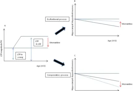
Paradoxically, aging has been associated to a progressive impairment of hippocampal dependent memory together with an increase in hippocampal LTP (Costenla et al., 1999; Diogenes et al., 2011). These data are intriguing and could be a consequence of an overactivation of NMDA receptors, an already postulated hypothesis for the mechanism of several disorders such as Alzheimer´s Disease (Danysz and Parsons, 2003). We thus proposed to decipher if the increased LTP, detected in old animals, was an outcome of an exacerbated activation of NMDA receptors and whether it was a compensatory or a dysfunctional phenomenon (Figure 2.1). To achieve this we evaluated 1) whether an NMDA receptor antagonist, memantine, was able to restore hippocampal dependent memory in old animals, 2) if this was accompanied, by a reestablishment of the increased LTP and finally 3) which would be the molecular consequences of the chronic memantine treatment for NMDA (subunit 2B) and AMPA (subunit GluR1) receptors.
Figure 2.1 – Possible effects of memantine upon aging. In A is represented what we have been observing regarding LTP magnitude, an increase on LTP magnitude with ageing. If this enhancement in LTP magnitude is a “dysfunctional LTP”, memantine would restore the normal (lower) LTP magnitude and improve learning and hippocampal dependent memory (B). If the higher LTP magnitude is a “compensatory LTP”, memantine would restore the normal (lower) LTP magnitude and aggravate the impairments in learning and hippocampal dependent memory (C).
13
3 - Techniques and Methods
Experiments were performed with two age groups of Wistar rats (male, Wistar, Harlan Interfauna Iberica, SL, Barcelona, Spain): young (10–15 weeks), age at which reproductive behavior is fully established and old (70–80 weeks old) (Havenaar, 1993). The animals were kept under standardized temperature, humidity and lighting conditions, and had access to water and food ad libitum. All animal procedures were carried out according to the European Community Guidelines for Animal Care (European Communities Council Directive - 86/609/EEC).
3.1 – Drugs
1-amino-3, 5-dimethyladmantane (memantine) was diluted in a saline vehicle (0.9%). Memantine was generously provided by Grünenthal (Germany).
3.2 - Intraperitoneal injection
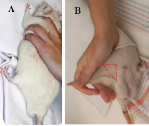
Memantine 10, 5 or 1 mg/kg/day was blindly administrated intraperitonealy (Figure 3.2.1) as previously described (Anderson et al., 2004; Meisner et al., 2008; Reus et al., 2010).
Figure 3.2.1 – Intraperitoneal injection: the immobilization procedure (A) and the local of the injection (B).
14
3.3- Behaviour Assays:
3.3.1 - Open Field Test
Background
The Open Field test (OF) was originally described for the study of emotionality in rats (Hall, 1934). Nowadays it is mostly used to assess locomotor, exploratory and anxiety-like behaviour in laboratory animals (rats/mice).
Open field is based on two concepts: individual testing (the animal is separated from its social group) and agoraphobia (as the arena is very large when comparing to the animal’s breeding or natural environment) (Prut and Belzung, 2003). The procedure consists in subjecting an animal, to an unknown environment (Walsh and Cummins, 1976) exploring the duality between the innate fear that rodents have of novel open and bright spaces versus their desire to explore new environments (Prut and Belzung, 2003).
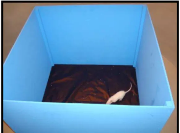
Different protocols of the open field are available (Prut and Belzung, 2003) both concerning the apparatus and the procedure. The Open Field area consists of an empty and bright square arena (67 x 67 x 52 cm), surrounded by walls to prevent animal from escaping (Figure 3.3.1.1). The animal is usually placed at the center of the arena and the behaviour recorded over a chosen period of time (from 3 to 15 min) (Prut and Belzung, 2003), in one trial or multiples trials (Walsh and Cummins, 1976). Ambulatory locomotion in the horizontal plane is quantified by an observer or software that records the number of lines crossed or squares entered and this can be used to access anxiety. When anxious, the natural tendency of rodents is to stay preferably close to the walls (thigmotaxis). In this context, anxious-related behaviour is evaluated by the degree to which the rodent avoids the center of the OF (Crawley, 1985; Stone, 1932), measuring the percentage of time spent in the central of the arena (Prut and Belzung, 2003). Freezing behaviour, defined as the absence of movement, can also be assessed and is usually taken as indicative of a high-stress state.
The spontaneous activity in the open field is the most standardized measure of motor function (Crawley, 1985). Changes in the locomotor performance can be evaluated by measuring the average speed or the travelling distances during a defined time interval. Behaviour of rodents
15
in the open field depends mainly on: tactile sensory factors as vibrissae (Prut and Belzung, 2003), lighting conditions and the light-dark cycle, field size (Blizard, 1971), apparatus colour and material (Delbarre et al., 1970; Oldham, 1970; Satinder, 1968), environmental odours and also on the start positions in the apparatus (Clark C. V. H., 1970; Clark, 1970; Satinder, 1969).
Figure 3.2.1.1 – Open Field apparatus and zones defined with the software.
Procedure:
Animals were gently placed in the center of the arena and the behaviour recorded for 5 minutes. Travelling distance and average speed were recorded using PanLab software to evaluate the locomotor activity. Light, noise, smell and temperature were controlled.
16
3.3.2 - Elevated Plus Maze
Background
The elevated plus maze has its origin in the Y-shaped apparatus (Montgomery, 1958) and was latter modified into an elevated maze with four arms (two open and two closed) that are arranged to form a plus shape (Handley, 1984). This task is based on the conflict the rodent has between his preferences for staying in the closed, protected areas (thigmotaxis) and his innate motivation to explore the novel, open environments. This maze allows the evaluation of both anti-anxiety behaviour, manifested as an increased time in the open arms, and motor activity, determined simultaneously by measuring spontaneous motor activity (total arm entries). Other ethological behaviours that can be measured in this maze are the number of rears, head dips, fecal boli, freezing or stretched-attend postures (Walf and Frye, 2007). This maze was validated by demonstrating that anxiogenic drugs reduce the time spent on the open arms and anxiolytic drugs increase the time spent on the open arms of the elevated plus maze (Pellow et al., 1985). Moreover, it has been shown that the increased open arms activity observed in rodents is correlated with increased central square entries in a brightly lit open field (Frye et al., 2000) and that the plasma corticosterone levels increase with open arms exposure and are positively correlated with risk assessment behaviour in the elevated plus maze (File et al., 1994; Rodgers et al., 1999).
Pre-exposure to another testing environment does not alter subsequent behaviour of rats or mice in elevated plus maze (Walf and Frye, 2007). On the other hand recent reports suggest that the pre-exposure to the plus maze itself induces test decay and alters the subsequent response of the animal (File et al., 1990; Lister, 1987; Pellow et al., 1985), there are differences when rodents are exposed to the plus maze on more than one occasion (Adamec et al., 2004; Adamec et al., 2006). Besides this the behaviour of rats and mice in the elevated plus maze can be influence by different parameters: circadian rhythms / light cycle (Andrade et al., 2003; Carobrez and Bertoglio, 2005; Jones and King, 2001); handling of the animals such as prior experience with handling (Andrews and File, 1993; Brett and Pratt, 1990), stress (Steenbergen et al., 1990) or injections (Lapin, 1995); age (Boguszewski and Zagrodzka, 2002); sex/gender (Imhof et al., 1993); strain (Rodgers and Cole, 1993); breeding line differences (Bert et al., 2001); estrous cycle (Marcondes
17
et al., 2001) and also differences in housing conditions and temperature of the room (21 ± 1ºC) (Zhu et al., 2006).
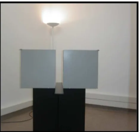
Figure 3.2.2.1 – Elevated Plus Maze apparatus.
Procedure:
The maze consists of four arms (two open without walls and two enclosed) (Walf and Frye, 2007) 120 cm above the floor, each arm has 100 cm length, 10 cm width and 50 cm height (Figure 3.3.2.1).
Animals were placed in the intersection of the arms facing an open arm and the behaviour was recorded during 5 minutes. The time spent in the open arms and the number of total entries were measured and used to evaluate the anxious behaviour and locomotion, respectively. The light, noise, smell and temperature were controlled.
18
3.3.3- Morris Water Maze
Background
The Morris water maze (MWM) task was described 30 years ago to study spatial learning and memory in laboratory rats (Morris, 1981). Learning is the acquisition of an altered behaviour due to an environmental stimulus; the animal changes its behaviour in response to an experience and stores a memory (Sweatt, 2003).
Spatial learning is an example of hippocampal-dependent learning, both in humans and lower animals. Animals must learn to navigate their environment and learn to associate particular places with particular items or events (Sweatt, 2003) generating a “cognitive map” (allocentric visuospatial cues) (Hodges et al., 1995). This type of learning has been defined to be hippocampal dependent. A wide variety of different studies have shown that molecular or anatomical lesions of the hippocampus lead to spatial learning deficits in both humans and lower animals. Also, direct measurements of a wide variety of molecular and physiologic changes have been shown to correlate with hippocampal dependent spatial learning (Sweatt, 2003), such as Long-term Potentiation and NMDA receptor function (Bannerman et al., 1995; Morris et al., 1986; Moser and Moser, 1998).
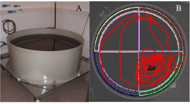
The Morris water maze is a hippocampal-dependent spatial learning task in which animals are required to learn how to locate an escape platform in a swimming pool, using visual cues that are surrounding the maze (Sweatt, 2003). The device consists of a large circular pool filled with opaque water that is positioned in a room with a sufficient amount of external cues visible to the swimming animal.
During the acquisition days, animals learn how to find the platform and escape from the pool (D'Hooge and De Deyn, 2001) using a random or semi-random set of start locations (Vorhees and Williams, 2006). This period consist at a minimum of 4 days, in each day the animals perform 4 trials separated by at least 15 minutes (Diogenes et al., 2011) in which they are allowed to search the platform for 60 seconds. If the animal finds the platform, stays there for 10 seconds, otherwise is guided to the platform remaining there during 20 seconds to acquire the spatial location (Diogenes et al., 2011). The acquisition days should be performed until the control condition is in steady phase different of learning, having all the same latency (time to find the platform).
19
Variation to the test can include a visible platform or different inter trail intervals (Rick et al., 1996).
To determine if the animals are using a spatial learning strategy to locate the escape platform, they are subjected to a “probe” trial after the train is completed. This test can be performed in the last day of the acquisition training or in following day. In the probe test the platform is removed and the animal is allow to swim freely for 60 seconds while recording the percentage of time spent in each quadrant.
Behaviour of animals in the Morris Water Maze can be influence by different parameters such as: body weight (Wenk, 2004), gender or strains/species (D'Hooge and De Deyn, 2001; Roof et al., 1993), age (Brandeis et al., 1989; D'Hooge and De Deyn, 2001; Gallagher and Nicolle, 1993; Geinisman et al., 1995), nutrition (Pitsikas et al., 1990), stress (Grauer and Kapon, 1993) or infectious agents and parasites (Gibertini et al., 1995). Differences in the apparatus and training procedures can also modifie the behaviour of animals. Alterations of the basic training protocols (Stewart, 1993; Wenk et al., 1998) such as introducing a Visible-platform during acquisition training (e.g. by putting a flag on top of the platform) (Hauben et al., 1999; Rick et al., 1996; Vorhees and Williams, 2006) or performing a second probe trial (Vorhees and Williams, 2006) are often made. The presence and amount of the external cues (Stewart, 1993), the opacifier and the size of pool (Morris, 1984) can also change the animal behaviour.
Figure 3.2.3.1 – Morris Water Maze apparatus (A) and representative tracing of probe test of one animal that learn where the platform was (B).
20
Procedure:
The Morris water maze apparatus consists in a circular pool (150cm diameter and 62 cm height) with a platform (12cm diameter and 42 cm height) submerged 1.5 cm beneath the water surface (Figure 3.3.3.1). The platform location was selected randomly for each animal but was kept constant throughout the training phase (Martinez-Coria et al., 2010).
The rats were subjected to four training trials a day for 4 days with a intra trial interval at least 15 minutes. Retention of the spatial training was assessed 24 hours after the last training session (long-term memory), by measuring the time spent in the platform quadrant. The light, noise, temperature of the room and the water, colour of the water and constant distribution of the cues were controlled.
21
3.4- Extracellular electrophysiology recordings
3.4.1 - Hippocampal slice preparation
Background
McIlwain and collaborators were pioneers in developing methods for ex vivo CNS preparations to perform biochemical studies (Mc et al., 1951). In 1957, Li and McIlwain published the first electrophysiological study performed in cortex slices (Li and Mc, 1957). In spite of these early reports, these biological preparations were only believed to maintain their normal physiological properties after the studies by Yamamoto and McIlwain (1966) and Richards and McIlwain (1967), showing that hippocampal slices, sectioned perpendicularly to the long axis of the hippocampus, maintained synaptic activity and that the evoked responses were similar to those recorded in vivo. Since then, slices of CNS are commonly used as experimental models in pharmacological, biochemical and neurophysiological studies. The hippocampus is used extensively in these techniques, (Anderson and Collingridge, 2001) since the arrangement of the neurons allows this brain structure to be sectioned such that most of the relevant circuitry is left intact presenting a unique laminated organization. The cell bodies of the pyramidal neurons lie in a single packed layer that is easily visualized and is possible to distinguish the different areas that constitute the hippocampal circuit. The hippocampus can be divided in three main areas that communicate mainly through an unidirectional circuit that projects from the Dentate Gyrus to CA3 and this to CA1.
22
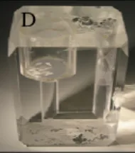
Figure 3.3.1.1 – A) Illustration of rat brain, B) hippocampus dissection, C) Chopper used to cut hippocampal slices and D) camera where the slices recovered energetically before recording.
Procedure:
Animals were sacrificed by decapitation under deep halothane anaesthesia. The hippocampi were isolated in ice-cold Krebs solution (Figure 3.4.1.1 A, B) with the following
composition (mM): NaCl 124, KCl 3, NaHCO3 26, Na2HPO4 1.25, MgSO4 1, CaCl2 2, glucose 10,
gassed with a 95% O2 + 5% CO2 mixture with pH 7.4. One hippocampus was used for
electrophysiological recordings (Figure 3.4.1.1 C, D) and the other for receptor quantification through Western blot.
23
3.4.2 - LTP induction
Background
The first theories regarding the cellular basis of learning were postulated by Hebb, suggesting that information storage is achieved by activity-dependent alterations at the synapse (Hebb, 1949). Latter in 1973 Bliss and Lomo described a phenomenon they termed “Long-Term Potentiation” (LTP) (Bliss and Lomo, 1973). They reported that brief periods of high-frequency (100Hz) synaptic stimulation could lead to a permanent enhancement of synaptic transmission in hippocampal recordings from rabbits.
LTP can be induced by different protocols (Sweatt, 2003) and in different synapses. In the hippocampus, most of the experiments to study the basic processes underlying LTP have been performed at the synapse connections between axons from CA3 pyramidal neurons that extend into CA1, the schaffer-collateral synapses (Sweatt, 2003), by recording in the dendrite regions of CA1.
In extracellular recordings a field excitatory post-synaptic potential (fEPSP) is obtained. It results from the depolarization of the recording cells as a consequence of the glutamate release, induced by the electrical stimulation. The typical waveform of the fEPSP consists of a “fiber volley” and after the excitatory postsynaptic potential (EPSP) itself. The “fiber volley results from the presynaptic action potential arriving at the recording site, while the EPSP is the manifestation of the synaptic activation (depolarization) in CA1 pyramidal neurons. To evaluate changes in the fEPSPs, the parameter typically measured is the initial slope of the EPSP waveform. Absolute peak amplitude of the fEPSP can also be measured, but the initial slope is the preferred index since it is less prome to contamination from other sources of current flow in the slice (Sweatt, 2003).
S1 S2 DG CA3 CA1 S1 S2 DG CA3 CA1 S1 S2 DG CA3 CA1 A B C
0.5 mV
5 mV
24
Figure 3.4.2.1 – A) Photography of the recording chamber where slices are placed, B) Illustration of the hippocampus slice and where the stimulation (S1 and S2) and recording electrodes are localized. C) Tracing obtained after stimulation composed by the stimulus artifact, followed by the presynaptic volley and the fEPSP.
Procedure:
Individual slices were transferred into a recording chamber continually superfused with Krebs solution at a constant flow (3 ml/min) and temperature (31ºC) with the same gassed solution
(Figure 3.4.2.1 A). fEPSPs were recorded thought a microelectrode filled with NaCl 4M (2-6 MΩ
resistance) and placed in CA1 stratum radiatum. Stimulation (rectangular 0.1-ls pulses, once every
15 s) was delivered through a concentric electrode placed on the Schaffer collateral-commissural fibbers, in the stratum radiatum near the CA3-CA1 border (Figure 3.4.2.1 B) (Diogenes et al., 2007). The protocol for LTP induction was a θ-burst stimulation (4x4: 4 bursts, 100Hz, 4 stimuli, separated by 200 ms) as previously described (Costenla et al., 2011). The initial intensity of the
stimulus was that eliciting 50% of the maximal response. Recordings were obtained with an
Axoclamp 2B amplifier and digitized (Axon Instruments, Foster City, CA). Individual responses were monitored, and averages of eight consecutive responses (Figure 3.4.2.1 C) were continuously stored on a personal computer with the LTP program (Anderson and Collingridge, 2001).
25
3.4.3 - Basal Synaptic Transmission - Input / Output Curve
Input-output curves were performed to evaluate the effect of memantine on basal synaptic transmission.
The underlying principle of this protocol is to evaluate the changes in synaptic responses that result from a step by step increase in stimulus intensity. It starts with a stimulation that elicits no synaptic response and ends with one that cannot further increase the fEPSP slope. The output results allow the evaluation of the maximal response that can be elicited and also to compare the ability to increase the synaptic response to each intensity of stimulation.
Procedure:
fEPSP were recorded through an extracellular microelectrode (4M NaCl, 2-6 MΩ resistance) placed in stratum radiatum of CA1 area. Stimulation (rectangular 0.1 ms pulses, once every 15 s) was delivered through a concentric electrode placed on the Schaffer collateral-commissural fibers, in stratum radiatum near CA3-CA1 border. The intensity of stimulus (80-200µA) was initially adjusted to obtain a large fEPSP slope with a minimum population spike contamination (Diogenes et al., 2011).
After obtaining a stable baseline for at least 15 min, the stimulus delivered to the slice was decreased until no fEPSPs was elicited. The stimulus was then successively increased by 20 µA steps. For each stimulation, data from three consecutive averaged fEPSP (each average fEPSP is the computerized mean of eight individual fEPSP) were stored. The range of stimulation was from 60 µA until 360 µA. The input-output curve was plotted as the relationship of the fEPSP slope vs stimulus intensity (e.g, fiber volley amplitude), which provides a measure of synaptic efficiency.
26
3.5 - Western Blotting
Background:
The method was originally described in the laboratory of George Stark at Stanford (Burnette, 1981).
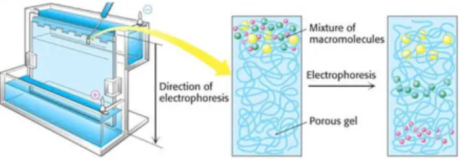
The first step in a Western blotting procedure is to separate the macromolecules using gel electrophoresis (Figure 3.5.1). After separated, proteins are transferred or blotted onto a second matrix, generally a nitrocellulose or polyvinylidene difluoride (PVDF) membrane (Figure 3.5.2). The membrane is then blocked, to prevent any nonspecific binding of antibodies to the surface of the membrane, and incubated with a primary antibody that recognizes the desired protein. The detection is made through an enzyme-labelled secondary antibody that recognizes specifically the primary antibody. An appropriate substrate is then added to the enzyme producing a detectable product such as light or a chromogenic precipitate. The intensity of the signal correlates with the
abundance of the antigen on the membrane.
Figure 3.5.1 - Discontinuous SDS-PAGE electrophoresis procedure. Samples are first loaded into wells in the gel, with one lane being usually reserved for a marker or ladder (commercially available mixture of proteins of defined molecular weights, stained to form visible, coloured bands). When voltage is applied, proteins migrate into the gel it at different speeds, according to molecular weight, causing smaller proteins to progress further along the gel. Image source: imb-jena.de.
27 Cassette holder Foam pad Filter paper Gel Membrane Filter paper Foam pad (+) Anode (-) Cathode Cassette holder Foam pad Filter paper Gel Membrane Filter paper Foam pad (+) Anode (-) Cathode
Figure 3.5.2 - Protein Blotting procedure. In a typical wet blot, a sandwich structure is placed in a tank filled with transfer buffer. Proteins migrate from the negative (gel) to the positive (membrane) pole. Picture source: komabiotech.co.kr.
Procedure:
Hippocampi were disrupted in sucrose-Tris solution (0.32M sucrose solution with 50 mMTris supplemented with protease inhibitor (ROCHE) with a Teflon pestle and protein concentrations were determined using the Bio-Rad protein assay (Bradford, 1976). Total hippocampal proteins (60 or 45 µg) were denatured in sample buffer (10% (w/v) SDS, 600 mM DTT, 350mM Tris pH
6.8, 30% (v/v) Glycerol and 0.012% Bromophenol Blue) at 95ºC for 5 minutes, fractionated on 8%
SDS-polyacrylamide electrophoresis gels and transferred onto polyvinylidene difluoride
membranes (Millipore). Ponceau S staining was carried out to check the equal loading of the
protein as well as the transfer to the membrane after blotting. After blocking with 5% non-fat dry
milk in TBS-T 0,1% (Tris buffer saline solution, 200nM Tris, 1.5 NaCl with 0.1% Tween-20) for 1h, the blots were washed in TBS-T and incubated overnight at 4ºC with primary antibodies diluted in BSA 3% in TBS-T (reactive to NMDA (2B subunit) and AMPA (GluR1 subunit) receptors and α-tubulin-loading control). After washing again with TBS-T membranes were incubated with the secondary antibodies in 5% non-fat dry milk in TBS-T for 1 hour at room temperature. Chemoluminescent detection was performed with ECL-PLUS western blot detection reagent (GE Healthcare) using X-Ray films (Fujifilm). Optical density was determined with Image-J software and normalized to the respective α-tubulin density.
28
Table 3.5.1 – Primary and secondary antibodies and related conditions used in Western Blot experiments for individual proteins. All primary antibodies were diluted in 3% Bovine Serum Albumin with 0.1% NaN3 and secondary antibodies in 5% non-fat dry milk.
Protein Protein Loading (µg)
Primary antibody
Animal Dilution Secondary antibody
Dilution
GluR1 45 Milipore
(05-855)
Rabbit 1:5000 Sta. Cruz Biotechno logy (goat anti-rabbit) 1:10000 NMDA (2B) receptor 60 Cell Signalling (D15B3) Rabbit 1:5000 1:10000
3.6 - Statistics
The values presented are mean ± SEM of n experiments. To test the significance of the differences between vehicle and memantine groups, an ANOVA was used, with age group (young, old) and memantine dosage (vehicle, 1, 5, 10) as between-subject factors. Values of P<0.05 were considered to be statistically significant.
29
4 - Results
4.1 - Behaviour assays:
To study the behaviour alterations induced by memantine, the open field arena, the elevated plus maze and the Morris water maze test were performed.
4.1.1 - Open Field
Motor control is mainly integrated by striatal areas that receive multiple glutamatergic inputs. Given that NMDA receptor antagonists can influence motor control (Kang et al., 2011; Lemay-Clermont et al., 2011), the open field test was performed to evaluate the impact of memantine on locomotor behaviour of the animals treated with this drug. Animal average speed was evaluated for 5 minutes and the behaviour compared between different groups.
The locomotor activity was significantly decreased (F(1.18) = 25.6, p< 0.001, ηp
2 = 0.6)
in old animals treated with saline vehicle (OFOldvehicle: 9.7 ± 0.4 cm/sec, n=10) when compared to
the younger animals (OFYoungvehicle 13.4 ± 0.6 cm/sec, n=10, figure 4.1.1.1).
Overall, memantine significantly affected locomotor activity in young (F(2.17) = 4.9,
p<0.05, ηp2 = 0.4) and in old (F(2.17) = 16.3, p<0.001, ηp2 = 0.7) animals. In younger animals,
memantine (10 mg/kg/day) significantly increased locomotor behaviour (95% C.I.]0.5; 5.8[; p<
0.05 by increasing the average speed in open field test in younger rats (OFYoungmemantine10mg/Kg: 16.5
± 0.9 cm/sec, n=5 versus OFYoungvehicle: 13.4 ± 0.6 cm/sec, n=10). In old animals memantine 10
mg/Kg/day also produced the same significant effect (95% C.I. ]0.4;4.2[; p< 0.05;
OFOldmemantine10mg/Kg: 12.0 ± 0.6 cm/sec, n=5 versus OFOldvehicle: 9.7 ± 0.4 cm/sec, n=10; figure
4.1.1.1).
Regarding the dosage of 5 mg/Kg/day, memantine only affected older animals (95% C.I.]0.6;4.5[; p<0.01). Old animals treated with 5 mg/Kg/day of memantine had a decreased
average speed when compared to old vehicle treated animals (OFOldmemantine: 7.2±0.6 cm/sec; n=5
30 0 10 20 30 * * * * Young Old 5 m g /K g 1 0 m g /K g v e h ic le 5 m g /K g 1 0 m g /K g v e h ic le A v e ra g e s p e e d ( c m /s e c )
Besides the effects in motor behaviour, the higher dose memantine (10 mg/kg/day) also induced alterations of "natural behaviour" in young rats characterized by standing on hind legs, open-mouthed tooth display, long ‘eeeps’ without biting other rats.
Figure 4.1.1.1 – Average speed evaluated in the locomotor test of the open field arena of young (black) and old rats (blue) treated with different doses of memantine (10 and 5mg/Kg/day (solid bars)) or vehicle (open bars) for 14 days. Results are mean ± SEM of the number of experiments (n): n youngvehicle = 10, nyoung5mg/kg = 5,
nyoung10mg/kg = 5, noldvehicle = 10, nold5mg/kg = 5, nold10mg/kg = 5. The data were analyzed using a 2 X 2 ANOVA,
31 0 20 40 60 80 Young Old V e h ic le V e h ic le 1 m g /K g 5 m g /K g 1 0 m g /K g 1 m g /K g 5 m g /K g 1 0 m g /K g T im e i n o p e n a rm s (% o f s e c )
4.1.2 - Elevated Plus Maze
Glutamate is ubiquitous within the central nervous system and has been shown to play important roles in many brain processes, including stress response and anxiety disorders (Drevets et al., 1997). Exposure to severe stress has been associated with glutamate excitotoxicity. In fact, there are data demonstrating that antagonists of NMDA receptors have anti-anxiety effects (Campeau et al., 1992; Fendt et al., 1996). Therefore, we considered important to assess whether this pharmacological treatment had any impact on anxious behaviour. Consequently the animals were submitted to the elevated plus maze test and the time spent in the open arms was measured and compared between animals.
The anxiety behaviour was not affected by ageing (Figure 4.1.2.1). Moreover, both young and old animals treated with memantine (10, 5, 1 mg/kg/day) did not show significant anxiolitic effects (Figure 4.1.2.1). However, in old animals treated with memantine, a strong tendency for an anti-anxiogenic behaviour was detected.
Figure 4.1.2.1 – Time in open arms evaluated in the anxiety test of the Elevated plus maze aparratus of young (black) and old rats (blue) treated with different doses of memantine (10, 5 and 1 mg/Kg/day (solid bars)) or vehicle (open bars) for 14 days. Results are mean ± SEM of the number of experiments (n): nyoungvehicle = 13,
nyoung1mg/kg = 5, nyoung5mg/kg = 12, nyoung10mg/kg = 5, noldvehicle = 13, nold1mg/kg = 7, nold5mg/kg = 9, nold10mg/kg = 5. The
data were analyzed using a 2 X 3 ANOVA, with Age Group (young, old) and Memantine Dosage (0, 1, 5, or 10 mg) as between-subject factors. *P<0.05 between the groups.
32 1 2 3 4 0 10 20 30 40 50 60 Days A) * Y-Vehicle O-Vehicle L a te n c y ( s e c )
4.1.3 - Morris Water Maze
Memantine or saline vehicle were administered chronically for 14 days and the effects on learning and hippocampal dependent memory were evaluated using the Morris water maze test. Learning was assessed during acquisition, while the animals were taught to find the platform. Memory retrieval was evaluated at the probe test, by the time spent in platform quadrant.
Old animals showed an impairment on hippocampal dependent memory when compared
with young animals F(1.23) = 10.9, p < 0.005, ηp2 = 0.3 (Figure 4.1.3.1 A).
When given in the higher dose, memantine (10 mg/kg/day), impaired learning in old animals (95% C.I. ]4.03; 24.0[; p< 0.05, n=13). That is visible during acquisition where old animals with memantine treatment take longer to find the platform comparing to vehicle treated rats (Figure 4.1.3.1 C). For lower doses of memantine (5 or 1mg/kg/day) no significant changes are observed in the learning performance of either young or old animals (Figure 4.1.3.1 D-G)
33 1 2 3 4 0 10 20 30 40 50 60 Days B) Y-Vehicle Y-Memantine 10 mg/kg/day L a te n c y ( s e c ) 1 2 3 4 0 10 20 30 40 50 60 Days C) * O-Vehicle O-Memantine 10mg/kg/day L a te n c y ( s e c ) 1 2 3 4 0 10 20 30 40 50 60 Days D) Y-Vehicle Y-Memantine 5mg/kg/day L a te n c y ( s e c ) 1 2 3 4 0 10 20 30 40 50 60 Days E) O-Vehicle O-Memantine 5mg/kg/day L a te n c y ( s e c ) 1 2 3 4 0 10 20 30 40 50 60 F) Days Y-Vehicle Y-Memantine 1mg/kg/day L a te n c y ( s e c ) 1 2 3 4 0 10 20 30 40 50 60 G) Days O-Vehicle O-Memantine 1mg/kg/day L a te n c y ( s e c )
Figure 4.1.3.1 – Latency evaluated in Morris Water Maze test of the pool apparatus of young (black) and old rats (blue) treated with different doses of memantine (10, 5 and 1 mg/Kg/day (solid bars)) or vehicle (open bars) for 14 days. Results are mean ± SEM of the n of experiments: nyoungvehicle = 13, nyoung1mg/kg/day = 6,
nyoung5mg/kg/day = 14, nyoung10mg/kg/day = 5, noldvehicle = 13, nold1mg/kg/day = 7, nold5mg/kg/day = 14, nold10mg/kg/day = 5. The
data were analyzed using a 2 X 3 X 4 mixed ANOVA, with Age Group (young, old) and Memantine Dosage (0, 1, 5, or 10 mg) as between-subject factors and Day (1, 2, 3,4) as a within-subject factor. *P<0.05.
The probe test was performed after acquisition to assess the hippocampal dependent memory retrieval. In the probe test the platform is removed and the time spent in the platform quadrant is measured. No changes were detected in the time spent in platform quadrant for treated groups when compared with vehicles (Figure 4.1.3.2).
34
Overall, it is possible to conclude that only in old animals and in the higher dose (10 mg/kg/day) memantine impacts on the learning acquisition in the Morris Water Maze.
Figure 4.1.3.2 – Time spent in quadrant in hidden platform in Morris Water Maze test of the pool apparatus
of young (black) and old rats (blue) treated with different doses of memantine (10, 5 and 1 mg/Kg/day (solid bars)) or vehicle (open bars) for 14 days. Results are mean ± SEM of the n of experiments: nyoungvehicle = 11,
nyoung1mg/kg = 5, nyoung5mg/kg = 13, nyoung10mg/kg = 5, noldvehicle = 10, nold1mg/kg = 7, nold5mg/kg = 11, nold10mg/kg = 5. The
data were analyzed using a 2 X 3 ANOVA, with Age Group (young, old) and Memantine Dosage (0, 1, 5, or 10 mg) as between-subject factors. 0 20 40 60 80 1 0 m g /k g 5 m g /K g 1 m g /k g 1 0 m g /k g 5 m g /k g 1 m g /k g
Young
Old
V e h ic le V e h ic le T im e i n p la ta fo rm q u a d ra n t (% o f s e c )35
4.2 – Extracellular electrophysiology recordings
4.2.1 – Input/output curve
Input Output curves were performed to evaluate changes in basal synaptic transmission that could result from the chronic blockade of NMDA receptors.
As previously demonstrated (Diogenes et al., 2011), no differences were observed in the input output curves between young and old animals (Figure 4.2.1.1 A). Regarding memantine treatment, no significant changes were detected in the basal synaptic transmission of neither young nor old animals (Figure 4.2.1.1 B-G).
0 1 2 3 0 1 2 3 4 5 6 O-Vehicle Y-Vehicle A)
Fiber volley amplitude (mV)
fE P S P s lo p e ( m V /m s)
36 0 1 2 3 0 1 2 3 4 5 6 B) Y-MEM 10 mg/kg/day Y-Vehicle
Fiber volley amplitude (mV)
fE P S P s lo p e ( m V /m s) 0 1 2 3 0 1 2 3 4 5 6 O-MEM 10 mg/kg/day O-Vehicle C)
Fiber volley amplitude (mV)
fE P S P s lo p e ( m V /m s) 0 1 2 3 0 1 2 3 4 5 6 Y-MEM 5 mg/kg/day Y-Vehicle D)
Fiber Volley Amplitude (mV)
fE P S P s lo p e ( m V /m s) 0 1 2 3 0 1 2 3 4 5 6 O-MEM 5 mg/kg/day O-Vehicle E)
Fiber Volley Amplitude (mV)
fE P S P s lo p e ( m V /m s) 0 1 2 3 0 1 2 3 4 5 6 Y-MEM 1 mg/kg/day Y-Vehicle F)
Fiber Volley Amplitude (mV)
fE P S P s lo p e ( m V /m s) 0 1 2 3 0 1 2 3 4 5 6 O-MEM 1 mg/kg/day O-Vehicle G)
Fiber Volley Amplitude (mV)
fE P S P s lo p e ( m V /m s)
Figure 4.2.1.1 – Input–output curves obtained from young and old rats. Input–output curves are displayed as the relationship between fEPSP slope (ordinates) and stimulus intensity (measured as the amplitude of the pre-synaptic volley, in the abscissa) in the two age groups. After obtaining a stable baseline for at least 15 min, the input delivered to the slice was decreased until the slope of the fEPSP was zero. Afterwards, the current delivered to the slice was increased by steps of 20 mA at the time, with three data points collected at
37
each stimulation amplitude (each data point being the average of eight individual fEPSP). The range of all the input delivered to the slice was typically from 60 mA to a supra-maximum intensity amplitude of 360 mA. For each age group, the data are mean ± SEM (both of fiber volley and fEPSP slope in each data point) of minimum n=3 slices taken from different animals. In A) are shown the fEPSP slopes in function of the fiber volley amplitudes over the stimulation measure the basal synaptic transmission, which was performed after the acquisition periodfor young (open dark (n=7) dots) and old (open blue (n=8) dots) animals vehicles. In B) and C) are shown the fEPSP slopes in function of the fiber volley amplitudes over the stimulation measure the basal synapse transmission, which was performed after the acquisition periodfor young (B) and old (C) animals, treated with 10 mg/kg/day of memantine (black (n=5) and blue (n=3) dots) or with vehicle (open dark (n=7) and open blue (n=8) dots). In D) and E) are shown the fEPSP slopes in function of the fiber volley amplitudes over the stimulation measure the basal synapse transmission, which was performed after the acquisition periodfor young (D) and old (E) animals, treated with 5 mg/kg/day of memantine (black (n=3) and blue (n=4) bars) or with vehicle (open dark (n=7) and open blue (n=8) dots). In F) and G) are shown the
fEPSP slopes in function of the fiber volley amplitudes over the stimulation measure the basal synapse transmission, which was performed after the acquisition periodfor young (F) and old (G) animals, treated with 1 mg/kg/day of memantine (black (n=3) and blue (n=4) dots) or with vehicle (open dark (n=7) and open blue (n=8) dots). Results are mean ±SEM of the number of experiments. The data were analyzed using a 2 X 3 X 4 mixed ANOVA, with Age Group (young, old) and Memantine Dosage (0, 1, 5, or 10 mg) as between-subject factors and Day (1, 2, 3,4) as a within-between-subject factor. *P<0.05 .
38
4.2.2 – Long term Potentiation (LTP)
As mentioned before, data from the laboratory have been demonstrating an increase on LTP magnitude in old animals (Costenla et al., 1999, Diógenes et al., 2011). The present results corroborate the previous results, LTP magnitude recorded from hippocampal slices taken from old
animals was significantly higher F(1.13) = 38.5, p< 0.001, ηp2 = 0.7 (LTPoldvehicle: 69.3 ± 3.7%,
n=7) than LTP recorded in slices from young rats (LTPyoungvehicle: 36.9 ± 3.2% , n=5, Figure 4.2.2.1
A).
Memantine (10mg/kg/day) decreased LTP magnitude in hippocampal slices from old rats
(95% C.I. ]16.2; 65.5[; p< 0.001; LTPold10mg/kg: 28.4 ± 6.2%; LTPoldvehicle: 69.3 ± 3.7%; Figure
4.2.2.1 B-E). For the intermediate dose, 5mg/kg/day, memantine only had a significant impact in
LTP magnitude in old animals (95% C.I. ]2.3; 41.0 [; p< 0.05; LTPold5mg/kg: 47.6 ± 4.8% ;
LTPoldvehicle: 69.3 ± 3.7; Figure 4.2.2.1 F-G). While for the lower dose (1 mg/kg/day), memantine
had no significant effect on LTP magnitude neither in young nor in old rats (Figure 4.2.2.1 I-L).
-20 -10 0 10 20 30 40 50 60 -20 0 20 40 60 80 100 120 θ-burst Y-Vehicle O-Vehicle A) * Time (min) 0.5 mV 5 ms 1 2 0.5 mV 5 ms 1 2 1 2 2 fE P S P S lo p e ( % o f c h a n g e )
39 0 20 40 60 80 100 H) * * * Young Old 1 0 m g /k g 5 m g /K g 1 m g /k g 1 0 m g /k g 5 m g /k g 1 m g /k g V e h ic le V e h ic le L T P m a g n it u d e ( % ) -20 -10 0 10 20 30 40 50 60 -20 0 20 40 60 80 100 120 θ-burst B) Y-Vehicle Y-MEM 10 mg/kg/day Time (min) 0.5 mV 5 ms 1 2 0.5 mV 5 ms 1 2 1 2 2 fE P S P S lo p e ( % o f c h a n g e ) -20 -10 0 10 20 30 40 50 60 -20 0 20 40 60 80 100 120 Time (min) θ-burst fE P S P s lo p e (% c h an g es ) O-MEM 10 mg/kg/day C) * 0.5 mV 5 ms 1 2 0.5 mV 5 ms 1 2 1 2 2 O-Vehicle -20 -10 0 10 20 30 40 50 60 -20 0 20 40 60 80 100 120 Time (min) θ-burst Y-Vehicle Y-MEM 5 mg/kg/day 0.5 mV 5 ms 1 2 0.5 mV 5 ms 1 2 D) 1 2 fE P S P s lo p e (m V /m s) -20 -10 0 10 20 30 40 50 60 -20 0 20 40 60 80 100 120 Time (min) θ-burst O-Vehicle O-MEM 5 mg/kg/day * 0.5 mV 5 ms 1 2 0.5 mV 5 ms 1 2 E) 1 2 2 fE P S P s lo p e (m V /m s) -20 -10 0 10 20 30 40 50 60 -20 0 20 40 60 80 100 120 Time (min) θ-burst Y-Vehicle F) 1 2 Y-MEM 1 mg/kg/day 0.5 mV 5 ms 1 2 0.5 mV 5 ms 1 2 2 2 fE P S P s lo p e (m V /m s) -20 -10 0 10 20 30 40 50 60 -20 0 20 40 60 80 100 120 θ-burst Time (min) O-Vehicle O-MEM 1 mg/kg/day 0.5 mV 5 ms 1 2 0.5 mV 5 ms 1 2 G) 1 2 2 fE P S P s lo p e (% c h an g es )
Figure 4.2.2.1 – θ-Burst induced long-term potentiation (LTP). In (A-G) are shown the averaged time courses changes in field excitatory post-synaptic potential (fEPSP) slope induced by a θ-burst stimulation. The
40
ordinates represent normalized fEPSP slopes, where 0% corresponds to the averaged slopes recorded for 14 min before θ-burst stimulation and the abscissa represents the time of recording. In A) field excitatory pos-synaptic potentials over the time, for young (open dark (n=6)) and old (open blue, (n=9) dots) treated with saline vehicle are shown. In B) and C) field excitatory pos-synaptic potential over the time for young (B) and old (C) animals, treated with 10 mg/kg/day of memantine (black (n=3) and blue (n=4) dots) or with vehicle (open dark (n=6) and open blue (n=9) dots) are represented. In D) and E) field excitatory pos-synaptic potential over the time for young (D) and old (E) animals, treated with 5 mg/kg/day of memantine (black (n=6) and blue (n=9) dots) or with vehicle (open dark (n=6) and open blue (n=9) dots) are shown. In F) and G) field excitatory pos-synaptic potential over the time for young (F) and old (G) animals, treated with 1 mg/kg/day of memantine (black (n=5) and blue (n=8) dots) or with vehicle (open dark (n=6) and
open blue (n=9) dots) are shown. Representative traces from representative experiments are shown
in the right panels in (A-G); each trace is the average of eight consecutive responses obtained before (1) and 46–60 min after (2) LTP induction, and is composed of the stimulus artifact, followed by the pre-synaptic volley and the fEPSP. In H) The Average of the last 13.3 minutes of the field excitatory pos-synaptic potential of the all groups of ages and treatment. Results are mean ±SEM of the number of experiments. The data were analyzed using a 2 X 3 X 5 mixed ANOVA, with Age Group (young, old) and Memantine Dosage (0, 1, 5, or 10 mg) as between-subject