HELENA MARIA RODRIGUES GONÇALVES
A
A
n
n
a
a
l
l
y
y
t
t
i
i
c
c
a
a
l
l
A
A
p
p
p
p
l
l
i
i
c
c
a
a
t
t
i
i
o
o
n
n
s
s
o
o
f
f
F
F
l
l
u
u
o
o
r
r
e
e
s
s
c
c
e
e
n
n
t
t
C
C
a
a
r
r
b
b
o
o
n
n
D
D
o
o
t
t
s
s
DEPARTAMENTO DE QUÍMICA E BIOQUÍMICA
FACULDADE DE CIÊNCIAS DA UNIVERSIDADE DO PORTO
HELENA MARIA RODRIGUES GONÇALVES
A
A
n
n
a
a
l
l
y
y
t
t
i
i
c
c
a
a
l
l
A
A
p
p
p
p
l
l
i
i
c
c
a
a
t
t
i
i
o
o
n
n
s
s
o
o
f
f
F
F
l
l
u
u
o
o
r
r
e
e
s
s
c
c
e
e
n
n
t
t
C
C
a
a
r
r
b
b
o
o
n
n
D
D
o
o
t
t
s
s
Dissertação submetida à Faculdade de Ciências da Universidade do Porto para a obtenção do grau de Doutor em Química
DEPARTAMENTO DE QUÍMICA E BIOQUÍMICA
FACULDADE DE CIÊNCIAS DA UNIVERSIDADE DO PORTO
“Research is the immersion into
the unknown. (...)
The “stupidity” is an existential
fact, inherent in our efforts to push
our way into unfamiliar
knowledge’s. The more
comfortable we become with
being “stupid”, the deeper we will
wade into the unknown and the
more likely we are to make big
discoveries”.
FCUP Analytical Applications of Fluorescent Carbon Dots III
Agradecimentos
Antes de mais gostaria de agradecer às pessoas sem as quais jamais teria realizado um percurso académico, nomeadamente: à minha mãe, ao meu irmão, ao Pedro, à D. Marieta e ao Sr. Manuel, por todo o seu amor e apoio incondicional que me permitiu ultrapassar todos os obstáculos inerentes a este percurso.
Não poderia deixar de agradecer ao INESC-Porto, em particular ao Dr. Pedro Jorge e ao Dr. Ramiro Fernandes pela participação na produção das nanopartículas.
Gostaria ainda de agradecer a todos os meus amigos e amigas pelo apoio e amizade ao longo de todos estes anos. O seu apoio e capacidade de discussão foi fundamental. De todos os que foram e são especiais para mim, não poderia deixar de mencionar: Abel Duarte, Diana Crista, Emanuel Ferreira, Isabel Tavares, Natércia Teixeira, Simone Marques e Sónia Salomé.
Agradeço ainda a todos os que de uma forma directa ou indirecta participaram neste meu percurso. A todos o meu muito obrigada.
Não poderia ainda deixar de agradecer à FCT pela atribuição da bolsa de doutoramento concedida, com a referência: SFRH/BD/46406/2008.
FCUP Analytical Applications of Fluorescent Carbon Dots IV
Abstract
The development in the nanochemistry over the past few years is overwhelming. Indeed, what was considered somewhat futuristic a few years ago is now a common reality in this area.
The work that will be presented is focused on a new type of carbon-based nanoparticles – Carbon Dots (Cdots). This new class of nanomaterials are being pointed out as the solution to overcome the toxicity issues inherent to the traditional Cadmium-based Quantum Dots (QDs). Traditional QDs posses outstanding fluorescence properties that are quite helpful in the nanosensor area, however their core is based on heavy metals, which limits its applicability for in vivo sensing. Cdots posses the same interesting features of their counterparts QDs, with the great advantage of non-toxicity. This is one of the main reasons why Cdots are so interesting for bioimaging and sensing applications. In this sense several synthesis strategies are now being developed in an area where it is easy to become obsolete.
In this work Cdots were produced by direct laser ablation [UV pulsed laser irradiation (248 nm, KrF)] of a carbon target immersed in water. The Cdots produced this way were functionalized initially with PEG200, since it has been proven that the
presence of this polymer on the nanoparticles surface helps prevent cytotoxicity responses. Additionally they were further functionalized with different molecules, in order to render the Cdots a given specificity towards an analyte. As such they were functionalized with N-acetyl-L-cysteine and mercaptosuccinic acid, for Hg(II) and iodine sensing, respectively.
The results showed that the Cdots functionalized with N-acetyl-L-cysteine are sensitive to Hg(II) in a micromolar range and suffer from Cu(II) interference in the same concentration. Additionally these Cdots are also quite sensitive to the media pH. This can be viewed as an advantage, indeed this sensor can be used for these three analytes, provided that adequate precautions are taken to guarantee that only one analyte is present at each time.
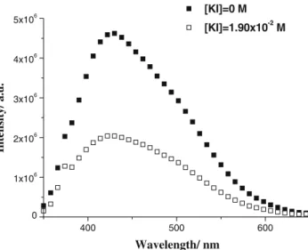
On the other hand the Cdots functionalized with mercaptosuccinic acid are quite sensitive to milimolar concentrations of iodine. This sensitivity was measured as a decrease in the fluorescence intensity at the maximum emission wavelength.
FCUP Analytical Applications of Fluorescent Carbon Dots V
Furthermore this Cdots are also sensitive to the pH media thereby presenting the double usage advantage.
Additionally it was determined that the Hg(II) sensing Cdots could be immobilized in adequate matrixes, as such they were immobilized in the tip of an optical fiber using the sol-gel technique. The immobilization of the Cdots is quite difficult since the objective is to maintain the photophysical properties and sensitivity and the same time that the nanoparticles are now part of another solid system. In a common scenario the Cdots tend to be less fluorescent and less sensitive since not all the nanoparticles are able to interact with the analyte due to its unavailability related with the characteristics of the solid matrix. Indeed, when the synthesized Cdots were immobilized in the sol-gel matrix there was a decrease in the quenching effect by 10%, which was expectable. Nonetheless the Cdots remained sensitive to Hg(II), Cu(II) and the solution pH. Furthermore, the sensing system responded in less than a second and it was completely reversible. The film was quite homogenous and had a thickness of about 700 nm. As such, the immobilization was successful and provided interesting results.
In an attempt to improve this sensing system in both time response and sensitivity, a new immobilization technique was applied to the same nanoparticles: the layer-by-layer immobilization. This technique allows the deposition of discrete layers of nanoparticles onto an adequate surface. In this sense, the Cdots were again immobilized in the tip of an optical fiber, that suffered the same pre-treatment as for the sol-gel immobilization, in order to establish a comparison between the two methods. The results obtained for this new sensing system were quite promising for the development of new lab-on-a-chip system based on Cdots. Indeed, it was determined that the quenching effect was more pronounced as the number of layers increased, reaching the best result at 6 mono-layers. Moreover, even with just one layer, the quenching effect was superior to the one observed using the sol-gel technique. This sensitivity with the number of layers is probably due to two main effects: the etching of the fiber leaves its surface quite irregular so as the number of layers increases the roughness of the fiber decreases and the Cdots deposition is more homogenous. On the other hand, this technique does not immobilized the Cdots in a solid matrix where its porosity needs to be adapted for both analyte and nanoparticles. In the solid matrix, the reaction is limited by the diffusion of the analyte through the matrix, on the contrary to the layer-by-layer deposition, where the Cdots are on the surface ready to interact. As such, it is easy to understand that by using the layer-by-layer technique the system response is only limited by the capacity of the acquisition equipment.
FCUP Analytical Applications of Fluorescent Carbon Dots VI
In all the sensing system described not all the obtained data are totally explored. Indeed, it is possible to use the Cdots Excitation Emission Matrixes (EEM) and chemometrics analysis to establish a different size population interaction with the analyte. As such, by using the Cdots functionalized with PEG200 and
N-acetyl-L-cysteine it was possible to distinguish between two different size populations that were responding to both pH and Hg(II).
Up until now the Cdots have been used as sensors for different analytes by taking advantage of the quenching effect on its fluorescence intensity. However this sensing system is limited by the capacity of the equipment to distinguish between the background noise and the quenching effect. In this sense the use of fluorescence enhancement is quite advantageous, however it is not easy to develop nanoparticles that will respond this way in the presence of an analyte. In an attempt to create such a system the Cdots were located near a Plasmon supporting material – silver islands, and the enhancement effect obtained is quite remarkable. This effect can be used for example when the immobilization of the Cdots results in a dramatic decrease in the fluorescence signal that will ultimately inhibit their further application.
There is an entire new world of applications were it is possible to use Cdots to improve numerous systems and create new ones. Since their discovery the interest in these nanoparticles has been growing and it is my believe that they will be a part of our daily life.
FCUP Analytical Applications of Fluorescent Carbon Dots VII
Resumo
O desenvolvimento da nanoquímica ao longo dos últimos anos é impressionante. Na verdade, o que foi considerado futurista há alguns anos, agora é uma realidade comum.
Este trabalho focar-se-á num novo tipo de nanopartículas à base de carbono –
Carbon Dots (Cdots). Esta nova classe de nanomateriais está a ser apontada como a
possibilidade para a superação dos problemas de toxicidade inerentes aos tradicionais
Quantum Dots (QDs). Os QDs possuem excelentes propriedades de fluorescência,
sendo portanto bastante úteis na área dos nanosensores, no entanto o seu núcleo é constituído por metais pesados, o que limita a sua aplicabilidade para a detecção in
vivo. Os Cdots possuem as mesmas características interessantes dos seus homólogos
QDs, com a grande vantagem de não-toxicidade. Esta é uma das principais razões pelas quais os Cdots são tão interessantes para aplicações em bioimagem. Neste sentido, estão a ser desenvolvidas várias estratégias de síntese numa área onde é fácil tornar-se obsoleto.
Neste trabalho os Cdots foram produzidos por ablação laser directa [irradiação com laser pulsado de UV (248 nm, KrF)], de um alvo de carbono imerso em água. Os Cdots produzido desta forma foram inicialmente funcionalizados com PEG200, uma vez
que tenha sido provado que a presença deste polímero sobre a superfície das nanopartículas ajuda a evitar respostas de citotoxicidade. Adicionalmente, os Cdots foram funcionalizadas com outras moléculas, de forma a torna-los sensores específicos para um determinado analito. Deste modo, os Cdots foram funcionalizados com a N-acetil-L-cisteína e o ácido mercaptossuccínico, para detecção do Hg (II) e do iodo, respectivamente.
Os resultados mostraram que os Cdots funcionalizados com N-acetil-L-cisteína são sensíveis a Hg (II) numa gama micromolar e sofrem a interferência de Cu (II) na mesma gama de concentrações. Estes Cdots são muito sensíveis ao pH do meio, sendo por isso possível utilizá-los como sensor de pH. Isto pode ser visto como uma vantagem, na verdade, este sensor pode ser utilizado para os três analitos, desde que sejam tomadas as devidas precauções para garantir que apenas um analito está presente em cada momento.
Por outro lado, os Cdots funcionalizados com o ácido mercaptossuccínico são bastante sensíveis a concentrações milimolar de iodo. Esta sensibilidade foi medida
FCUP Analytical Applications of Fluorescent Carbon Dots VIII
como uma redução na intensidade de fluorescência no comprimento de onda de emissão máxima. Adicionalmente, os Cdots são também sensíveis ao pH do meio, apresentando assim a vantagem da dupla aplicação.
Os Cdots funcionalizados com PEG200 e N-acetil-L-cisteína foram imobilizados
na ponta de uma fibra óptica usando a técnica de sol-gel. A imobilização dos Cdots é bastante difícil, pois o objetivo é manter as propriedades fotofísicas e sensibilidade ao mesmo tempo que as nanopartículas são agora parte de um outro sistema sólido. Num cenário comum os Cdots tendem a ser menos fluorescentes e menos sensíveis, uma vez que nem todas as nanopartículas são capazes de interagir com o analito, alguns. estão indisponíveis devido às características da matriz sólida. Com efeito, quando os Cdots sintetizados foram imobilizados na matriz de sol-gel, ocorreu uma diminuição no efeito de quenching de 10%, o que era expectável. No entanto, os Cdots permaneceram sensíveis ao Hg (II), Cu (II) e o pH da solução. Adicionalmente, o sistema de detecção respondeu em menos de um segundo e foi completamente reversível. A película era completamente homogénea e tinha uma espessura de cerca de 700 nm.
Numa tentativa de melhorar este sistema de sistema de detecção tanto no tempo de resposta como na sensibilidade, foi aplicada uma nova técnica de imobilização: imobilização camada por camada. Esta técnica permite a deposição de camadas distintas de nanopartículas sobre uma superfície adequada. Neste sentido, os Cdots foram novamente imobilizadas na ponta de uma fibra óptica, que sofreu o mesmo pré-tratamento usado na imobilização de sol-gel, de modo a ser possível estabelecer uma comparação. Os resultados obtidos para este novo sistema de detecção foram bastante promissores para o desenvolvimento de um sistema
lab-on-a-chip novo baseado em Cdots. De facto, determinou-se que o efeito de quenching foi
mais pronunciado à medida que o número de camadas aumentava, atingindo o melhor resultado com 6 monocamadas. Além disso, mesmo com apenas uma camada, o efeito de quenching foi superior ao observado usando a técnica de sol-gel. Esta sensibilidade com o número de camadas é provavelmente devido a dois efeitos: o pré-tratamento da fibra deixa a sua superfície bastante irregular, deste modo, à medida que o número de camadas aumenta a rugosidade das fibras diminui o que leva a uma deposição mais homogénea dos Cdots. Por outro lado, esta técnica não imobiliza os Cdots numa matriz sólida em que a sua porosidade deve ser adaptada tanto para o analito como para as nanopartículas. Na matriz sólida, a reacção é limitada pela difusão do analito através da matriz, ao contrário do que acontece na deposição de camada por camada, em que os Cdots estão na superfície prontos para interagir.
FCUP Analytical Applications of Fluorescent Carbon Dots IX
Como tal, é fácil compreender que, usando a técnica de deposição de camada por camada, a resposta do sistema é limitada apenas pela capacidade de aquisição do equipamento.
Em todos os sistema de detecção descritos até agora, não são utilizados todos os dados disponíveis. Com efeito, é possível utilizar as Matrizes de Excitação Emissão (EEM) dos Cdots para estabelecer uma correlação entre as diferentes populações (em tamanho) com as substâncias a analisar. Deste modo, usando os Cdots funcionalizados com PEG200 e N-acetil-L-cisteína, foi possível distinguir entre duas
populações de tamanhos diferentes que respondiam ao pH e Hg (II).
Até agora os Cdots foram usados como sensores para diferentes analitos, usando como propriedade aproveitando o efeito de quenching. No entanto, este sistema de detecção é limitado pela capacidade do equipamento de distinguir entre o ruído de fundo e o efeito de quenching. Neste sentido, o uso do aumento de fluorescência em vez da diminuição é muito vantajoso, no entanto, não é fácil desenvolver nanopartículas que respondam dessa forma. Numa tentativa de criar um sistema deste tipo os Cdots foram localizados perto um material com efeito Plasmónico - ilhas de prata, e o aumento da fluorescência obtido é bastante notável. Este efeito pode ser usado, por exemplo, quando a imobilização das Cdots leva a uma diminuição da fluorescência e este facto é limitativo na aplicação desejada.
Há todo um mundo de novas aplicações onde é possível usar os Cdots para melhorar sistemas já existentes, bem como criar novos sistemas. Desde a sua descoberta o interesse por essas nanopartículas tem crescido e é minha convicção que eles serão uma parte integrante da nossa vida diária.
FCUP Analytical Applications of Fluorescent Carbon Dots X
Contents
Agradecimentos ... II
Abstract ... III
Resumo ... VI
Contents ... IX
Figure Contents ... XI
Abbreviations and Acronyms ... XII
Preface ... XV
Thesis Layout ... XVI
Chapter 1- Introduction ... 17
1.1. Overview ... 18
1.1.1. Electrochemical Shocking of Multi-Walled Carbon Nanotubes ... 19
1.1.2. Electrochemical Exfoliation of Graphite ... 20
1.1.3. Arc-Discharge Soot ... 20
1.1.4. Laser Ablation ... 21
1.1.5. Candle or Natural Gas Burner Soot ... 21
1.2. Fluorescence Mechanism of Cdots ... 23
1.2.1. Dynamic Quenching ... 25
1.2.2. Static Quenching ... 26
1.3. Sensing Application of Cdots ... 27
1.3.1. Chemical and Bioanalytical sensing ... 27
1.3.2. Sensors based on Cdots ... 28
1.3.3. In vitro sensing and Tags using Cdots... 29
1.4. Toxicity assays of Cdots ... 31
1.5. Conclusions and Future Perspectives ... 34
1.6. References ... 36
Chapter 2- A New Insight on Silicon Dots ... 40
2.1- State of the Art ... 41
2.2. References ... 42
2.3- Personal Contribution to this Work ... 43
Paper ... 44
Chapter 3-
Hg(II) Sensing based on Carbon Dots Obtained By Direct Laser
Ablation ... 56
3.1- State of the Art ... 57
3.2. References ... 63
3.3- Personal Contribution to this Work ... 65
Paper ... 66
Chapter 4- Cdots for Iodine Sensing ... 72
4.1- State of the Art ... 73
4.2. References ... 77
4.3- Personal Contribution to this Work ... 80
FCUP Analytical Applications of Fluorescent Carbon Dots XI
Chapter 5-Optical Fiber Sensing for Hg(II) based on Carbon Dots ... 87
5.1- State of the Art ... 88
5.2. References ... 92
5.3- Personal Contribution to this Work ... 93
Paper ... 94
Chapter 6- Layer-by-Layer Immobilization of Carbon Dots Fluorescent
Nanomaterials on Single Optical Fiber ... 99
6.1- State of the Art ... 100
6.2. References ... 103
6.3- Personal Contribution to this Work ... 104
Paper ... 105
Chapter 7- Parallel Factor Analysis of EEM of the Fluorescence of Carbon Dots
Nanoparticles ... 111
7.1- State of the Art ... 112
7.2. References ... 113
7.3- Personal Contribution to this Work ... 114
Paper ... 115
Chapter 8- Metal Enhanced Photoluminescence from Carbon Nanodots ... 125
8.1- State of the Art ... 126
8.2. References ... 127
8.3- Personal Contribution to this Work ... 129
Paper ... 130
Chapter 9- Conclusions ... 133
Conclusions ... 136
FCUP Analytical Applications of Fluorescent Carbon Dots XII
Figure Contents
Fig. 1.1- – Synthetic pathways for Cdots production using Top-Down
approaches, adapted from [18]. ... 19
Fig. 1.2- Synthetic pathways for Cdots production using Bottom-Up approaches,
adapted from [18]. ... 22
Fig. 1.3- Simplified diagram illustrating the bimolecular process between the
luminescent molecule, M*, and an external molecule - quencher, Q,
adapted from [26]. ... 25
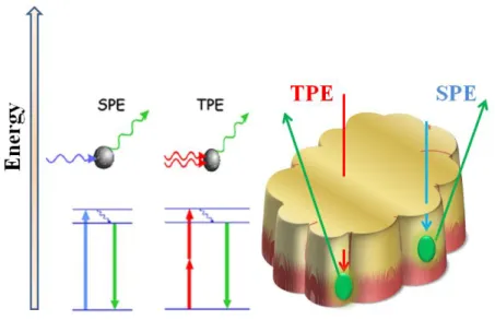
Fig. 1.4- Schematic representation the penetration depth of Two-Photon
Excitation (TPE) vs. Single-Photon Excitation (SPE). ... 31
Fig. 3.1- Representation of the mercury cycle in upon their release into the
atmosphere. ... 58
Fig. 3.2- Biological pathways of elemental mercury, mercuric chloride and
methyl mercury. ... 60
Fig. 6.1- Scheme representing the layer-by-layer deposition method with all the
steps required in order to obtain a good and homogenous film of discrete
Abbreviations and Acronyms
A
Abs. - Absorption.
C
Cdots – Carbon Dots.
H
HOMO - Highest Occupied Molecular Orbital.
L
LUMO - Lowest Unoccupied Molecular Orbital.
M
MWCNT – Multi-walled Carbon Nanotubes
N
NAC – N-acetyl-L-cysteine
Q
QDs - Quantum Dots.
P
PAGE – Polyacrylamide gel electrophoresis
PEG - Polyethyleneglycol
PBS - Phosphate Buffered Saline.
R
ROS – Reactive Oxygen Species.
S
FCUP Analytical Applications of Fluorescent Carbon Dots XIV
T
TEPA - tetraethylenepentamine pentahydrochloride
TEOS - Tetraethyl ortosilicate.
U
FCUP Analytical Applications of Fluorescent Carbon Dots XV
Preface
Nanoparticles are the theme of the century. Their application has expanded to almost all fields of research and the results are visible in daily life. In fact, nanoparticles are being used to change the properties of a given material, as advanced drug delivery systems, in therapeutics, electronics, environmental sensors, among others. As such, a question arises: What makes this nanoparticles so special? One can say that the main difference lies in the size. Indeed as the size decreases the properties of the material becomes different. It is possible to see changes in colour, reactivity, strength, thermal properties, electronical properties, magnetic properties, optical properties, among others.
The appearance of the semiconductor Quantum Dots (QDs) can be viewed as a changing point in the nanochemistry. These nanoparticles present outstanding properties, has led their application in fields, such as, environment, pharmaceutical, solar energy conversion, optoelectronic devices, molecular and cellular imaging and ultrasensitive detection. Despite the numerous QDs applications a problem arisen from their inherent toxicity. In fact, the traditional QDs, are heavy metal core-based, which for in vivo applications represents a toxicity problem. Some studies have been performed to evaluate their toxicity in biological media and it has been described an increase in Reactive Oxygen Species (ROS) due to their interaction with organelles. Other studies have concluded that part of the toxicity is due to bioaccumulation of the nanoparticles which lead to toxicity due to their heavy metal nature. Therefore the search for nanoparticles, that have the outstanding properties of QDs and at the same time do not represent a toxicological issue in biological media, began. As a solution to this problem silicon and carbon dots where found.
Silicon Dots (Sidots) and Carbon Dots (Cdots) are fluorescent nanoparticles that possess unique light emitting properties, such as, biocompatibility, high photoluminescence quantum efficiency, stability against photobleaching, and the non-blinking. Additionally the emission wavelength of these nanoparticles can be adjusted by size selection and/or functionalization with several molecules. In a similar manner of the traditional cadmium based QDs the functionalization can be performed, in theory, with any molecule, and this molecule can be chosen in such a way that it turns the nanoparticle into a sensor for a given analyte. Furthermore, both Sidots as Cdots can be use in Single Photon Excitation (UV: 330-400 nm), as well as, Two Photon Excitation (near infrared: 720-850 nm) that is considered biologically friendly.
FCUP Analytical Applications of Fluorescent Carbon Dots XVI
Thesis Layout
Throughout the three years of the PhD numerous experiments were performed, some gave rise to interesting results and some definitely did not work. The interesting results were published in international, peer review, journals and the bad results were considered a platform for the achievement of better ones.
This thesis results from an organized compilation of the main papers produced during these three years. In this sense the thesis layout is as follow:
Chapter 1 is an Introduction to the nanoparticles theme that is mainly focused on the Carbon Dots and the fluorescence mechanism by which these nanoparticles interact with the analyte.
Chapter 2 is a review on Silicon Dots that was published in the Current Analytical Chemistry with the reference: Vol. 8, 2012, page 67.
Chapter 3 is a compilation of results obtained for Cdots that were adequately functionalized for Hg(II) and pH sensing. These functionalized nanoparticles were extensively tested over more than three months and the results were published in Sensors and Actuators B with the reference: Vol. 145, 2010, page 702.
Chapter 4 represents a system developed for iodine sensing based on Carbon Dots. This system resulted from the selection of the most interesting fluorescence nanoparticles obtained from direct laser ablation. These results were published in the Journal of Fluorescence with the reference: Vol. 20, 2010, page 1023.
Chapter 5 is the follow up of the results obtained in Chapter 3. The nanoparticles functionalized obtained and tested in Chapter 4 were immobilized in the tip of an optical fiber using the sol-gel technique. The results were published in Biosensors and Bioelectronics with the reference: Vol. 26, 2010, page 1302.
The results obtained in Chapter 5 were consistent with the current immobilization procedures, where there is a decrease in the fluorescence intensity due to the nanoparticles entrapment. Chapter 6 represents a new immobilization method was tested and the objective was achieved. The results were published in Analytica Chimica Acta with the reference: Vol. 26, 2010, page 1302.
FCUP Analytical Applications of Fluorescent Carbon Dots XVII
Chapter 7 represent a compilation of the results obtained through chemometrics for the Cdots functionalized for Hg(II) and pH sensing. The results were published in the Journal of Chemometrics with the reference: Vol. 24, 2010, page 655.
Chapter 8 represents the enhancement of the fluorescence intensity due to the plamonic effect of silver islands that were put in contact to the Cdots produced by laser ablation. The results were published in Chemical Communications with the reference: Vol. 47, 2011, page 5313.
Chapter 9 is a general conclusion of all the data obtained and discussed throughout the thesis.
Finally the thesis finishes with some final remarks on the work developed in the three years of PhD.
FCUP Analytical Applications of Fluorescent Carbon Dots 2
1.1. Overview
Carbon Dots (Cdots) are the newest class of fluorescent nanoparticles. Ever since it appearance the number of papers on the theme has been rapidly increasing. These fluorescent nanoparticles present some outstanding properties, such as, high photostability, tuneable emission and excitation wavelength, ability to be functionalized with different molecules according to their desired application, high stability over time, among others. These properties have made them quite interesting in numerous areas, for example, biosensing, bioimaging, pharmaceuticals and fuel cells [1-4].
Cdots are viewed as the new member of the Quantum Dots (QDs) family. Indeed, they share some of the physical properties that have made QDs one of the most relevant propellant in the nanochemistry area, however the traditional QDs have a heavy metal core, that prevents its application for in vivo assays [5]. In this sense some authors have mentioned that Cdots are the most promising alternative to these traditional QDs. In fact, some studies have showed that Cdots are competitive agents for bioimaging studies [6].
Ever since their serendipitously discovery in 2004 there has been a great interest in this new carbon-based material. Cdots present themselves as a non-toxic alternative to the traditional Cadmium-based QDs and, as such, the synthetic pathways for their production are increasing rapidly. Nowadays there are several top-down and bottom-up approaches [7-17] for the Cdots synthesis. Nonetheless they can be grouped into nine main production methods. All the synthetic methods have advantages and disadvantages that need to be taken into account before starting their production. This methods differ mainly in the starting material, as such, for top-down approaches (Fig.1.1) it is possible to use: Multi-walled Carbon Nanotubes (MWCNT), Graphite and candle soot.
FCUP Analytical Applications of Fluorescent Carbon Dots 3
Fig.1.1. – Synthetic pathways for Cdots production using Top-Down approaches, adapted from [18].
1.1.1. Electrochemical Shocking of Multi-Walled Carbon Nanotubes
The electrochemical methods are becoming popular since it allows the production of blue luminescent Cdots in a simple path, on the contrary of the other Top-Down methods that requires an activation procedure for the Cdots to become fluorescent. This method uses as a starting material MWCNT, that are insoluble in water and it allows the production of highly luminescent, water stable, Cdots [11].
Zheng L. et al., (2009) [12] reported the production of well-defined spherical Cdots with an average size of ∼20 nm where the smallest population has an average size of ∼2 nm. In order to obtain such results they used MWCNT immersed in acetonitrile and realized despite the solvent used there were no change into the effective area of the working electrode (graphite rod). This led them to the discovery that the Cdots produced where initially immobilized into the porous graphite electrode and when exposed to Phosphate Buffered Saline (PBS) and electrochemically
FCUP Analytical Applications of Fluorescent Carbon Dots 4
oxidized, they became water soluble and released into the water phase. The main disadvantage of this method is the size dispersion of the nanoparticles, however it has the advantage of producing blue luminescent Cdots in a single step, which allows the method to be faster than others and, therefore most cost effective.
1.1.2. Electrochemical Exfoliation of Graphite
The electrochemical exfoliation of graphite can be viewed as a process where small particles are removed from a graphite electrode by using an adequate current. Li H. et al., (2010) [17] reported the synthesis Cdots with sizes ranging from 1.2 to 3.8 nm. This was performed using graphite rods as both anode and cathode, and NaOH/EtOH as electrolyte. Additionally they evaluated the necessity of having a alkali vs. acid media (NaOH:EtOH/H2SO4:EtOH) as an electrolyte solution for the Cdots
production. Their results led to the discovered that for the electrochemical oxidation of graphite to result in Cdots it is necessary to have an alkali media.
1.1.3. Arch-Discharge Soot
This can be considered the original method. In fact, the discovery of Cdots were made by chance upon the purification of Single-Walled Carbon Nanotubes (SWCNT) obtained by this method [8]. The production of Cdots from this method presents a great challenge in the separation step. Indeed this is one of the methods that generates more impurities, which makes it more difficult to remove. Common techniques of separation, such as, dialysis, fails since the impurities rapidly bock the pores of the membranes. In this sense the technique that has prove to be quite good for separating Cdots from impurities produced upon their production is electrophoresis. In fact the use of Polyacrylamide gel electrophoresis (PAGE) or agarose worked quite well in two senses: it allowed the separation of Cdots from the impurities, as well as, the separation of the different size populations of Cdots.
FCUP Analytical Applications of Fluorescent Carbon Dots 5
1.1.4. Laser Ablation
Laser ablation of carbon targets to obtain surface controlled Cdots is one of the most popular methods of production. However it is necessary to have specialized personnel operating the laser and the equipment itself is rather expensive. Nonetheless this method, since it allows to have a gas controlled atmosphere upon the Cdots production, it promotes a higher control over the chemical surface of the nanoparticles. Additionally it is possible to perform the laser ablation with the target immersed in an appropriate solvent. This has the advantage of dispersing the nanoparticles in the solvent of interest. When using this immersion technique is it necessary to have into account the absorption wavelength of the solvent, since it will be responsible for decreasing the energy for extracting nanoparticles from the carbon target. Moreover if the solvent used is not water it is probable that it will decompose and that the surface groups of the Cdots will affected by it. This also can be viewed as an advantage, since the surface groups are partially responsible for the fluorescence properties of these nanoparticles. One of the main disadvantages of the laser ablation is the high size dispersion of the Cdots, however this can be overcome using separation methods like dialysis and electrophoresis that allows the separation of the different size Cdots populations.
1.1.5. Candle and Natural Gas Burner Soot
Many research groups have been using this method, mainly due to the simplicity of acquiring the starting material [13]. In fact, this method provides a new use for a complicated by-product. However it has its disadvantages, namely the broad size dispersion, the uncontrolled chemical surface and the production of many products that can be dangerous to the human health. Nonetheless it is possible to separate the different nanoparticles using, for example, electrophoresis, but this still leaves the problem of dealing with the other by-products that can be dangerous to the researchers health.
On the other hand for bottom-up methods the starting material can simply be suitable carbon-based molecules (Fig.1.2). It is on this method that the researchers have been focusing lately, where the biggest innovation is the possibility of using
FCUP Analytical Applications of Fluorescent Carbon Dots 6
biomass for the Cdots production [19]. This is in fact quite remarkable since it allows the possibility of using, for example, industrial waste/by-products.
When using bottom-up approaches for producing Cdots it is possible to use three different physical methods: Ultrasonic treatment, Acid dehydration and Thermal carbonization of adequate carbon-based molecules. In this sense it is possible to use both acidic and alkali media, as well as, high temperatures to obtain either raw Cdots (that require an activation step in HNO3 reflux for them to become luminescent) or blue
luminescent Cdots, as it is possible to observe in Fig.1.2.
Fig.1.2. – Synthetic pathways for Cdots production using Bottom-Up approaches, adapted from [18].
Despite of the Cdots production method it is possible to cover their surface with adequate molecules according to their intended application, this step is called functionalization. For this step to elapse in the best way it is of the outmost importance to have a well characterized Cdots surface. As long as the functional groups in the surface are well known, it is possible to use soft chemistry to attach different molecules onto the nanoparticles exterior. These procedures have consequences into the luminescent properties of the Cdots. In fact, any operation that results into a change into the nanoparticles will have an influence on its optical properties. This can be used as an advantage by the researcher, since by knowing the effect that the surface change will produce into the nanoparticle optical properties it is possible to tune them just by introducing such changes. Sun Y-P. et al., 2008 [20], for example, introduced a ZnS and a ZnO shell onto the Cdots surface in order to enhance its Quantum Yield. This operation is usually called doping and is quite common for the traditional
FCUP Analytical Applications of Fluorescent Carbon Dots 7
Cadmium-based QDs. With this procedure it is possible to overcome one of the major drawbacks of the Cdots, that is a relative low Quantum Yield, when compared to the traditional QDs. In fact their work proved that Cdots doped with ZnS and ZnO are competitive with the common CdSe/ZnO QDs.
Since these surface changes have an influence on both Sidots and Cdots optical properties, it is safe to say that they represent a significant part in the fluorescence mechanism.
1.2. Fluorescence Mechanism of Cdots
There has been several attempts over the years to explain the fluorescence mechanism of the nanoparticles [21]. The most common theory is related to the Quantum Confinement. This theory states that all particles are confined in size between the band gap formed by the Highest Occupied Molecular Orbital (HOMO) and the Lowest Unoccupied Molecular Orbital (LUMO) [22]. In fact, as the particles get smaller the energy gap between these two orbitals becomes larger, hence the electrons in the HOMO orbital need more energy to be excited into the LUMO orbital. Upon excitation the electron relaxes and returns to the ground state with emission of light. As such, it is possible to say that the energy gap determines the emission wavelength of the nanoparticle [23].
The fluorescence mechanism of these new nanoparticles is not clearly defined, however it seems to be dependent of two main factors: the surface state defects and the quantum confinement. Indeed as mentioned before, almost all modifications onto the surface of these nanoparticles are followed by a change in their optical properties (fluorescence intensity, radiative lifetime and excitation/emission wavelength). This seems easy to understand since the nanoparticles are constituted by only a small amount of atoms that are mainly on the surface, therefore any change in these atoms results into a modification in the nanoparticle properties.
Since this is a quite interesting and important topic many researchers have attempt to prove these theories. Initially the theory was developed for Silicon Dots (Sidots), that are silicon-based nanoparticles. These nanoparticles were discovered long before Cdots, and since both nanoparticles are not intrinsic semiconductors, the theory for the fluorescence mechanism developed for Sidots is now accepted for Cdots. Some authors have concluded that the most important factor in quantum
FCUP Analytical Applications of Fluorescent Carbon Dots 8
confinement [22] (as their family counterparts - QDs). However these nanoparticles are not intrinsic semiconductors and, since this theory mainly applies to semiconductors, some researchers believe that the fluorescence mechanism of Sidots and Cdots is determined by the surface state defects [7]. In this sense there has been some experimental proves, as well as, theoretical calculations [24]. An example is the work of Yang et al. in 1999 [23], on Sidots with different surface groups. Their results led them to conclude that the optical differences between the Sidots are consistent with the quantum confinement theory, however it was not taken into account that the method of producing larger nanoparticles was by annealing smaller nanoparticles at different temperatures. The temperature itself can be responsible for changing the surface of the Sidots, as they proved by FT-IR, nevertheless this factor was not taken into account on their conclusions about the fluorescence mechanism.
The fluorescence mechanism of these nanoparticles needs to take into account two main factors: surface state defects and quantum confinement. This theory of the two factors influencing the fluorescence mechanism was proposed, for the Sidots, by Putzer A. et al., in 2003 [25]. They based the theory in computational studies where they evaluated the difference in ionic rearrangements and electronic relaxations upon absorption and emission, as well as, the resulting Stokes shift, with different surface molecules. From this study it became clear that all these factors were extremely sensitive to the surface groups, thereby supporting the surface state defects influence on the fluorescence mechanism. In fact, a 1 nm cluster can change ± 0.9 eV depending on the surface oxygen configuration – double bonded or bridged. Additionally they studied different clusters size with the same surface groups, and again there was a different in the mentioned parameters, even though the surface groups were the same, hence supporting the quantum size dependence of the fluorescence mechanism of Sidots. These results can carefully be extrapolated to Cdots, even though up to know there are no studies performed in this sense. Nevertheless and, since they share luminescent properties and there are experimental data that support this two factors theory for the fluorescence mechanism, it seems easy to accept that Cdots and Sidots share the same fluorescence mechanism dependence.
Common fluorescence sensors are based on the decrease of the fluorescence intensity in the presence of the analyte, this process is called quenching. The
quenching phenomenon was first observed and its relation to fluorescence sensing
determined in 1919 by Stern and Volmer [26]. This process occurs through a bimolecular reaction between an excited luminescent molecule, M*, and an external molecule quencher, Q. The fluorescent molecule absorbs light and passes to an
FCUP Analytical Applications of Fluorescent Carbon Dots 9
excited state. When this two molecules interact, the fluorescent molecule decays by non-radiative mechanisms (e.g.: electronic transfer, molecular rearrangements and others.), with a rate of kq (Fig.1.3). The passage of M* to the ground-state also happens
in the absence of a quencher but when this molecule is present and interacts with M*, the fluorescence intensity decreases more rapidly.
Fig.1.3- Simplified diagram illustrating the bimolecular process between the luminescent molecule, M*, and an external molecule - quencher, Q, adapted from [27].
Quenching mechanisms are photophysical, i.e., after all the deactivation processes the fluorophore returns to the ground-state unaltered, M. Depending on the type of interaction between the quencher and the luminescent molecule it is possible to establish different models to better understand the kinetics of these processes. In fact, the analysis of this phenomena gives important quantitative information on the surroundings of a fluorescent molecule (if a kinetic analysis is possible in the sequence of a kinetic competitive model), or at least qualitative information [27].
1.2.1. Dynamic Quenching
Quenching occurs when an excited fluorophore reacts with a quencher molecule. In order for these two molecules to react it is necessary for them to meet. These diffusion controlled reactions are time dependent. Indeed, when the excited fluorophores, M*, are at a shorter distance from a quencher, Q, the decay to the ground state, on average, occurs at shorter times than those that are more distant. These transient effects are not significant for moderate concentrations of quenchers in fluid solvents but they are quite relevant in viscous media.
M + Q
M
*
+ Q
k
q1/
h
"products"
= kMFCUP Analytical Applications of Fluorescent Carbon Dots 10
This diffusion controlled mechanism affects the beginning of the fluorescence decay curves. In steady-state experiments, for example, it is responsible for deviations from the well known and widely used Stern–Volmer equation.
(Equation 1.1)
where is the Stern-Volmer constant, I0 and I are the steady-state fluorescence intensities in the absence and in the presence of a quencher, respectively. Generally the ratio I0 /I are plotted against the quencher concentration (Volmer plot). If the variation is found to be linear, the slope gives the Stern-Volmer constant [26].
1.2.2. Static Quenching
Static quenching is not dependent from the diffusion processes and occurs in
two different situations: when there the formation of a sphere of effective quenching or when the formation of a ground-state non-fluorescence complex occurs.
The quenching mechanism that passes through the formation of an sphere of effective quenching was proposed by Perrin in 1924 [28]. The model describes the quencher as being inside a sphere around the fluorophore, and its effect on the dye is only when the quencher is inside this sphere. Additionally it is proposed that both M* and Q cannot change their positions in space, relative to one another, during the excited-state lifetime of M*. An example of this is when the quenching occurs in viscous media or rigid matrixes. In this sense, it is necessary to introduce some changes in the Stern-Volmer equation for it to properly represent the phenomena that is occurring is these cases.
(Equation 1.2)
where is the Avogadro’s number and represent the volume of the sphere. In contrast to the linear trend found in Equation 1.1, the static quenching by the
FCUP Analytical Applications of Fluorescent Carbon Dots 11
formation of a sphere plot can have an upward curvature when the quencher concentration is high or a linear trend for low quencher concentrations.
On the other hand the static quenching can also occur through the formation of a ground-state non-fluorescent complex. The formation of this non-fluorescent complex leads to the typical decay in the fluorescence intensity, however in this case, the excited-state lifetime of the uncomplexed fluorophore is not affected. Considering that the Stern-Volmer Equation shows a linear dependence between the fluorescence intensities and the quencher concentration, this equation is valid for this static quenching mechanism [26].
1.3. Sensing Applications of Cdots
1.3.1. Chemical and Bioanalytical sensing
Ever since the appearance of nanoparticles the interest in producing specific nanosensors has not stopped and, as such, there are actually numerous applications of different nanoparticles [29]. But the question remains: “Why the interest in nanoparticles for specific sensing?”. This question can be answered with two different answers: (i) There are now available numerous possibilities to functionalize different nanoparticles [30-33], which makes it possible to turn them into a specific sensor to a given analyte. (ii) The use of nanoparticles allows the miniaturization of the complex sensing systems, thereby it is possible to produce the so called – lab-on-a-chip [34]. This seemed futuristic a couple of years ago, however due to the development to the informatics technology and the incorporation of nanosensors, this is now a reality. In fact, there are some of these new miniaturized sensing systems already in the market.
Despite the simple explanation of the functionalization of the nanoparticle to become sensitive to a given target, in practice this is not easy to accomplish. There are several drawbacks that need to be overcome in order to say that the nanosensor is actually useful in comparison to others already in the market. First it is necessary to choose a molecule that is sensitive to a given analyte in order to functionalize the nanoparticle with it. Then some precautions must be made in the soft chemistry of functionalization so it will not affect the ability of the molecule to be sensitive to the analyte. If all this turns up well, there is still necessary that when the surface molecule interacts with the analyte it does so in a way that the fluorescence properties of the
FCUP Analytical Applications of Fluorescent Carbon Dots 12
nanoparticle changes (Emission shift, Lifetime and Fluorescence intensity decrease/increase). After all these steps are overcome it reaches the tricky part of the specific sensing, that is: functionalized nanoparticles must only interact with the desired analyte, therefore it must become inert to all other possible interferents. This is not easy to perform, since generally the chemical mechanism of interaction is trough affinity and there are some analyte that share the same affinity. One example of this is mercury sensing. It is quite difficult to get a Hg(II) sensor that does not react with Cu(II), therefore Cu(II) is a common interferent on mercury sensing [35].
Here it will be discussed some of the sensing applications were Sidots and Cdots have demonstrated an outstanding performance. Researchers in biomedicine area have been paying much attention to nanosensing and, as such, they are responsible for developing some sensors for important biomedical substances, like for example: dopamine and glucose. Developing sensors for in vivo and in vitro sensing it quite difficult, since, among other characteristics, these sensors need to be non-toxic, biocompatible and there are a number of parameters that can act as interferents.
1.3.2. Sensors based on Cdots
The use of Cdots is fairly recent, nevertheless there are some sensors developed based on these carbon nanomaterials. One example of a Cdots-based sensing is the Hg(II) sensor based on Cdots functionalized with Polyethylenoglicol 200 (PEG200) and N-acetyl-L-cysteine (NAC) [35]. This sensor was used in a suspension
form and also immobilized into different solid matrixes. In every case it showed a good response to Hg(II) and also the solution pH. Even though this Cdots-based sensor was tested in solution and after immobilization it was found that in all cases the main interferent for the system was Cu(II). The immobilized sensor showed a good reversibility. Additionally when the sensor was immobilized with poly(ethyleneimine), using the layer-by-layer technique, the sensitivity also increased, thereby allowing the detection of Hg(II) in a concentration range of (0.01–2.69)×10−6M.
Another sensor based on Cdots functionalized with PEG200 and
tetraethylenepentamine pentahydrochloride (TEPA) is for Fe(II). This sensor when in presence of a concentration range of (5.00x10⁻⁷-1.00x10⁻⁴)M of Fe(II) suffers a decrease in fluorescence (quenching) of about 55%. Additionally from all the known interferents of this type of sensor, only Fe(III) and Cu(II) had a measurable effect (50% and 35%).
FCUP Analytical Applications of Fluorescent Carbon Dots 13
1.3.3. In-vivo sensing and Tags using Cdots
Nowadays there are numerous organic dyes used for in vivo sensing and as fluorescence tags. Most of these molecules are synthetic mimics of natural occurring proteins that are involved in a process of interest. However these fluorescent dyes present some disadvantages, such as, photobleaching, blinking, short fluorescence lifetime, among others, that lead researchers to search for new alternatives.
Cdots are the new class of nanoparticles that present themselves as alternatives to the traditional organic dyes. These nanoparticles overcome the major drawbacks of the organic dyes and present the advantage of functionalization of specific targeting, are non-toxic and have a higher quantum yield and photostability. As such, it is without surprise the number of imaging applications that are now using these nanoparticles.
The number of assays in bioimaging using Cdots has been increasing, mainly due to three factors: (i) their non-toxicity nature, (ii) the easiness of functionalization for specific targeting and, (iii) their competitive fluorescence properties.
One of the main areas that is currently being explored is the specific targeting to cancer cells. Nevertheless before studying its internalization process and specific targeting of interesting biomolecules it is necessary to better understand how they act without any functionalization. In this sense studies like the one performed by Ray S.C.
et al. in 2009 [36] is important. They determined that when Cdots are incubated with
cells they are internalized by natural occurring mechanisms even without functionalization. This means that it is possible to track this nanoparticles using a conventional fluorescence microscope.
When the objective is specific targeting the functionalization is required. One interesting work of this matter is the one performed by Li Q. et al., in 2010 [37]. This research groups focused on the study of the internalization process and the specific targeting of Cdots functionalized initially with polyethylene glycol (PEG) chains, polyethylenimide-co-polyethyleneglycol-co-polyethylenimide copolymer, and 4-armed PEG molecules and, then with human transferring (Tf). Tf has been used before in targeting assays, since it was proven that cancer cell membranes over express the TF receptors. Therefore the use of this glycoprotein on the Cdots surface should be a guarantee of specific targeting. Lin Q. et al., found that Cdots functionalized with polyethylenimide-co-polyethyleneglycol-co-polyethylenimide copolymer and Tf had a higher passive cellular uptake when compared to the other functionalized and
non-FCUP Analytical Applications of Fluorescent Carbon Dots 14
functionalized Cdots. The authors indicate that this may be due to the charge on the Cdots surface, that in the case of this Cdots is positive, thereby allowing a higher binding ability with the cell membrane through electrostatic interactions.
When the fluorescence intensity is not adequate for the study or even if the excitation wavelength is too low, it is also possible to functionalize the Cdots with a shell of ZnO or ZnS. A study on these functionalized nanoparticles using bioimaging was performed by Yang S-T in 2009 [20]. The main conclusions of this work were: (i) despite the place where the Cdots were injected into the mouse, they remained strongly fluorescence throughout the experimental assay, (ii) upon the 3h of the intravenous injection of 440 μg in 200 μL, the urine exhibit strong fluorescence, thereby indicating that the pathway of excretion of these nanoparticles is through urine.
However when the particle size is critical, it may not be possible to use a shell just to increase the fluorescence intensity or to change the excitation wavelength, then it is necessary to use a new imaging technique: Two-Photon Excitation technique (TPE). In fact, ever since its appearance their application on Cdots has grown. The TPE technique allows the possibility of exciting the nanoparticle with two photons and, as such, it is possible to use a fluorophore that absorbs (is excited) in the UV with two photons of NIR. What happens is that it is possible to excite a fluorophore that typically is excited in the UV (i.e., 400 nm) with two photons of NIR (800 nm). By doing this the main advantage is to excite the fluorophore with a non-harmful energy for cells at the same time that emission wavelength remains the same. This system has two main advantages: it allows the possibility of studying cellular phenomena for longer times (since the cells will not be exposed to damaging wavelengths) and a higher penetration depth when compared to the traditional Single-Photon Excitation technique (SPE), since NIR is less absorbed by the tissue (Fig. 1.4).
FCUP Analytical Applications of Fluorescent Carbon Dots 15
Fig. 1.4. – Schematic representation the penetration depth of Two-Photon Excitation (TPE) vs. Single-Photon Excitation (SPE) [18].
1.4. Toxicity assays of Cdots
Due to the interest that these nanoparticles raise in the in vitro and in vivo area the need for toxicity assays was imperious. The traditional Cadmium-based QDs were a landmark in nanochemistry and they found numerous applications in bioimaging due to their outstanding fluorescence properties. However the inherent toxicity of using such materials rise and was proven by several studies [38,39]. There are two main toxicity issues in QDs: (i) their heavy metal core, that upon contact with cells are known to produce ROS that will eventually lead to cell death, and (ii) the fact that they suffer bioaccumulation, i.e., on lymphatic nodules. Some solutions have been proposed to deal with this problem, namely the use of a shell that completely covers the QDs surface. This shell can even be though in such a way that it increases the fluorescence intensity or shifts the emission wavelength to a more desirable one. However this solution only deals with the problem of exposing cells to the heavy metal-based core. In order to try to resolve the bioaccumulation problem it has been proposed the functionalization of the QDs with biocompatible molecules (i.e., PEG) that are recognized by the cell as innocuous, thereby allowing the cell to dispose the QDs using its natural methods [40,41]. This in fact can avoid tissue inflammation due to recognition of a foreign material, nevertheless the possibility of bioaccumulation of heavy metals is still a issue to be concerned.
In order to overcome this toxicity issues the search for new nanomaterials that have the same outstanding fluorescence properties of QDs but are biocompatible and
FCUP Analytical Applications of Fluorescent Carbon Dots 16
non-toxic, lead to the appearance of Sidots and Cdots. Cdots have been presented as the non-toxic alternative to traditional Cadmium-based QDs. This is only possible due to the toxicity assays that have been performed so far by different investigation groups [4, 42].
One of the first toxicological assays performed in Cdots was the one presented by Yang S.T. in 2009 [20]. In this assay they intended to see if the presence of a ZnS shell on the Cdots surface had a toxicological difference from the raw Cdots (non-functionalized). The mice were injected subcutaneously with a solution of Cdots functionalized with PEG1500N (30μg carbon-core equivalent in 30μL) or a solution of
Cdots/ZnS with PEG1500N (65μg in 30μL) (Fig.1.5). It was found that after 24h the
fluorescence was only residual. Additionally after the 24h pos-injection the axillary lymph nodes were harvested and dissected and it was observed no fluorescence. This results is quite important since nanoparticles, like carbon nanotubes, have a tendency to suffer bioaccumulation, which is readily seen in the lymph nodes.
On another set of experiences this investigation group injected a Cdots solution of 440μg in 200μL intravenously. After 4h of the injection the organs were harvested and dissected to detect fluorescence. It was observed that only the kidneys and the liver presented a fluorescence intensity compatible to the Cdots, however this intensity was larger in the case of the kidneys. These results suggest not only that the main way of excretion of the nanoparticles is the urine, but also that they do not suffer bioaccumulation. Additionally the fluorescence detected in the liver is consistent to the results observed for other nanoparticles, nonetheless the presence of Cdots was in a lower concentration of the other nanoparticles. The authors suggest that this result may be related to the PEG functionalization that reduced the protein affinity and made the Cdots stealthy with respect to hepatic uptake.
Another interesting toxicity assay was performed by Li Q. et al., in 2010 [37] that points out an interesting feature, the relationship between the nanoparticle size and the degradation/excretion process. Indeed it is difficult for the organism to excrete trough the kidneys nanoparticles larger them 10nm. Additionally particles over this size are hardly degraded by normal biological mechanisms, which can then lead to bioaccumulation. The study performed by Li Q. et al., resorted the use of Cdots in a size range of 1.5-3nm. These Cdots were functionalized with polyethylene glycol (PEG) chains, polyethylenimide-co-polyethyleneglycol-co-polyethylenimide copolymer, and 4-armed PEG molecules, thereby producing CD2, 3 and 4, respectively. These Cdots were further functionalized with human transferrin and exposed to HeLa cell cultures. Their cytotoxicity was evaluated using twice the concentration necessary for imaging
FCUP Analytical Applications of Fluorescent Carbon Dots 17
studies. The results clearly demonstrate that the Cdots present low cytotoxicity when compared to the traditional imaging agents.
In order to safely use the nanoparticles in biological media for applications like drug delivery systems, pharmaceuticals, among others, it is necessary to completely understand the biological path and responses that these nanoparticles produce once in contact with live biological organelles and tissues. Despite the toxicity assays already performed, mechanism beneath the cytotoxicity mechanism and the inflammatory response is still to be uncovered, as such, it is still necessary to perform additional studies focusing of these subjects.
1.5. Conclusions and Future Perspectives
The Era of the nanoparticles is upon us and their application in almost all areas of science is a clear evidence of this. One can say that this nanoparticle revolution had as star participants the traditional Cadmium-based QDs. These nanoparticle present outstanding optical properties that has lead to their success in micro-electronics, fluorescence sensors for a wide analyte application, bioimaging, among others. One of the most interesting features of QDs was precisely the imaging ability, without blinking and photobleaching problems that are common to the imaging agents mostly used – organic dyes. However, despite this clear advantages, there is a major problem in the use of QDs for in vivo and in vitro applications: the inherent toxicity due to the heavy metal core.
In order to overcome this drawback of QDs the rush for other imaging agents began. Sidots and Cdots appeared in this context. Nowadays it was already proven that not only they have a contrast imaging ability competitive with QDs, but also the non-toxicity feature that was so necessary.
Since their appearance the interest in Sidots and Cdots has grown and, as such, now they can be produced by several synthetic pathways. Additionally, and by using soft chemistry, it is possible to functionalize them with different molecules in order to increase their fluorescence, change the emission wavelength and also for targeting applications. In this sense these nanoparticles have found numerous applications in sensing/biosensing of specific analytes. However all these steps must be considered carefully for the intended application, since their toxicity can increase due to the presence of certain functional groups on the dots surface.
FCUP Analytical Applications of Fluorescent Carbon Dots 18
The toxicity of the nanoparticles is a current line of investigation, mainly due to the possibility of applying them into live organisms for in vivo imaging and as advanced drug delivery systems. From the studies performed so far it was found that both Sidots and Cdots have a toxicity level lower than the traditional QDs. Additionally it was found that the use of PEG as a functionalization molecule helps to reduce their toxicity. Nevertheless it is still necessary to perform more studies in order to completely understand the mechanism beneath their toxicity.