Could heart rate variability be associated with weight
bearing asymmetries in cerebrovascular diseases?
Rogério Batista Balthazar, Pedro Henrique Côrtes de Sousa,
Paulo Henrique Ferreira de Araujo Barbosa, Lidiane Teles de Menezes,
Abraão Souza Costa, Danilo Veloso Alves Carneiro, Vera Regina
Fernandes da Silva Marães, Emerson Fachin Martins
ABSTRACT
Introduction: Cerebrovascular diseases result in sensorymotor deficits disturbing postural control that is observed by weightbearing asymmetries commonly named as hemiparesis. Besides hemiparetic impairments, first observed after stroke, many studies have pointed cardiac failure and risk of sudden death as the main factors responsible for death of stroke survivors. This case series characterized weightbearing asymmetries and heart rate variability, and describes relationships between these parameters in hemiparesis. Case Series: Five Brazilian male subjects with chronic hemiparesis acquired after ischemic stroke in the middle cerebral artery were selected to study heart rate variability obtained by Root
Mean Square Successive Difference. Also, weightbearing asymmetries were measured by Symmetry Ratio calculated by weightbearing recorded between each foot. The Symmetry Ratio was 1.1±0.43 for all cases presenting a symmetry case (n = 1) and different types of asymmetries cases (n = 4) during upright position. Root Mean Square Successive Difference was 9.9±3.4, presenting strong and significant (p < 0.05) positive correlation with age and a strong but not significant (0.05 < p < 0.10) negative correlation with hemiparesis chronicity. A strong but not significant negative correlation was observed between the Root Mean Square Successive Difference and the Symmetry Rate values. Conclusion: A characteristic pattern of heart rate variability for patients with cerebrovascular disease was observed in these cases, associated significantly with age. Still, this behavior seems to be influenced by chronicity and by different types of asymmetries in the distribution of weight bearing that could be investigated in more appropriate clinical research designs.
Keywords: Cardiovascular, Neurological, Stroke, Posture, Physical therapy
*********
Balthazar RB, Sousa PHCd, Barbosa PHFdA, Menezes LTd, Costa AS, Carneiro DVA, Marães VRFdS, Martins EF. Could heart rate variability be associated with weightbearing asymmetries in cerebrovascular diseases? International Journal of Case Reports and Images 2012;3(2):15.
*********
doi:10.5348/ijcri20120287CS1
CASE SERIES OPEN ACCESS
Rogério Batista Balthazar
1, Pedro Henrique Côrtes de
Sousa
2, Paulo Henrique Ferreira de Araujo Barbosa
2,
Lidiane Teles de Menezes
2, Abraão Souza Costa
2, Danilo
Veloso Alves Carneiro
2, Vera Regina Fernandes da Silva
Marães
3, Emerson Fachin Martins
4Affiliations:
1Physical Educator and Physical Therapy
student, Faculty of Ceilandia, University of Brasilia,
Brasilia, Federal District, Brazil;
2Physical Therapy
student, Faculty of Ceilandia, University of Brasilia,
Brasilia, Federal District, Brazil;
3Ph.D. in Physical
Therapy, Faculty of Ceilandia, University of Brasilia,
Federal District, Brazil;
4Ph.D. in Neuroscience and
Behaviour, Faculty of Ceilandia, University of Brasilia,
Brasilia, Federal District, Brazil.
Corresponding Author: Emerson Fachin Martins,
Universidade de Brasilia, Faculdade de Ceilandia,
Campus UnB Ceilandia, QNN 1 4 Area Especial,
Ceilandia Sul, Brasilia, Distrito Federal, Brasil,
72220-1 40; Ph: 55 672220-1 372220-1 0784072220-1 ; Fax: 55 672220-1 372220-1 078472220-1 6; E-mail:
[email protected]
Received: 1 4 June 2011
Accepted: 05 November 2011
Published: 28 February 201 2
INTRODUCTION
Cerebrovascular diseases (CVD) damaging one of the cerebral hemispheres commonly results in movement deficits in the opposite hemibody promoting weight bearing asymmetries commonly observed as hemiparesis [12]. Besides sensorymotor impairments, first observed after stroke, cardiovascular dysfunctions have also been reported for this population and many studies have pointed cardiac failure and sudden death risk as the main factors responsible for death of stroke survivors [36].
Cardiovascular dysfunction is present because the stroke can also damage brain areas related with autonomic control modifying sympatheticvagal balance [68]. A lot of evidences show that autonomic dysfunction can be identified by heart rate variability [46, 913] and specific heart rate profiles have been described as a predictor of sudden death [1415].
It has already been reported in literature during past decades that abnormal reduction of the heart rate variability reflects autonomic dysfunction and risk of sudden death, and that heart rate variability could be correlated with motor impairments in stroke survivals, there is no evidence to show if heart rate variability could be associated with weightbearing asymmetries in cerebrovascular diseases.
This case series aimed to characterize weight bearing asymmetries and heart rate variability, describing the relationships between these measurements in cases of hemiparesis.
CASE SERIES
A multiplecase descriptiveexploratory research design was selected for this study with measurements performed in a single session. We describe five Brazilian male subjects with chronic hemiparesis acquired after ischemic stroke in the middle cerebral artery were identified in the available records on files at the Ceilandia Regional Hospital. All cases had eligibility criteria to be submitted to procedures analysis to calculate parameter of heart rate variability [35, 13]. To be included in the study the participants had to: 1) have had a postbrain (CVD) injury period of over six months, 2) have spastic hemiparesis, 3) be able to maintain themselves in an upright stance posture long enough to register weightbearing on the digital scales, and 4) be able to walk themselves without assistance or support device. Study excluded, the participants who: 1) were smokers and/or alcoholic; 2) were on betablocker medication, and 3) were presenting other types of disability in addition to hemiparesis. All of them signed a consent form approved by the Research Ethics Committee issued by the Faculty of Health Science of the University of Brasília (protocol number 034/2009).
Heart rate was monitored during 6minutes walk test and the heart rate variability was obtained by comparisons between adjacent RR intervals and
calculated by Root Mean Square Successive Difference (RMSSD) [13].
Weightbearing distribution was evaluated by the ratios of the weight supported by each lower limb between the affected and nonaffected hemibodies. The measurements of the weight supported under each lower limb of the body were obtained with the use of two parallel calibrated scales with a digital display (Plenna®) with a maximum capacity of 150 kg. The subjects were placed barefoot, with their feet free and aligned on the scales with each foot about 20 cm away from the other, without any type of additional support, and the limbs placed separately on each scale. All subjects were instructed to maintain an upright position as comfortable as possible, always looking forward to a fixed point on the wall at a distance of three meters.
The display of each scale indicated integer values in kilograms (kg) with one decimal value representing tenths of a kg. Despite instability of the decimal value, once the examiner observed stability in the integer value indication of the displays of each scale, the bilateral reading was obtained and recorded (in integer values). In sequence, the equivalence between the total body weight and the sum of the values obtained for both scales was confirmed. In case the sum had been inferior or superior (> or <1 kg) to the total body weight, the reading would be performed again. The values obtained for each limb were registered as weightbearing values for the affected and nonaffected hemibodies.
Weightbearing asymmetries were calculated and classified by the Symmetry Ratio (SR), as described by Pereira and collaborators [1], SR was calculated using the formula: SR = a/na, in which RS is the dimensionless value of the symmetry ratios calculated by the division of the weightbearing values of the affected (a) by the nonaffected side (na). As such, the values of RS = 1 would represent the total weight bearing symmetry in the orthostatic position. Values of RS > 1 would represent weightbearing asymmetries towards the affected side and values of RS < 1 towards the nonaffected side.
We used descriptive statistical (mean ± standard deviation) and Spearman correlation test once the KolmogorovSmirnov test did not confirm Gaussian distribution. The significance level for all analyses was established at p < 0.05.
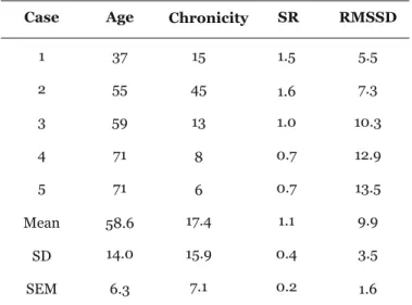
They had an average age of 58.6±14.03 years with an average chronicity of 17.4±15.85 months. The RMSSD and RS were 9.9±3.4 and 1.1±0.43 respectively and the values for each case are presented in the table 1.
The RS identifies three types of weightbearing distribution classified as symmetric (n = 1 and RS = 1) and asymmetric with overweight toward affected side (n = 2 and RS > 1) or nonaffected side (n = 2 and RS < 1).
The relationships between variables are presented by scatter graphs in the figures 1, 2 and 3. Strong and significant positive correlation was found between RMSSD and years old, showing that the oldest patients obtained the highest RMSSD (figure 1). Moreover, strong but not significant negative correlation between RMSSD and chronicity suggests that patients surviving
Figure 1: Dispersion graph of the variables correlated by the Spearman correlation tests. The level of correlation between root mean square successive difference (RMSSD) and age (years old) are indicated by the correlation indices (r2) and the
significance is indicated by the p value. Asterisk indicates strong correlation (p < 0.05).
Figure 2: Dispersion graph of the variables correlated by the Spearman correlation tests. The level of correlation between root mean square successive difference (RMSSD) and chronicity (months poststroke) are indicated by the correlation indices (r2) and the significance is indicated by the
p value. The letter t indicates strong tendency to correlate (0.1 > p > 0.05).
Figure 3: Dispersion graph of the variables correlated by the Spearman correlation tests. The level of correlation between root mean square successive difference (RMSSD) and symmetry ratio (SR) are indicated by the correlation indices (r2) and the significance is indicated by the p value. The letter t
indicates strong tendency to correlate (0.1 > p > 0.05). Discontinuous line indicates subjects with weightbearing symmetrically distributed (SR = 1) separating subjects with asymmetries toward affected hemibody (SR > 1) of those with asymmetries toward nonaffected hemibody (SR < 1).
poststroke for long time have the lowest RMSSD (figure 2).
Strong but not significant negative correlation also was found between RMSSD and RS suggesting that the RS > 1 are associated with the lowest RMSSD and the RS < 1 with the highest RMSSD (figure 3).
DISCUSSION
The values of SR and the RMSSD in the five cases were within the values commonly reported for
hemiparetic population [1, 3, 16]. As explained by Marães [13], the highest values of the RMSSD among them could indicate subjects with better cardiovascular adjustments by autonomic nervous system.
In this study, cases in which the subjects had more advanced ages were those where the highest values were obtained from RMSSD (figure 1), disagreeing with McLaren and collaborators [4]. However, Tulppo et al. [17] reported that the low values of RMSSD were observed in elderly during rest and as described the RMSSD was recorded during six minutes walk test.
Table 1: Description of the variables recorded for each case (n=5).
Case Age Chronicity SR RMSSD
1 37 15 1.5 5.5 2 55 45 1.6 7.3 3 59 13 1.0 10.3 4 71 8 0.7 12.9 5 71 6 0.7 13.5 Mean 58.6 17.4 1.1 9.9 SD 14.0 15.9 0.4 3.5 SEM 6.3 7.1 0.2 1.6
Abbreviations: Age in years old, Chronicity in months post stroke, Symmetry Ratio (RS) and Root Mean Square Successive Difference (RMSSD). Values were detailed for each case in the first five rows and the last three rows describe Mean, Standard Deviation (SD) and Standard Error of Mean (SEM).
Moreover, the vagal modulation of heart rate during exercise is also affected by physical fitness [17]. However, we did not record the level of physical fitness of the subjects, so it is in incorrect to state that our results disagree with McLaren et al. [4]. This is a limitation of this study.
In the five cases, chronicity seems to contribute to a loss of effectiveness of autonomic adjustment. Other researches investigating heart rate variability in stroke survivors during acute [3, 6] and chronic [4] phases found similar relationship, however, still no definite arguments to support a clear association between chronicity and heart rate variability is present.
It was interesting to note that subjects overweighting on the nonaffected side had the highest RMDSS values and those overweighting to the affected side had the lowest RMDSS values, suggesting that the compensatory strategy of overweighting nonaffected side seems to favor a better autonomic adjustment.
However, the present descriptiveexploratory study describes five patients with which is a small number of patients for drawing conclusions from statistical analysis. The results are not sufficient for generalizations that the asymmetric strategy toward the nonaffected side was responsible for increased heart rate variability, a drawback of this study, however it pointed towards evidences that could be investigated by others research designs.
More appropriate designs are necessary to confirm the findings described here, since the sample was small and the information previously collected were not sufficient to disregard the influences of other variables. Except the limitations outlined here, this study has generated new questions about the influences of postural asymmetry in heart rate variability.
CONCLUSION
It was concluded that a characteristic pattern of heart rate variability for patients with cerebrovascular disease was observed in these cases, associated strongly and significantly with age. Still, this behavior seems to be influenced by chronicity and by different types of asymmetries in the distribution of weight bearing that could be investigated in more appropriate clinical research designs.
*********
Acknowledgements
We thank the Núcleo Regional de Atenção Domiciliar (NRAD) staff for their contribution to data recording. This study was supported by grant for research from University of Brasilia (DPP/UnB – UnBDoc 99456/2009) and from Fundação de Empreendimentos Científicos e Tecnológicos (FINATEC, Edital 04/2009).
Author Contributions
Rogério Batista Balthazar – Acquisition of data, Analysis and interpretation of all data, Drafting the article, Final approval of the version to be published
Pedro Henrique Côrtes de Sousa – Acquisition of data and Analysis of weightbearing data, Final approval of the version to be published
Paulo Henrique Ferreira de Araujo Barbosa – Acquisition of data and Analysis of weightbearing data, Final approval of the version to be published
Lidiane Teles de Menezes – Acquisition of data and Analysis of weightbearing data, Final approval of the version to be published
Abraão Souza Costa – Acquisition of data and Analysis of weightbearing data, Final approval of the version to be published
Danilo Veloso Alves Carneiro – Acquisition of data and Analysis of heart rate variability data, Final approval of the version to be published
Vera Regina Fernandes da Silva Marães – Acquisition, Analysis and interpretation of heart rate variability data, Critical revision of the article, Final approval of the version to be published
Emerson Fachin Martins – Conception and design, Acquisition of data, Analysis and interpretation of data, Drafting the article, Critical revision of the article, Final approval of the version to be published
Guarantor
The corresponding author is the guarantor of Submission.
Conflict of Interest
The authors declare no conflict of interest.
Copyright
© Rogério Batista Balthazar et al. 2012; This article is distributed under the terms of Creative Commons attribution 3.0 License which permits unrestricted use, distribution and reproduction in any means provided the original authors and original publisher are properly credited. (Please see www.ijcasereportsandimages.com /copyrightpolicy.php for more information.)
REFERENCES
1. Pereira LC, Botelho AC, Martins EF. Relationships between body symmetry during weightbearing and functional reach among chronic hemiparetic patients. Rev Bras Fisioter 2010;14(3):2296. 2. Genthon N, Rougier P. Influence of an asymmetrical
body weight distribution on the control oundisturbed upright stance. Journal of Biomechanics 2005;38(10):20379.
3. Dutsch M, Burger M, Dorfler C, Schwab S, Hilz MJ. Cardiovascular autonomic function in poststroke patients. Neurology 2007;69(24):22495.
4. McLaren A, Kerr S, Allan L, et al. Autonomic function is impaired in elderly stroke survivors. Stroke 2005;36(5):102630.
5. KatzLeurer M, Shochina M. Heart Rate Variability (HRV) parameters correlate with motor impairment and aerobic capacity in stroke patients. Neurorehabilitation 2005;20(2):915.
6. Colivicchi F, Bassi A, Santini M, Caltagirone C. Cardiac autonomic derangement and arrhythmias in
rightsided stroke with insular involvement. Stroke 2004;35(9):20948.
7. Korpelainen JT, Huikuri HV, Sotaniemi KA, Myllyla VV. Abnormal heart rate variability reflecting autonomic dysfunction in brainstem infarction. Acta Neurologica Scandinavica 1996;94(5):3372.
8. Barron SA, Rogovski Z, Hemli J. Autonomic consequences of cerebral hemisphere infarction. Stroke 1994;25(1):1136.
9. Lewis MJ, Short AL. Exercise and cardiac regulation: what can electrocardiographic time series tell us? Scandinavian Journal of Medicine & Science in Sports 2010;20(6):7944.
10. Umetani K, Singer DH, McCraty R, Atkinson M. Twentyfour hour time domain heart rate variability and heart rate: Relations to age and gender over nine decades. Journal of the American College of Cardiology 1998;31(3):5931.
11. La Rovere MT, Bigger JT, Marcus FI, Mortara A, Schwartz PJ, Investigators A. Baroreflex sensitivity and heartrate variability in prediction of total cardiac mortality after myocardial infarction. Lancet 1998;351(9101):4784.
12. Yeragani VK, Sobolewski E, Kay J, Jampala VC, Igel G. Effect of age on longterm heart rate variability. Cardiovascular Research 1997;35(1):352.
13. Marães VRFS. Freqüência cardíaca e sua variabilidade: análises e aplicações. Revista Andaluza de Medicina del Deporte 2010;3(1):187. 14. Jouven X, Empana JP, Schwartz PJ, Desnos M,
Courbon D, Ducimetiere P. Heartrate profile during exercise as a predictor of sudden death. New England Journal of Medicine 2005;352(19):19518. 15. Tokgozoglu SL, Batur MK, Topcuoglu MA, Saribas
O, Kes S, Oto A. Effects of stroke localization on cardiac autonomic balance and sudden death. Stroke 1999;30(7):13071.
16. Chagas EF, Tavares M. A simetria e transferência de peso do hemiplégico: relação dessa condição com o desempenho de suas atividades funcionais Revista de Fisiterapia da Universidade de São Paulo 2001;8(1):4050.
17. Tulppo MP, Makikallio TH, Seppanen T, Laukkanen RT, Huikuri HV. Vagal modulation of heart rate during exercise: effects of age and physical fitness. American Journal of PhysiologyHeart and Circulatory Physiology 1998;274(2):H424H9.