Protective Effects of Baicalin on Experimental
Myocardial Infarction in Rats
Longfei Wang
1, MD; Yong Li
1, MD; Shenglan Lin
2, MD; Zhiqiang Pu
3, MD; Haiping Li
1, MD; Zhili Tang
1, MD
Abstract
Objective: This study aimed to investigate the protective effects of baicalin on myocardial infarction in rats and explore the related mechanisms.
Methods: Fifty Sprague Dawley rats were randomly divided into the control, model, and low-, medium- and high-dose baicalin groups. The latter 3 groups were intraperitoneally injected with baicalin, with a dose of 12.5, 25 and 50 mg/ kg, respectively. Then, the myocardial infarction model was established. The hemodynamic of rats was tested, the serum lactate dehydrogenase (LDH), creatine kinase-MB (CK-MB), prostacyclin (PGI2) and thromboxane A2 (TXA2) were determined, the myocardial superoxide dismutase (SOD) and malondialdehyde (MDA) levels were detected, and the myocardial B-cell lymphoma-2 (Bcl-2) and Bcl-2 associated X (Bax) protein expressions were determined.
Results: Compared with the model group, in the
high-dose baicalin group the ST segment height and LVEDP were significantly decreased (P<0.05), the LVSP was significantly increased (P<0.05), the serum LDH, CK-MB and TXA2 levels were significantly decreased (P<0.05), the PGI2 level was significantly increased (P<0.05), the myocardial SOD level was significantly increased (P<0.05), and the myocardial MDA level was significantly decreased (P<0.05); the myocardial Bcl-2 protein level was significantly increased, and the Bax protein level was significantly decreased (P<0.05).
Conclusion: Baicalin has protective effects on myocardial infarction in rats. The possible mechanisms may be related to its resistance to oxidative stress, and up-regulation of Bcl-2 protein expression and down-regulation of Bax protein expression in myocardial tissue.
Keywords: Myocardial Infarction. Oxidative Stress. Bcl-2-Associated x Protein. Flavonoids.
DOI: 10.21470/1678-9741-2018-0059
1Department of Pharmacy, Nanchong Central Hospital, Nanchong, China. 2High School Biology Group, Nanchong Senior High School, Nanchong, China. 3Department of Pharmacy, General Hospital of Chengdu Military Region,
Chengdu, China.
This study was carried out at Nanchong Senior High School, Nanchong, China.
No financial support. No conflict of interest.
Correspondence Address: Zhili Tang
Nanchong Central Hospital Department of Pharmacy
97 South Renmin Road, Shunqing District, Nanchong, 637000, China E-mail: [email protected]
Article received on April 14th, 2018.
Article accepted on May 18th, 2018.
Abbreviations, acronyms & symbols
Bax Bcl-2 CK-MB ELISA HPLC LDH LVEDP LVSP MDA PGI2
SD SOD TXA2
= Bcl-2 associated X = B-cell lymphoma-2 = Creatine kinase-MB
= Enzyme-linked immunosorbent assay = High performance liquid chromatography = Serum lactate dehydrogenase
= Left ventricular end diastolic pressure = Left ventricular systolic pressure = Malondialdehyde
= Prostacyclin = Sprague Dawley = Superoxide dismutase = Thromboxane A2
INTRODUCTION
Cardiovascular diseases are the major factors that threaten the people’s health. They mainly refer to the function disorders in heart and vascular system, including hypertension, coronary heart disease, congestive heart failure, stroke and congenital heart disease. The ischemic heart disease is a major etiology of cardiovascular diseases, in which the coronary atherosclerotic heart disease occupies a large proportion[1]. Myocardial infarction
is the most common cause of ischemic heart disease. It is mainly caused by myocardial ischemia due to coronary circulation disorder. Myocardial infarction is the common cause of death from cardiovascular diseases[2]. In addition, the incidence of
arrhythmias caused by myocardial infarction is very high, with the high mortality rate[3]. Radix Scutellariae is the dried root of
detected using the enzyme-linked immunosorbent assay (ELISA). The serum prostacyclin (PGI2) and thromboxane A2 (TXA2) levels
were determined using radioimmunoassay. The procedures were in accordance with the instructions of kits (Sigma-Aldrich Corp., MO, USA).
Determination of Myocardial Superoxide Dismutase and Malondialdehyde
Heart of rats was taken, immediately followed by rinsing with saline. The 10% myocardial homogenate was made from 100 mg myocardial tissue using 5 ml of Tris-HCl solution (pH 7.4). After centrifugation at 626 X g for 10 min, the supernatant was obtained. The superoxide dismutase (SOD) and malondialdehyde (MDA) levels were determined by ELISA. The procedures were in accordance with the instructions of kits (Sigma-Aldrich Corp., MO, USA).
Determination of Myocardial B-Cell Lymphoma-2 and Bcl-2 Associated X Protein Expression
The myocardial tissue of rats was homogenized and the protein was extracted. The expressions levels of B-cell lymphoma-2 (Bcl-2) and Bcl-2 associated X (Bax) protein were determined using Western blot assays. The procedures were in accordance with the instructions of kits (Fuzhou Maixin Biotechnology Development Co., Ltd., Fuzhou, China).
Statistical Analysis
Statistical analysis was carried out using SPSS 22.0 software (SPSS Inc., Chicago, IL, USA). The data were presented as mean ± SD and were compared using single-factor analysis of variance test with SNK-q test. P<0.05 was considered statistically significant.
RESULTS
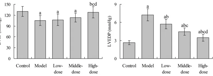
Effects of Baicalin on ST Segment Height in Rats
At the 12 hours after myocardial infarction modeling, the ST segment height in the control group, model group, and low-, middle- and high-dose baicalin groups was 0.15±0.03, 0.31±0.06, 0.28±0.04, 0.23±0.03 and 0.19±0.02 mV, respectively. The ST segment height in the model group was significantly higher than that in the control group (P<0.05), and that in the middle- and high-dose baicalin groups was significantly lower than that in the model group, respectively (P<0.05). However, the ST segment height in the three baicalin groups was significantly higher than that in the control group, respectively (P<0.05).
Effects of Baicalin on Hemodynamics of Rats
Compared with the control group, the LVSP of rats in the model group was significantly decreased (P<0.05). Compared with the model group, the LVSP in the high-dose baicalin group was significantly increased (P<0.05), with no significant difference with the control group (P>0.05). Compared with the control group, the LVEDP in the model group were significantly increased (P<0.05). Compared with the model group, the Chinese medicinal herbs in the Asia region and has a long history
of clinical application. Baicalin belongs to flavonoids. It is the main active component of Radix Scutellariae[4]. Baicalin has a wide
range of pharmacological effects, which are mainly presented in the antioxidant, free radical scavenging, inflammatory, anti-tumor, blocking calcium channel, inhibiting aldose reductase, antiviral and anti-allergic aspects[5-8]. In addition, baicalin has
protective effects on the immune, cardiovascular, digestive and nervous system[9-11]. This study investigated the protective effect
of baicalin on experimental myocardial infarction in rats and explored the related mechanisms. The objective was to provide a theoretical basis for the development of baicalin related medicines for mitigation and treatment of myocardial infarction.
METHODS
Animal Grouping and Treatment
Fifty Sprague Dawley (SD) rats (200±20 g; Laboratory Animal Center of Sichuan University, Chengdu, China) were randomly divided into 5 groups: control group, model group, and low-, middle- and high-dose baicalin groups, 10 rats in each group. The rats in low-, middle- and high-dose baicalin groups were intraperitoneally injected with baicalin [High performance liquid chromatography (HPLC) purity ≥ 98%; Shanghai Jingdu Biological Technology Co., Ltd., Shanghai, China], with a dose of 12.5, 25 and 50 mg/kg, respectively. The rats in the control and in the model group were intraperitoneally injected with normal saline. The injection was performed once per day and was continued for 10 days. On the 8th day, the rats in the
model group, low-, middle- and high-dose baicalin groups were subcutaneously injected with isoprenaline (Shanghai Hefeng Pharmaceutical Co., Ltd., Shanghai, China) (20 mg/kg) once per day, for continued 2 days, thus the rat model of myocardial infarction was established. The rats in the control group were subcutaneously injected with the equivalent volume of normal saline. Finally, the electrocardiography was performed, and the value of ST-segment elevation was used as the index to assess the myocardial ischemia.
Hemodynamic Test
At the 12 hours after the second injection of isoprenaline, a polyethylene plastic pipe with the diameter of 1 mm was inserted into the left ventricle of rats through the left carotid artery to perform the cardiac catheterization and was connected with the biological signal acquisition system. The left ventricular systolic pressure (LVSP) and left ventricular end diastolic pressure (LVEDP) were measured, which were used to evaluate the hemodynamic of rats.
Determination of Serum Lactate Dehydrogenase, Creatine Kinase-MB, Prostacyclin and Thromboxane A2
and middle-dose baicalin group were significantly higher than those in the control group, respectively (P<0.05) (Figure 2).
Effects of Baicalin on Serum PGI2 and TXA2 levels in Rats
Compared with the control group, the serum PGI2 level in
rats in the model group was significantly decreased (P<0.05). Compared with the model group, the PGI2 level in the three
baicalin groups was significantly increased, respectively (P<0.05). Compared with the control group, the serum TXA2 level in the
model group was significantly increased (P<0.05). Compared with the model group, the TXA2 level in the three baicalin groups
was significantly decreased, respectively (P<0.05). There was no LVEDP in the low-, middle- and high-dose baicalin groups was
significantly decreased, respectively (P<0.05). However, the LVEDP in the three baicalin groups was significantly higher than that in the control group, respectively (P<0.05) (Figure 1).
Effects of Baicalin on Serum LDH and CK-MB Levels in Rats
Serum LDH and CK-MB levels in rats in the model group were significantly increased when compared with the control group (P<0.05). Compared with the model group, the LDH and CK-MB levels in the middle- and high-dose baicalin groups were significantly decreased, respectively (P<0.05). However, the LDH level in the three baicalin groups and the CK-MB level in the low-
Fig. 1 – Effects of baicalin on hemodynamics of rats.
aP<0.05 compared with the control group; bP<0.05 compared with the model group; cP<0.05 compared with the low-dose group; dP<0.05
compared with the middle-dose group.
LVSP=left ventricular systolic pressure; LVEDP=left ventricular end diastolic pressure
Fig. 2 – Effects of baicalin on serum LDH and CK-MB level in rats.
aP<0.05 compared with the control group; bP<0.05 compared with the model group; cP<0.05 compared with the low-dose group.
model group, the myocardial MDA level in the middle- and high-dose baicalin groups was significantly decreased, respectively (P<0.05).
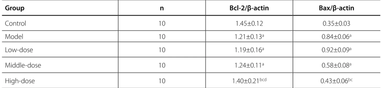
Effects of Baicalin on Myocardial Bcl-2 and Bax Protein Expression in Rats
Table 1 showed that, compared with the control group, the myocardial Bcl-2 protein level in rats in the model group, and low- and middle-dose baicalin groups was significantly decreased, respectively (P<0.05). Compared with the model group, the Bcl-2 protein level in the high-dose baicalin group was significantly increased (P<0.05). Compared with the control significant difference in each index between each baicalin group
and control group (P>0.05) (Figure 3).
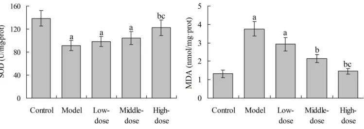
Effects of Baicalin on Myocardial SOD and MDA in Rats
As shown in Figure 4, compared with the control group, the myocardial SOD level in rats in the model group, low- and middle-dose baicalin groups was significantly decreased, respectively (P<0.05). Compared with the model group, the SOD level in the high-dose baicalin group was significantly increased (P<0.05). Compared with the control group, the myocardial MDA level in the model group and low-dose baicalin groups was significantly increased, respectively (P<0.05). Compared with the
Fig. 4 – Effects of baicalin on myocardial SOD and MDA in rats.
aP<0.05 compared with the control group; bP<0.05 compared with the model group; cP<0.05 compared with the low-dose group.
SOD=superoxide dismutase; MDA=malondialdehyde Fig. 3 – Effects of baicalin on serum PGI2 and TXA2 level in rats.
aP<0.05 compared with the control group; bP<0.05 compared with the model group. PGI
increased when compared with control group, respectively. Compared with the model group, the LDH and CK-MB levels in middle- and high-dose baicalin group were significantly decreased, respectively. This indicates that the pretreatment with baicalin can prevent the leakage of intracellular LDH and CK-MB into plasma, thus exerting the myocardial protection functions.
TXA2 can promote the platelet aggregation and the
contraction of the coronary arteries[16]. On the contrary, PGI 2
is known as the most potent inhibitor of platelet aggregation, with cytoprotective effect indirectly expanding coronary artery and inhibiting the production of oxygen free radicals[17]. In
myocardial infarction, the arterial endothelial is damaged, so the PGI2 synthesis in coronary artery endothelial cells is decreased.
Therefore, the platelet aggregates in the subendothelial collagen tissue, and excessively activates the production of a large number of TXA2[18]. In the present study, compared with
the control group, the serum PGI2 level in rats in model group
was significantly decreased, and the TXA2 level was significantly
increased. Compared with the model group, the PGI2 level in the
three baicalin groups was significantly increased, and the TXA2
level was significantly decreased. This suggests that baicalin can prevent the decrease of PGI2 level and the increase of TXA2
level in myocardial infarction rats, which may be related to its myocardial protective effects.
In isoprenaline-induced myocardial infarction model, the pathogenesis is related to lipid peroxidation, free radical production, membrane permeability changes and calcium overload in the myocardium[19]. SOD is an important antioxidant
enzyme in the body. It can catalyze the transformation of oxygen free radicals to hydrogen peroxide, thus avoiding the damage to cells. MDA is one of the final products of cell membrane lipid peroxidation. It indirectly reflects the degree of cell membrane peroxidation[20]. Results of this study showed that, compared
with the control group, the myocardial SOD level in the model group was significantly decreased, and the myocardial MDA level in the model group was significantly increased. Compared with the model group, the SOD level in the high-dose baicalin group was significantly increased, and the myocardial MDA level in the middle- and high-dose baicalin groups was significantly group, the myocardial Bax protein level in the model group, low-
and middle-dose baicalin groups was significantly increased, respectively (P<0.05). Compared with the model group, the myocardial Bax protein level in the high-dose baicalin group was significantly decreased (P<0.05).
DISCUSSION
It is found that the large-dose intravenous injection of isoprenaline can induce acute myocardial infarction, especially in the endocardium of the left ventricle and septum. The pathophysiological changes and myocardial morphological abnormalities induced by isoprenaline are similar to those of human myocardial infarction. Therefore, isoprenaline-induced myocardial infarction model is widely used to study the pathophysiological disturbances and morphological abnormalities of myocardial infarction and evaluate the effect of corresponding drugs[12]. Hemodynamic abnormalities often
occur during myocardial infarction, with decreased cardiac diastolic and systolic function and increased myocardial oxygen consumption[13]. Results of this study showed that, after injection
of with isoprenaline, the ST segment height and LVEDP in rats were significantly increased, respectively, and the LVSP was significantly decreased. This indicates that the myocardial infarction of rats has been successfully created. Compared with the model group, the ST segment height and LVEDP in groups with pretreatment using a certain dose of baicalin were significantly decreased, and the LVSP was significantly increased, respectively. This indicates that baicalin can improve the cardiac systolic and diastolic function, reduce the myocardial oxygen consumption, improve ventricular compliance, and mitigate the myocardial ischemia.
The activity of plasma LDH and CK-MB indirectly reflect the integrity of myocardial cell membrane and the degree of myocardial injury[14]. In myocardial cell necrosis, the integrity
of myocardial cell membrane is damaged, with increased permeability, which results in the leakage of intracellular LDH and CK-MB into plasma, leading to the increased LDH and CK-MB concentration in blood[15]. Results of this study showed that the
serum LDH and CK-MB levels in model group were significantly
Table 1. Effects of baicalin on myocardial Bcl-2 and Bax protein expression in rats.
Group n Bcl-2/β-actin Bax/β-actin
Control 10 1.45±0.12 0.35±0.03
Model 10 1.21±0.13a 0.84±0.06a
Low-dose 10 1.19±0.16a 0.92±0.09a
Middle-dose 10 1.24±0.11a 0.58±0.08a
High-dose 10 1.40±0.21bcd 0.43±0.06bc
aP<0.05 compared with the control group; bP<0.05 compared with the model group; cP<0.05 compared with the low-dose group; dP<0.05 compared with the middle-dose group
6. Lee W, Ku SK, Bae JS. Anti-inflammatory effects of baicalin, baicalein, and wogonin in vitro and in vivo. Inflammation. 2015;38(1):110-25. 7. Ma C, Ma Z, Fu Q, Ma S. Anti-asthmatic effects of baicalin in a mouse
model of allergic asthma. Phytother Res. 2014;28(2):231-7.
8. Ding Y, Dou J, Teng Z, Yu J, Wang T, Lu N, et al. Antiviral activity of baicalin against influenza A (H1N1/H3N2) virus in cell culture and in mice and its inhibition of neuraminidase. Arch Virol. 2014;159(12):3269-78. 9. Huang Y, Tsang SY, Yao X, Chen ZY. Biological properties of baicalein in
cardiovascular system. Curr Drug Targets Cardiovasc Haematol Disord. 2005;5(2):177-84.
10. Liu LL, Gong LK, Wang H, Xiao Y, Wu XF, Zhang YH, et al. Baicalin protects mouse from Concanavalin A-induced liver injury through inhibition of cytokine production and hepatocyte apoptosis. Liver Int. 2007;27(4):582-91.
11. Yang J, Yang X, Li M. Baicalin, a natural compound, promotes regulatory T cell differentiation. BMC Complement Altern Med. 2012;12:64. 12. Farvin KH, Anandan R, Kumar SH, Shiny KS, Mathew S, Sankar TV, et al.
Cardioprotective effect of squalene on lipid profile in isoprenaline-induced myocardial infarction in rats. J Med Food. 2006;9(4):531-6. 13. Bakkestrøm R, Andersen MJ, Ersbøll M, Bro-Jeppesen J, Gustafsson F,
Køber L, et al. Early changes in left atrial volume after acute myocardial infarction. Relation to invasive hemodynamics at rest and during exercise. Int J Cardiol. 2016;223:717-22.
14. Amani M, Jeddi S, Ahmadiasl N, Usefzade N, Zaman J. Effect of HEMADO on level of CK-MB and LDH enzymes after ischemia/reperfusion injury in isolated rat heart. Bioimpacts. 2013;3(2):101-4.
15. Chen YH, Wu XD, Yao ST, Sun S, Liu XH. Calcineurin is involved in cardioprotection induced by ischemic postconditioning through attenuating endoplasmic reticulum stress. Chin Med J (Engl). 2011;124(20):3334-40.
decreased, respectively. This suggests that baicalin has the ability of scavenging radical and reducing lipid peroxidation, thus playing a role in myocardial protection in rats.
Bcl-2 gene is a specific survival gene for inhibiting the apoptosis of cells. It plays an important role in the regulation of cell apoptosis[21]. Bcl-2 can resist various forms of cell death
and prolong the life-span of cells, which leads to the increase of cells[22]. Bax gene is the apoptosis-promotion gene. Bax gene
and Bcl-2 gene belong to the same gene family. Bax can not only inhibit the apoptosis inhibition effect of Bcl-2, but also directly promote the apoptosis of cells[23]. In this study, compared with
the control group, the myocardial Bcl-2 protein level in the model group was significantly decreased, and the myocardial Bax protein level was significantly increased. Compared with the model group, the Bcl-2 protein level in the high-dose baicalin group was significantly increased, and the Bax protein level was significantly decreased (P<0.05). This indicates that baicalin can up-regulate the expression of the Bcl-2 protein and down-regulate the expression of the Bax protein in rats, thus mitigating the myocardial infarction.
CONCLUSION
Baicalin has protective effects on myocardial infarction in rats. The possible mechanisms may be related to its resistance of oxidative stress, and up-regulation of the Bcl-2 protein expression and down-regulation of the Bax protein expression in myocardial tissue. This study has provided a theoretical basis for the development of baicalin related medicines for mitigation and treatment of myocardial infarction. This study still has some limitations. ST segment height, LVEDP and serum LDH in the baicalin groups still had significant differences with the control group (P<0.05). The reason may be that the baicalin doses used in this study are not enough to largely lower above indexes in the myocardial infarction rats to normal levels. Therefore, other suitable doses of baicalin should be further investigated. In addition, whether there are other mechanisms in the protective effects of baicalin on myocardial infarction needs to be further confirmed.
Authors’ roles & responsibilities
LW YL SL ZP HL ZT
Agreement to be accountable for all aspects of the work in ensuring that questions related to the accuracy or integrity of any part of the work are appropriately investigated and resolved; final approval of the version to be published Agreement to be accountable for all aspects of the work in ensuring that questions related to the accuracy or integrity of any part of the work are appropriately investigated and resolved; final approval of the version to be published Agreement to be accountable for all aspects of the work in ensuring that questions related to the accuracy or integrity of any part of the work are appropriately investigated and resolved; final approval of the version to be published Agreement to be accountable for all aspects of the work in ensuring that questions related to the accuracy or integrity of any part of the work are appropriately investigated and resolved; final approval of the version to be published Agreement to be accountable for all aspects of the work in ensuring that questions related to the accuracy or integrity of any part of the work are appropriately investigated and resolved; final approval of the version to be published Substantial contributions to the conception or design of the work; or the acquisition, analysis, or interpretation of data for the work; drafting the work or revising it critically for important intellectual content; final approval of the version to be published
REFERENCES
1. Shepard D, Vander Zanden A, Moran A, Naghavi M, Murray C, Roth G. Ischemic Heart Disease Worldwide, 1990 to 2013: Estimates from the Global Burden of Disease Study 2013. Circ Cardiovasc Qual Outcomes. 2015;8(4):455-6.
2. Pasupathy S, Air T, Dreyer RP, Tavella R, Beltrame JF. Systematic review of patients presenting with suspected myocardial infarction and nonobstructive coronary arteries. Circulation. 2015;131(10):861-70. 3. Francis Stuart SD, De Jesus NM, Lindsey ML, Ripplinger CM. The
crossroads of inflammation, fibrosis, and arrhythmia following myocardial infarction. J Mol Cell Cardiol. 2016;91:114-22.
4. Lu T, Song J, Huang F, Deng Y, Xie L, Wang G, et al. Comparative pharmacokinetics of baicalin after oral administration of pure baicalin, Radix scutellariae extract and Huang-Lian-Jie-Du-Tang to rats. J Ethnopharmacol. 2007;110(3):412-8.
production and postischemic myocardial reperfusion injury. Ann N Y Acad Sci. 1994;723:180-96.
20. Li J, Tang HL, Chen Y, Fan Q, Shao YT, Jia M, et al. Malondialdehyde and SOD-induced changes of gastric tissues in acute gastric mucosal injury under positive acceleration. Genet Mol Res. 2015;14(2):4361-8. 21. Karsan A, Yee E, Poirier GG, Zhou P, Craig R, Harlan JM. Fibroblast growth
factor-2 inhibits endothelial cell apoptosis by Bcl-2-dependent and independent mechanisms. Am J Pathol. 1997;151(6):1775-84. 22. Pepper C, Thomas A, Hoy T, Fegan C, Bentley P. Flavopiridol circumvents
Bcl-2 family mediated inhibition of apoptosis and drug resistance in B-cell chronic lymphocytic leukaemia. Br J Haematol. 2001;114(1):70-7. 23. Wang Y, Yin RF, Teng JS. Wogonoside induces cell cycle arrest and
mitochondrial mediated apoptosis by modulation of Bcl-2 and Bax in osteosarcoma cancer cells. Int J Clin Exp Pathol. 2015;8(1):63-72. 16. Higuchi W, Fuse I, Hattori A, Aizawa Y. Mutations of the platelet
thromboxane A2 (TXA2) receptor in patients characterized by the absence of TXA2-induced platelet aggregation despite normal TXA2 binding activity. Thromb Haemost. 1999;82(5):1528-31.
17. Gershlick AH, Spriggins D, Davies SW, Syndercombe Court YD, Timmins J, Timmis AD, et al. Failure of epoprostenol (prostacyclin, PGI2) to inhibit platelet aggregation and to prevent restenosis after coronary angioplasty: results of a randomised placebo controlled trial. Br Heart J. 1994;71(1):7-15.
18. Santilli F, Davì G, Basili S, Lattanzio S, Cavoni A, Guizzardi G, et al. Thromboxane and prostacyclin biosynthesis in heart failure of ischemic origin: effects of disease severity and aspirin treatment. J Thromb Haemost. 2010;8(5):914-22.
19. Kramer JH, Misík V, Weglicki WB. Lipid peroxidation-derived free radical