INTRODUCTION
Spontaneous bacterial peritonitis (SBP) is defined as the infec-tion of ascites fluid in patients with cirrhosis, in the absence of apparent abdominal focus of infection(1). Its prevalence in cirrhotic
patients with ascites on hospital admission ranges from 10% to 30%(2). The risk of developing SBP during hospitalization ranges
from 20% to 60%, with mortality from 20% to 40%(3). Treatment
failure occurs in about 10%, leading to a poor prognosis with hos-pital mortality from 50% to 80%(4). The probability of recurrence
is 43% in 6 months, and the 2-year survival rate is only 50%(1).
The culture of ascites fluid is negative in more than 60% of cases, when using conventional techniques, even in the presence of suggestive clinical manifestations. The use of blood culture bottles to inoculate the collected material allows an increase of up to 90% in the chance of obtaining a positive culture(4).
Over 60% of SBP episodes are caused by enteric gram-negative bacteria, and the most common germs isolated were Escherichia coli (E coli) and Klebsiella pneumoniae (K. pneumoniae). Gram-positive germs are present in 25% of SBP episodes, with streptococcal
spe-Impact of microbiological changes on
spontaneous bacterial peritonitis in
three different periods over 17 years
Paulo Roberto Lerias de
ALMEIDA
1,2, Gabriel Stefani
LEÃO
1, Charlles David Gonçalves
GONÇALVES
1,
Rafael Veiga
PICON
1,3and Cristiane Valle
TOVO
1,2Received 18/9/2017 Accepted 24/11/2017
ABSTRACT – Background – Spontaneous bacterial peritonitis is a serious complication in cirrhotic patients, and changes in the microbiological char-acteristics reported in the last years are impacting the choice of antibiotic used for treatment. Objective – The aim of the present study is to evaluate the changes in the epidemiology and bacterial resistance of the germs causing spontaneous bacterial peritonitis over three different periods over 17 years. Methods – All cirrhotic patients with spontaneous bacterial peritonitis and positive culture of ascites fluid were retrospectively studied in a reference Hospital in Southern Brazil. Three periods were ramdomly evaluated: 1997-1998, 2002-2003 and 2014-2015. The most frequent infecting organisms and the sensitivity in vitro to antibiotics were registered. Results – In the first period (1997-1998) there were 33 cases, the most common were: E. coli in 13 (36.11%), Staphylococcus coagulase-negative in 6 (16.66%), K. pneumoniae in 5 (13.88%), S. aureus in 4 (11.11%) and S. faecalis in 3 (8.33%). In the second period (2002-2003), there were 43 cases, the most frequent were: Staphylococus coagulase-negative in 16 (35.55%), S. aureus
in 8 (17.77%), E. coli in 7 (15.55%) and K. pneumoniae in 3 (6.66%). In the third period (2014-2015) there were 58 cases (seven with two bacteria), the most frequent were: E. coli in 15 (23.1%), S. viridans in 12 (18.5%), K. pneumoniae in 10 (15.4%) and E. faecium 5 (7.7%). No one was using antibi-otic prophylaxis. Considering all staphylococci, the prevalence increased to rates of the order of 50% in the second period, with a reduction in the third period evaluated. Likewise, the prevalence of resistant E. coli increased, reaching 14%. Conclusion – There was a modification of the bacterial population causing spontaneous bacterial peritonitis, with high frequency of gram-positive organisms, as well as an increase in the resistance to the traditionally recommended antibiotics. This study suggests a probable imminent inclusion of a drug against gram-positive organisms in the empiric treatment of spontaneous bacterial peritonitis.
HEADINGS – Peritonitis. Bacterial infections. Liver cirrhosis. Ascites.
Declared conflict of interest of all authors: none Disclosure of funding: no funding received
1 Hospital Nossa Senhora da Conceição (HNSC), Porto Alegre, RS, Brasil; 2 Universidade Federal de Ciências da Saúde de Porto Alegre (UFCSPA), RS, Brasil; 3 Universidade do Vale do
Taquari (UNIVATES), Lajeado, RS, Brasil.
Correspondence: Cristiane Valle Tovo. Rua Cel Aurelio Bitencourt 115, 201 – CEP: 90430-080 – Porto Alegre, RS, Brasil. E-mail: [email protected]
cies being the most frequently isolated(5). However, considerable
change in the epidemiology of germ causing SBP has occurred. This alteration has been attributed to increased survival, increased invasive procedures and prolonged use of antibiotics for intestinal decontamination(3,6).
The preponderance of infections caused by Gram-negative bacteria due to epidemiological changes has shifted to a higher prevalence of infections being caused by gram-positive cocci(6). The
use of systemic antibiotics within 30 days before SBP diagnosis and a lower Sequential Organ Failure Assessment (SOFA) score were independent predictors of SBP caused by Gram-positive bacteria in patients with cirrhosis(7). In the study by Alexopoulou et al.(8) and
Almeida et al.(9), the majority of isolated pathogens from patients
with SBP were gram-positive cocci.
Thus, changes in the microbiological characteristics reported in recent years may have an impact on the choice of empirical treatment.
METHODS
The evaluated population consists of patients hospitalized at the Hospital Nossa Senhora da Conceição – Porto Alegre, Brazil, a public tertiary hospital. All cases of cirrhotic patients with SBP in which culture of ascites fluid was positive were retrospectively evalu-ated. Three different periods randomly assessed were studied: 1997-1998, 2002-2003 and 2014-2015. The most frequent microorganisms and in vitro susceptibility to antibiotics were verified, as well as the presence or absence of chemoprophylaxis. All the bacteria that grew in the culture were reported in each period. Identification of the bacterial samples and the antimicrobial susceptibility test were performed by the automated Microscan® test (Dade Behring,
WalkAway-96). The use of previous quimioprophylaxis against SBP was also searched in the charts.
For statistical analysis, the chi-square test was used to com-pare the proportions between Gram-positive and Gram-negative microorganisms in the two evaluated periods, using the Epi-Info Program. Chi-square for non-linearity was used to assess non-linear trend between proportions of Gram-negatives and Gram-positives cultures across the three periods under study. Alpha was set at 5%.
RESULTS
In the first period evaluated (1997-1998) 33 cases of SBP with positive culture were identified, with 3 (9.1%) polymicrobial infections. The most frequent bacteria were E. coli in 13 (36.1%), coagulase-negative staphylococci in 6 (16.2%), K. pneumoniae in 5 (13.9%), Staphylococus aureus (S. aureus) in 4 (11.1%) and Ente-rococcus faecalis (E. faecalis) with 3 (8.3%) cases.
In the second period (2002-2003), 43 cases of SBP were identified, of which 2 (4.6%) were polymicrobial. The most common germs were coagulase-negative staphylococci in 16 (35.5%) cases, S. aureus in 8 (17.8%), E coli in 7 (15.5%) and K. pneumoniae in 3 (6.7%).
In the third period (2014-2015) 58 cases of SBP were identified, being 7 (12.1%) polymicrobial. The most frequent germs were E coli in 15 (23.1%) cases, Streptococcus viridans (S. viridans) in 12 (18.5%), K. pneumoniae in 10 (15.4%) and Enterococcus faecium (E. faecium) in 5 (7.7%).
The total number of Gram-negative germ was 20 (55.6%), 16 (35.6%) and 34 (52.3%) in the first, second and third study peri-ods, respectively. The number of Gram-positive patients was 16 (44.4%), 29 (64.4%) and 31 (47.7%) in the first, second and third study periods, respectively.
Other germs identified are described in TABLE 1.
The comparison of the three evaluated periods showed a de-crease in the proportion of Gram negative in the second period followed by a new increase in the third period (i.e. 55.6% in the first, 35.6% in the second and 52.3% in the third periods), representing a significant non-linear trend across the three periods (P=0.043).
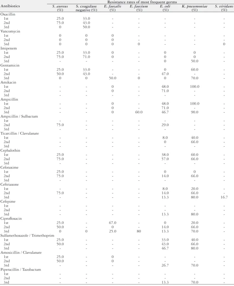
No patient was on prophylaxis for SBP at the time of hospi-talization. With respect to bacterial resistance rates, the complete spectrum in the three periods analyzed is shown in TABLE 2.
The prevalence of methicillin-resistant S. aureus (MRSA) in-creased from 25% to 75% between the first and second periods, which implies the same levels of resistance to other β-lactam antibiotics such as cephalosporins I and III generation. In the third period, there was no resistance of S. aureus to oxacxilin, although the resistance rates of coagulase-negative Staphylococci were progressively higher. Only the vancomycin remained 100% active against S. aureus and S. coagulase negative during the analyzed periods.
TABLE 1. Germs causing SBP in the first (1997-1998), second (2002-2003) and third (2014-2015) periods evaluated
Bacteria
1st Period 2nd Period 3rd Period
N (%) N (%) N (%)
Escherichia coli 13 (36.1) 7 (15.5) 15 (23.1)
Staphylococcus
coagulase-negative 6 (16.7) 16 (35.5) 2 (3.1)
Klebsiella pneumonia 5 (13.9) 3 (6.7) 10 (15.4)
Staphylococcus aureus 4 (11.1) 8 (17.8) 2 (3.1)
Enterococcus faecalis 3 (8.3) 0 - 4 (6.2)
Streptococcus viridans 0 - 2 (4.4) 12 (18.5)
Enterococcus faecium 0 - 0 - 05 (7.7)
Enterococcus avium 1 (2.8) 0 - 0
-Streptococcus bovis 1 (2.8) 0 - 0
-Pseudomonas
aeruginosa 1 (2.8) 0 - 2 (3.1)
Salmon/Arizona 1 (2.8) 0 - 0
-Klebsiella oxytoca 0 - 2 (4.4) 0
-Acinetobacter sp. 0 - 2 (4.4) 0
-Streptococcus
agalactiae 0 - 1 (2.2) 1 (1.5)
Enterobacter cloacae 0 - 1 (2.2) 1 (1.5)
Kluyvera ascorbata 0 - 1 (2.2) 0
-Enterococcus spp. 0 - 0 - 1 (1.5)
Leuconostoc
pseudomesentoroides 0 - 0 - 1 (1.5)
Hafnia alvei 0 - 0 - 1 (1.5)
Haemophilus specie 0 - 0 - 1 (1.5)
Pantoea specie 0 - 0 - 1 (1.5)
Citrobacter freundi 0 - 0 - 1 (1.5)
Streptococcus
pneumoniae 0 - 0 - 1 (1.5)
Streptococcus specie 0 - 0 - 1 (1.5)
Enterococcus durans 0 - 0 - 1 (1.5)
Enterobacter aerogenes 0 - 0 - 1 (1.5)
Sphingomonas
paucimobilis 0 - 0 - 1 (1.5)
Total 36* (100.0) 45** (100.0) 65*** (100.0)
SBP: spontaneous bacterial peritonitis. * 33 cases, 3 polymicrobial; ** 43 cases, 2 polymicrobial; *** 58 cases, 7 polymicrobial.
E. faecalis, another Gram-positive coccus with some frequency in SBP, presented a 50% resistance rate to gentamicin in the third period, however, it showed an improvement in the resistance pro-file for ciprofloxacin from the first to the third evaluated period, maintaining the sensitivity of 100% for vancomycin. Unlike E. faecalis, which showed 100% sensitivity to ampicillin at all periods, cases of E. faecium showed resistance to ampicillin by 60% and to ciprofloxacin by 80%.
TABLE 2. Bacterial resistance rates in SBP in the first (1997-1998), second (2002-2003) and third (2014-2015) periods evaluated
Antibiotics
Resistance rates of most frequent germs
S. aureus
(%) negative (%)S. coagulase E. faecalis(%) E. faecium (%) E. coli (%) K. pneumoniae(%) S. viridans(%)
Oxacillin
1st 25.0 33.0 - - - -
2nd 75.0 43.0 - - - -
3rd 0 50.0 - - - -
-Vancomycin
1st 0 0 0 - - -
2nd 0 0 0 - - -
3rd 0 0 0 0 - - 0
Imipenem
1st 25.0 33.0 0 - 0 0
2nd 75.0 71.0 0 - 0 0
3rd - - - - 0 50.0
-Gentamicin
1st 25.0 33.0 - - 0 60.0
2nd 50.0 43.0 - - 47.0 -
3rd 0 0 50.0 0 0 70.0
-Amikacin
1st - - 0 - 48.0 100.0
2nd - - 0 - 71.0 -
3rd - - -
-Ampicillin
1st - - 0 - 48.0 100.0
2nd - - 0 - 71.0 -
3rd - - 0 60.0 46.7 90.0
-Ampicillin / Sulbactam
1st - - -
2nd 75.0 - - - 29.0 -
3rd - - -
-Ticarcillin / Clavulanate
1st - - - - 8.0 40.0
2nd - - - - 0 66.0
3rd - - -
-Cephalothin
1st 25.0 - - - 38.0 60.0
2nd 75.0 - - - 57.0 66.0
3rd - - -
-Cefotaxime
1st 25.0 - - - 0 0
2nd 75.0 - - - 14.0 66.0
3rd - - -
-Ceftriaxone
1st - - - - 8.0 20.0
2nd 75.0 - - - 14.0 66.0
3rd - - - - 13.3 80.0 16.7
Cefepime
1st - - -
2nd - - -
3rd - - - - 13.3 80.0
-Ciprofloxacin
1st 25.0 - 67.0 - 0 20.0
2nd 50.0 - 0 - 14.0 66.0
3rd 0 0 25.0 80 13.3 70.0
-Sulfamethoxazole / Trimethoprim
1st 25.0 - - - 33.0 40.0
2nd 50.0 - - - 43.0 66.0
3rd - - - - 46.7 80.0
-Amoxicillin / Clavulanate
1st 25.0 - 0 - - -
2nd 50.0 - 0 - - -
3rd - - - - 26.7 70.0
-Piperacillin / Tazobactam
1st - - -
2nd - - -
3rd - - - - 13.3 70.0
DISCUSSION
The present study showed a modification of the bacterial flora causing SBP in the last 17 years in a reference hospital in Southern Brazil, detecting a significant alternating pattern of predominance between Gram-negative and Gram-positive organisms over time.
Bacterial infections are frequent in patients with decompensated cirrhosis, and may promote increased morbidity and mortality(3).
Management of cirrhotic patients with severe complications has spread, particularly with the expansion of liver transplantation pro-grams and invasive procedures used in this setting(2), which may also
be associated with infections(10). Similarly, another responsible factor
can be the selective intestinal decontamination with norfloxacin, that has been widely used in primary and secondary prophylaxis of SBP(3,5).
All these factors may have influenced the modifications in the flora and the resistance of SBP-causing germs in cirrhotic patients(2-4). This change in the profile of patients and pathogens
should be carefully monitored, since it has a direct implication in the empirical choice of antimicrobials.
In the present study, both ciprofloxacin and third-generation cephalosporins were observed to maintain good antimicrobial activity over some Gram-negatives, such as E. coli, which was re-sistant in only 14% of the sample in the second and third periods evaluated. It should be noted, however, that none of the patients were on prophylactic use of norfloxacin.
Many authors(11-13) have shown an increase in the prevalence
of infections caused by multi-resistant bacteria, especially in nosocomial episodes, presenting resistance to third-generation cephalosporins of 21.5% to 45%. Taking into account the increasing evidence for multiresistance among the bacteria that cause SBP and the lower efficacy of treatment with third-generation cephalospor-ins, new treatment recommendations have become necessary for the management of infections in cirrhotic patients. The distinction between community-acquired infectious episodes, healthcare-associated or nosocomial infections, and the identification of risk factors for multiresistant germs can help in the decision-making process for empirical antibiotic therapy choice(14).
Regarding Klebsiella, a Gram-negative bacteria also prevalent in this series, attention is drawn to the development of antimicro-bial resistance in the order of 66% to 80% for third generation cephalosporins in the second and maintaining in the third periods. In addition, resistance to imipenem appeared in the third period, which had not been observed in previous periods.
Piano et al.(15) presented a prospective randomized study which
compared the effectiveness of third-generation cephalosporin, ceftazidime, versus meropenem plus daptomycin. The authors discussed the possible concerns of starting highest broad-spectrum antibiotics as a first line treatment might possibly increase the risk of more resistant bacteria in the hospitalized patients. They rec-ommended that based on the early results of cultures, antibiotics may be tailored to narrow-spectrum drugs and this approach may partially overcome this problem.
There were also increased cases of SBP by Gram-positive cocci in the second period, with S. coagulase-negative and S. aureus be-ing relevant, although none of the patients had used prophylactic antibiotics prior to the occurrence of SBP. More significantly, its resistance profile increased from 25% to 50% to 75% for all drugs tested, including third generation cephalosporins, except for vanco-mycin. In the third period, staphylococci were not frequent germs.
Enterococci and streptococci, not frequent germs found in the first and second periods, were more prevalent in the third, cor-responding to 30.9% of SBP cases. It is worth noting that while the resistance of E. faecalis and Streptococcus is small relative to the antibiotics commonly tested, the resistance of E. faecium was 60% to ampicillin. In contrast to streptococcus species, enterococci are frequently resistant to penicillin with a minimum inhibitory concentration (MIC) of 1-8 μg/mL for E. faecalis and 16-64 μg/ mL for E. faecium(16). In a study by Friedrich K et al.(17), 88.6%
of all Enterococcus spp. agents were resistant to third-generation cephalosporins and 80.6% were resistant to carbapenems, 62.9% of the patients with Enterococcus-related SBP received ineffective empirical antibiotic treatment. These antimicrobial resistance patterns might explain the observed association for SBP infection with Enterococcus spp. and poor patient survival (P=0.048), which was not present for infection with Streptococci, Staphylococci, Enterobacteriaceae, or Candida.
The trend of increased participation of Gram-positive cocci in severe bacterial infections of hospitalized cirrhotic patients, not only in the form of SBP, but also and especially in the form of bacteremia, has been emphasized(3,10,18). The clinical value and
significance of species of coagulase-negative staphylococci obtained in the most diverse biological materials continues to increase as diagnostic and therapeutic strategies lead to a growing propor-tion of invasive procedures(19). Immunocompromised hospitalized
patients constitute the population most vulnerable to these types of pathogens. However, identifying them as etiological agents or contaminating flora continues as a strong challenge(19).
When addressing the progression of Gram-positive and Gram-negative bacteria across the three periods, we identified a significant non-linear variation over time. A possible hypothesis to explain these findings could be a seasonality to the alternation of predominance between Gram-negative and Gram-positive germs, which is consistent with the expected dynamic balance of nosocomial bacterial populations.
As possible limitations of the study, we can mention the ret-rospective design, which could limit the obtaining of some data. In addition, because it has been performed in a single center, the external validity may be limited, and similar studies in other hos-pitals should be performed.
In conclusion, there was a modification of the bacterial popula-tion causing SBP, with high frequency of Gram-positive organisms, as well as an increase in the resistance to the traditionally recom-mended antibiotics. Future research should focus on the clinical effectiveness of empiric Gram-negative and Gram-positive antibi-otic coverage for at-risk cirrhantibi-otic patients. An imminent extension in treatment protocols to include empiric Gram-positive antibiotic coverage for SBP in centers with similar flora may be considered. The precise timing of this change in therapeutics should be guided by local epidemiological surveillance.
Authors’ contributions
REFERENCES
1. Mowat C, Stanley A. Spontaneous bacterial peritonitis – diagnosis, treatment and prevention. Aliment Pharmacol Ther. 2001;15:1851-9.
2. Fernández J, Bauer TM, Navasa M, Rodés J. Diagnosis, treatment and prevention of spontaneous bacterial peritonitis. Baillieres Best Pract Res Clin Gastroenterol. 2000;14:975-90.
3. Fernández J, Navasa M, Gómez J, Colmenero J, Vila J, Arroyo V, Rodés J. Bac-terial infections in cirrhosis: epidemiological changes with invasive procedures and norfloxacin prophylaxis. Hepatology. 2002;35:140-8.
4. Rimola A, García-Tsao G, Navasa M, Piddock LJ, Planas R, Bernard B, Inadomi JM. Diagnosis, treatment and prophylaxis of spontaneous bacterial peritonitis: a consensus document. J Hepatol. 2000;32:142-53.
5. Such J, Runyon B. Spontaneous bacterial peritonitis. Clin Infect Dis. 1998;27:669-76. 6. Wiest R, Krag A, Gerbes A. Spontaneous bacterial peritonitis: recent guidelines
and beyond. Gut. 2012;61:297-310.
7. Kim JH, Jeon YD, Jung IY, Ahn MY, Ahn HW, Ahn JY, et al. Predictive factors of spontaneous bacterial peritonitis caused by gram-positive bacteria in patients with cirrhosis. Medicine (Baltimore). 2016;95:e3489.
8. Alexopoulou A, Papadopoulos N, Eliopoulos DG, Alexaki A, Tsiriga A, Toutouza M, Pectasides D. Increasing frequency of gram-positive cocci and gram-nega-tive multidrugresistant bacteria in spontaneous bacterial peritonitis. Liver Int. 2013;33:975–981.
9. Almeida PRL, Camargo NS, Arenz M, Tovo CV, Galperim B, Behar P. Peritonite bacteriana espontânea: impacto das mudanças da microbiologia. Arquivos de Gastroenterologia. 2007;44:68-72.
10. Wong F, Bernardi M, Balk R, Christman B, Moreau R, Garcia-Tsao G, Patch D, Soriano G, Hoefs J, Navasa M. Sepsis in cirrhosis: report on the 7th Meeting of the Internal Ascites Club. Gut. 2005;54:718-25.
11. Ariza X, Castellote J, Lora-Tamayo J, Girbau A, Salord S, Rota R, Ariza J, Xiol X. Risk factors for resistance to ceftriaxone and its impact on mortality in community, healthcare and nosocomial spontaneous bacterial peritonitis. J Hepatol. 2012;56:825-32.
12. Tandon P, Delisle A, Topal JE, Garcia-Tsao G. High prevalence of antibiotic-re-sistant bacterial infections among patients with cirrhosis at a US liver center. Clin Gastroenterol Hepatol. 2012;10:1291-1298.
13. Fernández J, Gustot T. Management of bacterial infections in cirrhosis. J Hepatol. 2012;56 (Suppl 1): S1-12.
14. Mattos AA, Costabeber AM, Lionço LC, Tovo CV. Multi-resistant bactéria in spontaneous bacterial peritonitis: a new step in the management? WJG. 2014;20:14079-86.
15. Piano S, Fasolato S, Salinas F, Romano A, Tonon M, Morando F, et al. The empirical antibiotic treatment of nosocomial spontaneous bacterial peritonitis: results of a randomized, controlled clinical trial. Hepatology. 2016; 63:1299-309. 16. Klibi N, Ben Slama K, Sáenz Y, Masmoudi A, Zanetti S, Sechi LA, Boudabous A, Torres C. Detection of virulence factors in high-level gentamicin-resistant Enterococcus faecalis and Enterococcus faecium isolates from a Tunisian hospital. Can J Microbiol. 2007;53:372-9.
17. Friedrich K, Nüssle S, Rehlen T, Stremmel W, Mischnik A, Eisenbach C. Micro-biology and resistance in first episodes of spontaneous bacterial peritonitis: impli-cations for management and prognosis. J Gastroenterol Hepatol. 2016;31:1191-5. 18. Campillo B, Dupeyron C, Richardet JP, Mangeney N, Leluan G. Epidemiology of severe hospital-acquired infections in patients with liver cirrhosis: effect of long–term administration of norfloxacin. Clin Infec Dis. 1998;26:1066-70. 19. Kloosi WE, Bannerman TL. Update on clinical significance of coagulase-negative
staphylococci. Clin Microbiol Rev. 1994;7:117-40.
Almeida PRL, Leão GS, Gonçalves CDG, Picon RV, Tovo CV. Impacto das mudanças da microbiologia na peritonite bacteriana espontânea em três diferentes períodos ao longo de 17 anos. Arq Gastroenterol. 2018;55(1):23-7.
RESUMO – Contexto – A peritonite bacteriana espontânea é uma complicação séria em pacientes cirróticos e as alterações nas características microbi-ológicas relatadas nos últimos anos podem afetar a escolha do antibiótico utilizado no tratamento. Objetivo – Os objetivos do presente estudo são avaliar as mudanças na epidemiologia e perfil de resistência bacteriana dos germes causadores de peritonite bacteriana espontânea em três períodos diferentes ao longo de 17 anos. Métodos – Todos os pacientes cirróticos com peritonite bacteriana espontânea e cultura positiva de fluido ascítico foram estudados retrospectivamente em um hospital de referência no Sul do Brasil. Foram avaliados três diferentes períodos selecionados de forma randômica: 1997-1998, 2002-2003 e 2014-2015. Os organismos infecciosos mais frequentes e a sensibilidade in vitro a antibióticos foram registados.
Resultados – No primeiro período (1997-1998) houve 33 casos; os mais comuns foram: E. coli em 13 (36,1%), Staphylococcus coagulase-negativo em 6 (16,7%), K. pneumoniae em 5 (13,9%), S. aureus em 4 (11,1%) e S. faecalis em 3 (8,3%). No segundo período (2002-2003), houve 43 casos, os mais frequentes foram: Staphylococus coagulase-negativo em 16 (35,5%), S. aureus em 8 (17,8%), E. coli em 7 (15,5%) e K. pneumoniae em 3 (6,7%). No terceiro período (2014-2015), houve 58 casos (sete com duas bactérias), os mais frequentes foram: E. coli em 15 (23,1%), S. viridans em 12 (18,5%),
K. pneumoniae em 10 (15,4%) e E. faecium 5 (7,7%). Nenhum paciente estava usando profilaxia antibiótica. Quando considerados todos os estafilo-cocos, a prevalência aumentou para taxas da ordem de 50% no segundo período, apresentando redução no terceiro período avaliado. Do mesmo modo, a prevalência de E coli resistente aumentou, chegando a 14%. Conclusão – Houve modificação da população bacteriana causadora de peritonite bacteriana espontânea, com alta frequência de organismos gram-positivos, bem como aumento da resistência aos antibióticos tradicionalmente recomendados. Este estudo sugere uma provável inclusão iminente de um medicamento contra organismos gram-positivos no tratamento empírico da peritonite bacteriana espontânea.