ABSTRACT.-
Zafalon L.F., Cunha M.L.R.S., Brandão H.M., Mosqueira V.C.F., Santana R.C.M.,
Barioni Júnior W., Martins K.B. & Pilon L.E. 2018.
Relationship between virulence factor
genes in coagulase-negative Staphylococcus spp. and failure of antimicrobial treatment
of subclinical mastitis in sheep
.
Pesquisa Veterinária Brasileira 38(4):579-585.
Embrapa
Pecuária Sudeste, Rodovia Washington Luiz Km 234, Cx. Postal 339, São Carlos, SP 13560-970,
Brazil. E-mail: luiz.zafalon@embrapa.br
Coagulase-negative
Staphylococcus
spp. (CNS) are the main microorganisms involved in
ovine mastitis. Treatment at the end of lactation can contribute towards cure and prevention
of subclinical cases during the subsequent lactation. However, virulence factors and resistance
mechanisms presented by CNS can decrease cure rates. The aims of the study were to identify
the species of CNS in milk of mastitic ewes with and without antimicrobial treatment, and to
investigate the presence of genes relating to resistance of β-lactam antimicrobials, formation
of biofilms, production of enterotoxins and production of the toxic shock syndrome toxin.
Cases of failure in the treatment were related with the presence/absence of the respective
genes. Sixty sheep were divided into three groups: G1, without treatment; G2, animals treated
via the intramammary route with 100mg of cloxacillin during drying off; and G3, sheep
treated via the intramammary route with 50 mg of nanoparticulate cloxacillin. Milk samples
were gathered during drying off and 15 and 30 days after the parturition of the subsequent
lactation. The analyses to identify the species of CNS were carried out by means of the internal
transcribe spacer technique and the investigation of the genes responsible for the virulence
factors and resistance to oxacillin was performed using the polymerase chain reaction (PCR)
technique. No sample was positive for the
mec
A gene. The only gene relating to production
of enterotoxins was
sec
. Among the genes relating to production of biofilm,
ica
D was the only
one identified in the three experimental groups.
Staphylococcus warneri
was the main species
of CNS isolated during the pre and post-partum periods of the sheep. The species carrying
genes relating to production of enterotoxins and biofilms were present in uncured sheep.
INDEX TERMS: Virulence factor genes, coagulase-negative, Staphylococcus spp., antimicrobial treatment, mastitis, sheep, biofilms, enterotoxins, bacterioses.Vet 2420 pvb-4984 LD
Relationship between virulence factor genes
in coagulase-negative
Staphylococcus
spp. and
failure of antimicrobial treatment of subclinical
mastitis in sheep
1Luiz F. Zafalon
2*, Maria L.R.S. Cunha
3, Humberto M. Brandão
4,
Vanessa C.F. Mosqueira
5, Raul C.M. Santana
2, Waldomiro Barioni Júnior
2,
Katheryne B. Martins
3and Lucas E. Pilon
61 Received on January 31, 2017.
Accepted for publication on April 3, 2017.
2 Embrapa Pecuária Sudeste, Rodovia Washington Luiz Km 234, Cx.
Postal 339, São Carlos, SP 13560-970, Brazil. E-mails: raul.mascarenhas@ embrapa.br, waldomiro.barioni@embrapa.br; *Corresponding author: luiz.zafalon@embrapa.br
3 Instituto de Biociências, Universidade Estadual Paulista (Unesp),
Distrito de Rubião Júnior, Botucatu, SP 18618-970, Brazil. E-mails: cunhamlr@ibb.unesp.br, katheryne_bm@yahoo.com.br
4 Embrapa Gado de Leite, Rua Eugênio do Nascimento 610, Juiz de Fora,
MG 36038-330, Brazil. E-mail: humberto.brandao@embrapa.br
5 Universidade Federal de Ouro Preto, Rua Costa Sena 171, Ouro Preto,
MG 35400-000, Brazil. E-mail: mosqueira@ef.ufop.br
6 Programa de Pós-Graduação em Medicina Veterinária Preventiva,
RESUMO.- [Relação entre genes de fatores de virulência
em Staphylococcus spp. coagulase-negativos e a falha
do tratamento antimicrobiano da mastite subclínica
ovina.]
Staphylococus
spp. coagulase-negativos (SCN) estão
entre os principais micro-organismos envolvidos na mastite
ovina. O tratamento ao final da lactação pode contribuir com
a cura e a prevenção de casos subclínicos durante a lactação
seguinte. Todavia, fatores de virulência e mecanismos de
resistência apresentados por SCN podem reduzir as taxas
de cura. Os objetivos desse estudo foram identificar as
espécies de SCN no leite de ovelhas com mastite com e sem
tratamento antimicrobiano e investigar a presença de genes
relacionados com resistência a antibióticos beta lactâmicos,
formação de biofilmes, produção de enterotoxinas e produção
da toxina da síndrome do choque tóxico. Casos de falhas no
tratamento foram relacionados com a presença/ausência
dos respectivos genes. Sessenta ovelhas foram divididas em
três grupos: G1, sem tratamento; G2, animais tratados via
intramamária com 100mg de cloxacilina antes da secagem;
e G3, ovelhas tratadas via intramamária com 50 mg de
cloxacilina nanoparticulada. Amostras de leite foram obtidas
durante a secagem e 15 e 30 dias depois do parto na lactação
seguinte. As análises para identificar as espécies de SCN
foram conduzidas por meio da técnica de
Internal transcribe
spacer
e a investigação dos genes responsáveis pelos fatores
de virulência e resistência à oxacilina foi realizada usando a
técnica reação em cadeia da polimerase. Nenhuma amostra
foi positiva para o gene
mecA
. O único gene relacionado
com a produção de enterotoxinas foi o
sec
. Dentre os genes
relacionados com a produção de biofilme,
icaD
foi o único
identificado nos três grupos experimentais.
Staphylococcus
warneri
foi a principal espécie de SCN isolada durante o pré e
pós-parto. As espécies que apresentaram genes relacionados
com a produção de enterotoxinas e biofilmes estavam presentes
nas ovelhas não curadas.
TERMOS DE INDEXAÇÃO: Genes, fatores de virulência, Staphylococcus spp., coagulase-negativos, tratamento antimicrobiano, mastite subclínica, ovinos, biofilmes, enterotoxinas, bacterioses.
INTRODUCTION
Mastitis is among the main sanitary problems in ovine
breed
ing. The disease can be classified as clinical or
subclinical. Unlike the clinical form, subclinical mastitis
does not present macroscopic alterations of the mammary
gland and is strongly related to financial losses for producers
(McDougall et al. 2001). Its etiology is quite broad, although
the pathogens of greatest occurrence are
Staphylococcus
spp., especially coagulase-negative
Staphylococcus
spp. (CNS)
(Bolsanello et al. 2009).
Intramammary therapy to control mastitis in sheep can be
performed using different antimicrobials. However, excessive
and indiscriminate use of antimicrobials increases the
resistance of the microorganisms. The appearance of CNS
that is resistant to oxacillin is determined by alteration of
the enzyme targeted by β-lactam antibiotics, encoded by
the
mec
A gene (Diekema et al. 2001). Virulence factors also
favor maintenance of CNS in hosts, and these factors include
production of biofilm, encoded through the genes
ica
A,
ica
C,
ica
D,
bap
and
bhp
, and production of enterotoxins and the
toxin responsible for toxic shock syndrome, encoded by the
genes
sea
,
seb
,
sec
,
sed
and
tsst-1
(Balaban & Rasooly 2001).
The aims of the present study were to identify the species
of CNS in milk of mastitic ewes with and without antimicrobial
treatment, and to investigate the presence of genes relating to
resistance of β-lactam antimicrobials, formation of biofilms,
production of enterotoxins and production of the toxic shock
syndrome toxin. Cases of failure in the treatment were related
with the presence/absence of the respective genes.
MATERIALS AND METHODS
The study was conducted using an experimental herd of 60 sheep of
the Santa Inês and Morada Nova breeds, located in São Carlos, São Paulo, Brazil. The animals underwent a general clinical examination and a specific examination of their mammary glands, following the routine of the farm. The sheep selected for inclusion in the experimental groups did not present any diseases or signs of clinical mastitis.Milk samples were gathered approximately 15 days before weaning and on the 15th and 30th day after parturition of the subsequent lactation. The mammary glands that presented somatic cell count (SCC) >2.5x10⁵ cells/mL of milk (Pengov 2001) and microbiological isolation, were considered to be positive for subclinical mastitis. A case of mastitis was defined when one or more colonies equal (the same morphology and size, pigmentation and type of hemolysis), until two different types were identified in the two samples (Harmon et al. 1990). Milk samples were collected for SCC after storage in plastic containers containing bronopol conservative, and were sent to a reference laboratory within the Brazilian Milk Quality Network, where counting procedures were performed using an electronic device (Somacount 300; Bentley Instruments).
Milk aliquots of 100µL were plating on blood agar with 5% defibrinated ovine blood and then incubated at 37°C for up to 72 hours, with readings every 24 hours. Genotype identification of CNS species was performed using conserved sequence primers for the genes 16S and 23S through the internal transcribed spacer polymerase chain reaction technique (ITS-PCR): G1 “GAAGTCGTAACAAGG” 16S and L1 “CAAGGCATCCACCGT” 23S (Couto et al. 2001).
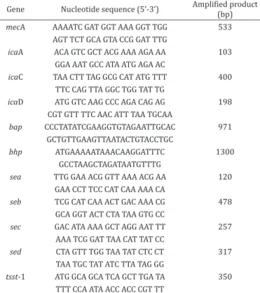
The presence of genes mecA (Murakami et al. 1991), icaA, icaC, icaD, bap and bhp (Cucarella et al. 2004, Arciola et al. 2001, Qin et al. 2007), sea, seb, sec, sed and tsst-1 (Johnson et al. 1991, Cunha et al. 2004) were assessed (Table 1).
Antimicrobial treatments were carried out before weaning, with two formulations of cloxacillin in a single dose: one in an oil-based formulation (100mg) and the other using a nanoparticulate structure (50mg), in water vehicle. The animals were treated after in vitro confirmation of susceptibility of the microorganisms towards the active ingredient. The sheep were randomly distributed into three experimental groups, while maintaining homogeneity of the groups according to weight, age and parity: G1 (n=21), composed of sheep with subclinical mastitis without intramammary antimicrobial treatment; G2 (n=19), sheep with subclinical mastitis treated with 100mg of intramammary cloxacillin–benzathine; and G3 (n=20), sheep with subclinical mastitis treated with 50 mg of nanoparticulate intramammary cloxacillin-benzathine, in accordance with the methodology described in patent WO2011150481A1 (Mosqueira et al. 2011).
was evaluated in combination with the SCC results, as previously mentioned.
The frequency distributions of the cases of subclinical mastitis were compared using the chi-square test. Significant values that were close to those of the significance reference were adjusted in accordance with Yates’s correction for continuity (p=0.05) (Sampaio 1998). The multiple correspondence analysis was carried out to determine the relationship between the classes of variables. It was also considered in this analysis the experimental three groups.
The experiment was approved by the Embrapa Southeast Livestock Ethics Committee on the Experiments with animals and register under the number PRT 04/2015.
RESULTS AND DISCUSSION
Table 2 shows the distribution of animals into the different
experimental groups before weaning, i.e. before treatment,
and also 15 and 30 days after the parturition of the subsequent
lactation. All the animals presented subclinical mastitis in one
of the mammary halves, while the other was healthy.
Three microorganisms isolated in the ewes before the
treatment in G2 were not recovered after storage under
frozen conditions, which made it impossible to conduct
analyses later on, to identify the species. As shown in Table 2,
15 days after parturition, the ewes of G3 treated with a half
of antibiotic dose presented greater occurrence of cure than
ewes treated with 100mg of cloxacillin-benzathine (G3)
and then the untreated sheep (G1) (p=0.0192). This finding
was probably due to the efficiency of the nanoparticulate
antimicrobial administered during the drying off, among
the sheep with subclinical mastitis. The nanoparticulate
system used in this experiment can deliver the antibiotic to
polymorph nuclear cell compartment (Mosqueira et al. 2011).
In addition, Brownian motion influence the small particle
diffusivity (Uma et al. 2011), that favored the mammary
biodistribution of the nonoestructured cloxacillin when
compared to oil
-
based cloxacillin.
Table 3 shows the species of CNS isolated before weaning the
distributed among the three experimental groups. The species of
greatest occurrence in the milk of these sheep with subclinical
mastitis before drying-off treatment were in agreement with
those isolated by other authors, also from subclinical cases
(Martins 2013, Pilon et al. 2014). Among the agents involved
in the infectious etiology of ovine subclinical mastitis, CNS
is the most prevalent agent and for this reason was chosen
for investigation in the present study. Veríssimo et al. (2010)
Table 1. Primers used in detection of genes relating to resistance to oxacillin and to encoding for biofilms, enterotoxins and the toxin responsible for toxic shock syndrome, in strains of
Staphylococcus spp. isolated from ovine milk
Gene Nucleotide sequence (5’-3’) Amplified product (bp)
mecA AAAATC GAT GGT AAA GGT TGG 533
AGT TCT GCA GTA CCG GAT TTG
icaA ACA GTC GCT ACG AAA AGA AA 103
GGA AAT GCC ATA ATG AGA AC
icaC TAA CTT TAG GCG CAT ATG TTT 400
TTC CAG TTA GGC TGG TAT TG
icaD ATG GTC AAG CCC AGA CAG AG 198
CGT GTT TTC AAC ATT TAA TGCAA
bap CCCTATATCGAAGGTGTAGAATTGCAC 971
GCTGTTGAAGTTAATACTGTACCTGC
bhp ATGAAAAATAAACAAGGATTTC 1300
GCCTAAGCTAGATAATGTTTG
sea TTG GAA ACG GTT AAA ACG AA 120
GAA CCT TCC CAT CAA AAA CA
seb TCG CAT CAA ACT GAC AAA CG 478
GCA GGT ACT CTA TAA GTG CC
sec GAC ATA AAA GCT AGG AAT TT 257
AAA TCG GAT TAA CAT TAT CC
sed CTA GTT TGG TAA TAT CTC CT 317
TAA TGC TAT ATC TTA TAG GG
tsst-1 ATG GCA GCA TCA GCT TGA TA 350
TTT CCA ATA ACC ACC CGT TT
Table 2. Distribution of sheep with subclinical mastitis into different groups according to whether treatment was
implemented, before weaning and 15 and 30 days after parturition
Periods
Groups
G1 G2 G3
N % N % N %
Before weaning 21 40.4 19 44.2 20 57.1
15 days post-partum 16a 30.8 13a 30.2 7b 20.0
30 days post-partum 15a 28.8 11a 25.6 8a 22.9
TOTAL 52 100.0 43 100.0 35 100.0
G1 = Sheep that did not receive intramammary antimicrobial, G2 = sheep that were treated with conventional antimicrobial (100mg of cloxacillin–benzathine), and G3 = sheep that were treated with nanoencapsulated antimicrobial (50mg of cloxacillin-benzathine). Values with different letters in the same line: p<0.05.
Table 3. Coagulase-negative Staphylococcus spp. isolated before weaning of sheep with subclinical mastitis
Species
Experimental groups
G1 G2 G3
N % N % N %
S. warneri 8 38.1 2 12.5 5 25.0
S. simulans 5 23.8 5 31.3 4 20.0
S. xylosus 4 19.0 2 12.5 2 10.0
S. epidermidis 2 9.5 3 18.8 4 20.0
S. lentus 1 4.8 - - -
-S. caprae 1 4.8 - - -
-S. haemolyticus - - 2 12.5 -
-S. hominis - - - - 1 5.0
S. cohnii - - - - 1 5.0
S. capitis - - - - 1 5.0
S. chromogenes - - 2 12.5 1 5.0
S. auricularis - - - - 1 5.0
TOTAL 21 100.0 16 100.0 20 100.0
reported a CNS occurrence rate in milk samples from Santa
Inês sheep of 64.3%. A similar rate was also reported by other
authors (Pereira et al. 2014, Santana et al. 2013). CNS can also
be isolated from animals with clinical mastitis (Lucheis et al.
2010). These microorganisms are considered to be colonizers
of the skin of the teat. Thus, any deficiency in managing these
animals can facilitate entry of these microorganisms into the
mammary gland (Laffranchi et al. 2001). Before weaning,
S. warneri
was the most prevalent species in G1 and G3.
In G2,
Staphylococcus simulans
presented greater occurrence,
followed by
S. epidermidis.
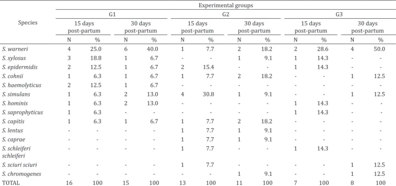
Table 4
shows the species of CNS identified in the sheep
15 and 30 days after parturition.
S. warneri
remained as the
species with greatest occurrence in G1 and G3, and in the latter,
even after the treatment at the end of the previous lactation.
In G2, 15 days after parturition,
S. simulans
was the species
with greatest occurrence, as it also was before treatment.
Table 5 presents the distribution of the
sec
gene in CNS
species before treatment, and also 15 and 30 days after
parturition. Among the enterotoxins genes investigated,
this was the only one identified. There are no reports in the
literature correlating the failure in the sheep mastitis treatment
with CNS strains and genes responsible for production of
enterotoxins or the toxin responsible for toxic shock syndrome.
On the other hand, a correlation has been made with public
health risks due to consumption of milk contaminated with
microorganisms that produce enterotoxins and the toxin
responsible for toxic shock syndrome (Mariano et al. 2007,
Nader Filho et al. 2007, Ferreira et al. 2014).
None of the microorganisms isolated presented the
mec
A
gene, which relates to resistance to oxacillin, and this was in
agreement with the findings of other authors (França et al.
2012, Silva 2012, Martins 2013). Other oxacillin resistance
mechanisms that are unrelated to
mec
A expression may be
present (Zafalon et al. 2012), but were not investigated in the
present study. The presence of homologous
mec
A genes or
other classes of penicillin-binding proteins may be related
to oxacillin resistance mechanisms (Mendonça et al. 2012).
In goats, microorganisms isolated from animals carrying
clinical mastitis presented greater virulence capacity than
microorganisms isolated from subclinical mastitis (Bezek
& Hull 1995). So, the lack of identification of the remaining
genes relating to production of enterotoxins may have been
because the samples were from sheep with subclinical and
not clinical mastitis.
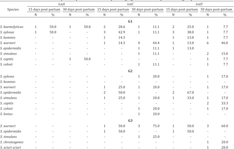
The distributions of the genes
ica
C,
ica
D and
bap
among
CNS species identified during weaning and the genes
ica
A,
ica
C
and
ica
D after parturition are presented in the Tables 6 and 7,
respectively. All the species of CNS studied were negative for
the genes
ica
A and
bhp
immediately before the drying-off
period. However, the gene icaC was present in CNS specimens
isolated from sheep from G1 and G3, while the genes
ica
D
and
bap
were found in CNS isolated from sheep distributed
in all three groups. CNS identified 15 and 30 days after the
parturition of the subsequent lactation did not present the
bap
gene.
Biofilm production can contribute towards maintaining
CNS within the mammary gland. It can be encoded by the
genes
ica
A,
ica
C,
ica
D,
bap
and
bhp
. The genes of the
ica
operon are responsible for synthesizing the production
of biofilm. Strains of CNS carrying
ica
genes can produce
biofilms, which may be related to the difficulty of attaining
a cure for the mammary gland after treatment (Arciola et al.
2001). Even with inactivation or absence of the
ica
operon,
Table 4. Coagulase-negative Staphylococcus spp. isolated from treated and untreated sheep, 15 and 30 days after parturition
and lactation subsequent to treatment
Species
Experimental groups
G1 G2 G3
15 days post-partum
30 days post-partum
15 days post-partum
30 days post-partum
15 days post-partum
30 days post-partum
N % N % N % N % N % N %
S. warneri 4 25.0 6 40.0 1 7.7 2 18.2 2 28.6 4 50.0
S. xylosus 3 18.8 1 6.7 - - 1 9.1 1 14.3 -
-S. epidermidis 2 12.5 1 6.7 2 15.4 - - 1 14.3 -
-S. cohnii 1 6.3 1 6.7 1 7.7 2 18.2 - - 1 12.5
S. haemolyticus 2 12.5 1 6.7 - - -
-S. simulans 1 6.3 2 13.0 4 30.8 1 9.1 - - 1 12.5
S. hominis 1 6.3 2 13.0 - - - - 1 14.3 -
-S. saprophyticus 1 6.3 - - - 1 14.3 -
-S. capitis 1 6.3 1 6.7 1 7.7 2 18.2 - - -
-S. lentus - - - - 1 7.7 1 9.1 - - -
-S. caprae - - - - 1 7.7 1 9.1 - - -
-S. schleiferi schleiferi
- - - - 1 7.7 - - 1 14.3 -
-S. sciuri sciuri - - - - 1 7.7 - - - - 1 12.5
S. chromogenes - - - 1 9.1 - - 1 12.5
TOTAL 16 100 15 100 13 100 11 100 7 100 8 100
Table 6. Distributions of the genes icaC, icaD and bap among coagulase-negative Staphylococcus spp. identified in sheep with
subclinical mastitis immediately before the drying-off period
Species icaC icaD bap
N % N % N %
G1
S. epidermidis 1 33.3 2 33.3 -
-S. xylosus 1 33.3 - - 1 100.0
S. simulans 1 33.3 1 17.0 -
-S. warneri - - 3 50.0 -
-G2
S. epidermidis - - -
-S. xylosus - - - - 1 100.0
S. simulans - - 1 50.0 -
-S. warneri - - 1 50.0 -
-G3
S. epidermidis 1 33.3 - - 1 100.0
S. xylosus - - -
-S. simulans 2 67.0 3 37.5 -
-S. warneri - - 4 50.0 -
-S. chromogenes - - 1 12.5 -
-G1 = Control group, composed of sheep that did not receive intramammary antimicrobials, G2 = sheep that were treated with conventional antimicrobials (100mg of cloxacillin–benzathine), and G3 = sheep that were treated with nanoencapsulated antimicrobials (50mg of cloxacillin-benzathine). icaC, icaD and bap = genes responsible for biofilm formation.
Table 5. Distribution of the sec gene in coagulase-negative Staphylococcus identified during weaning and after treatment at
15 and 30 days after parturition and subsequent lactation
Species
Experimental groups
G1 G2 G3
N % N % N %
Pretreatment
S. warneri 6 60.0 - - 1 33.3
S. simulans 3 30.0 1 50.0 -
-S. xylosus 1 10.0 - - 1 33.3
S. epidermidis - - 1 50.0 -
-S. chromogenes - - - - 1 33.3
15 days post-partum
S. warneri 2 100.0 - - 1 50.0
S. simulans - - 1 100.0 -
-S. epidermidis - - - - 1 50.0
30 days post-partum
S. warneri 2 50.0 1 25.0 4 57.1
S. epidermidis 1 25.0 - - 1 14.3
S. simulans 1 25.0 - - 1 14.3
S. cohnii - - 1 25.0 -
-S. lentus - - 1 25.0 -
-S. xylosus - - 1 25.0 -
-S. sciuri sciuri - - - - 1 14.3
G1 = Control group, composed of sheep that did not receive intramammary antimicrobials, G2 = sheep that were treated with conventional antimicrobials (100mg of cloxacillin–benzathine), and G3 = sheep that were treated with nanoencapsulated antimicrobials (50mg of cloxacillin-benzathine).
strains of
bap
-positive
S. aureus
continued to present
in vitro
biofilm synthesis (Cucarella et al. 2004), thereby becoming
10 to 1,000 times more resistant to antimicrobials than free
cells (Amorena et al. 1999). Genes
ica
C and
ica
D were found
in a greater number of species before the drying-off period.
Six untreated sheep presented
S. warneri
with virulence
factor genes before weaning. All of them continued to present
subclinical mastitis during the subsequent lactation. The multiple
correspondence analysis among the effects of treatment
before drying the sheep and the presence of microorganisms
and genes demonstrated that this species of staphylococci
when presented the
sec
and
icaD
genes was related to the
absence of spontaneous recovery of sheep, i.e. with the
permanence of cases of the disease when not effected the
treatment. In G2, three sheep presented reinfection 30 days
after parturition, after being treated with 100mg of
cloxacillin-benzathine. It was established
S. xylosus
carrying the gene
bap
was related with the reinfection. Bacteria inside biofilms
are subject to less action by neutrophils, which facilitates
bacterial growth for long periods (Rasmussen & Givskov
2006). Consequently, the antimicrobial may be inefficient.
Then, sheep that presented CNS strains carrying genes
responsible for formation of biofilm during the pre-partum
period can continue to present subclinical mastitis during
the drying off period and over the next lactation. In the sheep
treated with nanoparticulate antimicrobials, the cases of
lack of cure by 15 and 30 days after parturition occurred
in mammary halves infected by
S. epidermidis, S. warneri
and
S. simulans
. The absence of cure mastitis in this group
of animals was not related to the bacterial species or their
genes, probably by this type of treatment being more effective
than the use of conventional cloxacillin and the absence of
treatment, in the 15 days prepartum.
CONCLUSIONS
The identification of several CNS species in the milk of sheep
with mastitis is consistent with the widespread occurrence of
these microorganisms in the etiology of subclinical mastitis
in ewes. These bacteria have genes for virulence factors that
can negatively interfere with disease control methods.
The knowledge about the ability of CNS remaining in
the mammary halves of sheep, even after intramammary
treatment, provides information regarding the epidemiology
of the disease and contributes towards adoption of measures
for future control.
Acknowledgements.- This study was funded by grants of the Research Support Foundation of the State of São Paulo (Fundação de Amparo à Pesquisa do Estado de São Paulo, FAPESP) (FAPESP procedural no. 2012/23044-0).
Our acknowledgements to Danilo Flávio Moraes Riboli by collaboration in
laboratory activities for the identification of staphylococci.
Table 7. Distribution of the genes icaA, icaC and icaD in species of coagulase-negative Staphylococcus identified
15 and 30 days after parturition in sheep belonging to the three experimental groups
Species
icaA icaC icaD
15 days post-partum 30 days post-partum 15 days post-partum 30 days post-partum 15 days post-partum 30 days post-partum
N % N % N % N % N % N %
G1
S. haemolyticus 1 50.0 1 50.0 2 28.6 1 11.1 2 25.0 1 7.7
S. xylosus 1 50.0 - - 3 42.9 1 11.1 3 38.0 1 7.7
S. hominis - - - - 1 14.3 1 13.0 1 7.7
S. warneri - - - - 1 14.3 4 44.4 1 13.0 6 46.0
S. epidermidis - - - 1 11.1 1 13.0
S. simulans - - - 1 11.1 - - 2 15.0
S. capitis - - 1 50.0 - - - 1 7.7
S. cohnii - - - 1 11.1 - - 1 7.7
G2
S. xylosus - - - 1 20.0 - - 1 17.0
S. hominis - - -
-S. warneri - - - - 1 25.0 1 20.0 - - 1 17.0
S. epidermidis - - - - 2 50.0 - - 2 67.0 -
-S. simulans - - - - 1 25.0 1 20.0 1 33.0 1 17.0
S. capitis - - - 2 33.3
S. cohnii - - - 1 20.0 - - 1 17.0
S. lentus - - - 1 20.0 - - -
-G3
S. warneri - - - - 1 50.0 3 75.0 1 50.0 3 60.0
S. epidermidis - - - - 1 50.0 - - 1 50.0 -
-S. simulans - - - 1 25.0 - - -
-S. chromogenes - - - 1 20.0
S. sciuri sciuri - - - 1 20.0
REFERENCES
Amorena B., Gracia E., Monzón M., Leiva J., Oteiza C., Pérez M., Alabart J.L. & Hernández-Yago J. 1999. Antibiotic susceptibility assay for Staphylococcus
aureus in biofilms developed in vitro. J. Antimicrob. Chemother. 44(1):43-55.
http://dx.doi.org/10.1093/jac/44.1.43. PMid:10459809.
Arciola C.R., Baldassarri L. & Montanaro L. 2001. Presence of icaA and icaD genes and slime production in a collection of staphylococcal strains from catheter-associated infections. J. Clin. Microbiol. 39(6):2151-2156. PMid:11376050. Balaban N. & Rasooly A. 2001. Analytical chromatography for recovery of small amounts of staphylococcal enterotoxins from food. Int. J. Food Microbiol. 64(1/2):33-40. http://dx.doi.org/10.1016/S0168-1605(00)00439-6. PMid:11252509.
Bezek D.M. & Hull B.L. 1995. Peracute gangrenous mastitis and cheilitis associated with enterotoxin-secreting Staphylococcus aureus in a goat. Can. Vet. J. 36(2):106-107. PMid:7728725.
Bolsanello R.X., Hartman M., Domingues P.F., Mello Júnior A.Z. & Langoni H. 2009. Etiology of mastitis in Bergamacia sheep submitted in milking machine, raised in farm at Botucatu, SP. Vet. Zootec. 16:221-227.
Couto I., Pereira S., Miragaia M., Sanches I.S. & Lencastre H. 2001. Identification of clinical staphylococcal isolates from humans by Internal Transcribed Spacer PCR. J. Clin. Microbiol. 39(9):3099-3103. http://dx.doi.org/10.1128/ JCM.39.9.3099-3103.2001. PMid:11526135.
Cucarella C., Tormo M.A., Ubeda C., Trotonda M.P., Monzon M., Peris C., Amorena B., Lasa I. & Penades J.R. 2004. Role of biofilm: associated protein bap in the pathogenesis of bovine Staphylococcus aureus. Infect. Immun. 72(4):2177-2185. http://dx.doi.org/10.1128/IAI.72.4.2177-72(4):2177-2185.2004. PMid:15039341. Cunha M.L.R.S., Sinzato Y.K. & Silveira L.V.A.. 2004. Comparision of methods for identification of coagulase-negative staphylococci. Mem. Inst. Oswaldo Cruz 99(8):855-860. http://dx.doi.org/10.1590/S0074-02762004000800012. PMid:15761602.
Diekema D.J., Pfaller M.A., Schmitz F.J., Smayevsky J., Bell J., Jones R.N. & Beach M. 2001. Survey of infections due to Staphylococcus species: frequency of occurrence America, Europe, and the Western Pacific region for the SENTRY Antimicrobial Surveillance Program, 1997-1999. Clin. Infect. Dis. 32(Suppl.2):114-132. http://dx.doi.org/10.1086/320184. PMid:11320452. Ergun Y., Aslantas O., Kireçci E., Ozturk F., Ceylan A. & Boyar Y. 2012. Antimicrobial susceptibility, presence of resistant genes and biofilm formation is coagulase negative staphlococci isolated from subclinical sheep mastitis. Kafkas Univ. Vet. Fak. Derg. 18:449-456.
Ferreira D.H., Carvalho M.G.X., Nardelli M.J., Sousa F.G.C. & Oliveira C.J.B. 2014. Occurrence of enterotoxin-encoding genes in Staphylococcus aureus causing mastitis in lactating goats. Pesq. Vet. Bras. 34(7):633-636. http://dx.doi. org/10.1590/S0100-736X2014000700004.
França C.A., Peixoto R.M., Cavalcante M.B., Melo N.F., Oliveira C.J.B., Veschi J.L.A., Mota R.A. & Costa M.M. 2012. Antimicrobial resistence of Staphylococcus
spp. from small ruminant mastitis in Brazil. Pesq. Vet. Bras. 32(8):747-753. http://dx.doi.org/10.1590/S0100-736X2012000800012.
Harmon R.J., Eberhart R.J., Jasper D.E., Langlois B.E. & Wilson R.A. 1990. Microbiological Procedures for the Diagnosis of Bovine Udder Infections. 3rd ed. National Mastitis Council, Arlington. p.4-6.
Johnson W.M., Tyler S.D., Ewan E.P., Ashton F.E., Pollard D.R. & Rozee K.R. 1991. Detection of genes for enterotoxins, exfoliative toxins, and toxic shock syndrome toxin 1 in Staphylococcus aureus by the polymerase chain reaction. J. Clin. Microbiol. 29(3):426-430. PMid:2037659.
Laffranchi A., Müller E.E., Freitas J.C., Pretto-Giordano L.G., Dias J.A. & Salvador R. 2001. Aetiology of mammary infections in primiparous during the first four months of lactation. Ciência Rural 31(6):1027-1032. http://dx.doi. org/10.1590/S0103-84782001000600018.
Lucheis S.B., Hernandes G.S. & Troncarelli M.Z. 2010. Microbiological monitoring of the region of ovine mastitis Bauru, SP. Arqs Inst. Biológico, São Paulo, 77:305-403.
Mariano F.A., Folly M.M., Teixeira G.N., Carmo L.S. & Vieira-Da-Mota O. 2007. Enterotoxins production by Staphylococcus isolated from milk of goats from Rio de Janeiro state. Revta Bras. Ciênc. Vet. 14(2):105-110.
Martins K.B. 2013. Caracterização do perfil clonal, fatores de virulência e determinação da resistência em Staphylococcus spp isolados de leite ovino. Dissertação de Mestrado, Programa de Biologia Geral e Aplicada, Instituto de Biociências, Universidade Estadual Paulista, Botucatu, SP. 96p. McDougall S., Murdough P., Pankey W., Delaney C., Barlow J. & Scruton D.
2001. Relationship among somatic cell count, California mastitis test, impedance and bacteriological status of milk in goats and sheep in early lactation. Small Rumin. Res. 40(3):245-254. http://dx.doi.org/10.1016/ S0921-4488(01)00185-7. PMid:11323209.
Mendonça E.C.L., Marques V.F., Melo D.A., Alencar T.A., Coelho I.S., Coelho S.M.O. & Souza M.M.S. 2012. Phenogenotypical characterization of antimicrobial resistence in Staphylococcus spp. isolated from bovine mastitis. Pesq. Vet. Bras. 32(9):859-864. http://dx.doi.org/10.1590/S0100-736X2012000900008. Mosqueira V.C.F., Brandão H.M. & Araujo R.S. 2011. Patent No WO2011150481A1. Nanoparticulate composition containing antibiotics for intramammary administration in animals. Wipo (World Intellectual Property Organization), Genebra.
Murakami K., Minamide W., Wada K., Nakamura E., Teraoka H. & Watanabe S. 1991. Identification of methicillin-resistant strains of staphylococci by polymerase chain reaction. J. Clin. Microbiol. 29(10):2240-2244. PMid:1939577. Nader Filho A., Ferreira L.M., Amaral L.A., Rossi Junior O.D. & Oliveira R.P. 2007. Production of enterotoxins and toxic shock syndrome toxin by Staphylococcus
aureus strains isolated from bovine mastitis. Arq. Bras. Med. Vet. Zootec.
59(5):1316-1318. http://dx.doi.org/10.1590/S0102-09352007000500032. Pengov A. 2001. The role of coagulase-negative Staphylococcus spp. and
associated somatic cell counts in the ovine mammary gland. J. Dairy Sci. 84(3):572-574. http://dx.doi.org/10.3168/jds.S0022-0302(01)74509-2. PMid:11286408.
Pereira P.F.V., Stotzer E.S., Pretto-Giordano L.G., Müller E.E. & Lisbôa J.A.N. 2014. Fatores de risco, etiologia e aspectos clínicos da mastite em ovelhas de corte no Paraná. Pesq. Vet. Bras. 34(1):1-10. http://dx.doi.org/10.1590/ S0100-736X2014000100001.
Pilon L.E., Zafalon L.F., Santana R.C.M., Fim Junior G.A., Manieri F.Z. & Lopes N.S.S. 2014. Coagulase-negative Staphylococcus species isolated from the mammary gland of sheep with subclinical mastitis. Revta Bras. Hig. Sanid. Anim. 8(5):34-39. http://dx.doi.org/10.5935/1981-2965.20140038. Qin Z., Yang X., Yang L., Jiang J., Ou Y., Molin S. & Qu D. 2007. Formation and
properties of in vitro biofilms of ica-negative Staphylococcus epidermidis
clinical isolates. J. Med. Microbiol. 56(Pt 1):83-93. http://dx.doi.org/10.1099/ jmm.0.46799-0. PMid:17172522.
Rasmussen T.B. & Givskov M. 2006. Quorum-sensing inhibitors as anti-pathogenic drugs. Int. J. Med. Microbiol. 296(2-3):149-161. http://dx.doi. org/10.1016/j.ijmm.2006.02.005. PMid:16503194.
Sampaio I.B.M. 1998. Estatística Aplicada à Experimentação Animal. Fundação de Ensino e Pesquisa em Medicina Veterinária e Zootecnia, Belo Horizonte. p.108-117.
Santana R.C.M., Zafalon L.F., Esteves S.N., Tanaka E.V., Pilon L.E. & Massa R. 2013. Occurrence of etiologic agentes causing subclinical mastites in Morada Nova and Santa Inês ewes. Ars Vet. 29(3):148-152. http://dx.doi. org/10.15361/2175-0106.2013v29n3p148-152.
Silva G.V. 2012. Evaluation of the species and antimicrobial susceptibility profile
of Staphylococcus isolated from sheep milk. Dissertação de Mestrado, Programa
de Ciências, Universidade Federal Rural do Rio de Janeiro, Seropédica, RJ. 89p. Uma B., Swaminathan T.N., Radhakrishnan R., Eckmann D.M. & Ayyaswamy
P.S. 2011. Nanoparticle Brownian motion and hydrodynamic interactions in the presence of flow fields. Phys Fluids 23(7):73602-7360215. http:// dx.doi.org/10.1063/1.3611026. PMid:21918592.
Verissímo C.J., Zafalon L.F., Otsuk I.P. & Nassar A.F.C. 2010. Damages caused by mastitis in Santa Inês Brazilian sheep breed. Arqs Inst. Biológico, São Paulo, 77:583-591.