UNIVERSIDADE TÉCNICA DE LISBOA
FACULDADE DE MOTRICIDADE HUMANA
Optimizing patient selection for Cardiac
Resynchronization Therapy: The role of
cardiopulmonary exercise testing
Dissertação elaborada com vista à obtenção do Grau de Mestre em Exercício e Saúde
Orientador: Professora Doutora Maria Helena Santa-Clara Pombo Rodrigues
Júri:
Presidente
Professor Doutor Fernando Manuel da Cruz Duarte Pereira Vogais
Professora Doutora Maria Helena Santa-Clara Pombo Rodrigues Doutora Ana Maria Ferreira das Neves de Abreu
Maria Rita da Silva Alexandre Pinto 2012
2
Acknowledgments
I would like to express my gratitude to my supervisor Professor Helena Santa Clara for all the support and clear guidance that provided me through all my work. Her enthusiasm, knowledge and motivation were fundamental for me and made me better understand how to design, organize and develop a scientific project.
To Dr. Ana Abreu from Sta Marta Hospital who made possible all the practical component of my thesis. She was crutial not only for me to get access to the patients but for her enthusiasm and support that provided me through all my work. Also all the team in Sta Marta Hospital, including technicians and nurses, were essential for the good outcome of my work.
I also want to thank Margarida Carrolo and Mafalda Gonçalves for all their assistance and support on the elaboration of this scientific project.
I would like to thank all of my closer friends for their help, support and interest at this journey since the beginning until the end.
Last but not the least, I would like to thank my family, specially my sister Ana, my brother Fausto, my father and my mother. Thank you for supporting me all the time.
3
List of abbreviations
ACC: American College of Cardiology AHA: American Heart Association AV: atrial ventricular
BMI: body mass index Bpm: beats per minute
CARE-HF: cardiac resynchronization-heart failure CHF: chronic heart failure
COMPANION: comparison of medical therapy pacing and defibrillation in heart failure
CPET: cardiopulmonary exercise testing CRT: cardiac resynchronization therapy
CRT-P: cardiac resynchronization therapy pacemaker DHF: diastolic heart failure
HF: heart failure HR: heart rate
ICD: implantable cardioverter defibrillator
LBBB: left bundle branch block LV: left ventricular
LVEF: left ventricular ejection fraction LVESV: left ventricular end systolic volume
MIRACLE: multicenter insync randomized clinical evaluation MUSTIC: multisite simulation in cardiomyopathies
NASPE: North American Society for Pacing and Electrophysiology NYHA class: New York Heart Association class
RER: respiratory exchange ratio SBP: systolic blood pressure SCD: sudden cardiac death SHF: systolic heart failure
VAT: ventilator anaerobic threshold
VE/VCO2: volume of expired gas to carbon dioxide production
VO2: oxygen consumption
4
Contents
1. ABSTRACT ... 6
2. THEME’S PRESENTATION ... 7
2.1 INTRODUCTION ... 7
2.2 OBJECTIVES’ OF THE STUDY ... 8
2.3 LIMITATIONS ... 9
3 LITERATURE REVIEW ... 11
3.1 HEART FAILURE ... 11
3.2 CARDIAC RESYNCHRONIZATION THERAPY ... 13
3.3 CARDIOPULMONARY EXERCISE TESTING ... 20
HEART RATE RESPONSE ... 21
OXYGEN UPTAKE ... 23
SLOPE OF THE VENTILATORY RESPONSE ... 25
VENTILATORY ANAEROBIC THRESHOLD ... 26
4 METHODOLOGY ... 27 4.1 INTRODUCTION ... 27 4.2 STUDY DESIGN ... 27 4.3 PARTICIPANTS ... 27 RESPONSE CRITERIA... 28 4.4 VARIABLES IN STUDY ... 28 4.4.1 DEPENDENT VARIABLES ... 28
EQUIPMENTS AND PROTOCOLS OF ASSESSMENT ... 28
4.4.2 INDEPENDENT VARIABLES ... 30
4.5 STATISTICAL ANALYSIS ... 30
5 RESULTS AND DISCUSSION ... 32
5
7 FUTURE RECOMMENDATIONS ... 42 8 REFERENCES ... 44
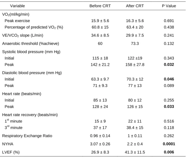
Table 1: NYHA functional classification (Severity based on symptoms and physical activity)31 ... 17 Table 2: Seventeen Different Response Criteria Identified From the Fornwalt (2010) review6 ... 18 Table 3: Clinical and demographic characteristics at baseline for the sample in the study (n=15) ... 32 Table 4: Comparisons of baseline demographic, clinical, and functional characteristics in responders vs. non responders to CRT considering echocardiographic parameters ... 33 Table 5: Comparisons of baseline demographic, clinical, and functional characteristics in responders vs. non responders to CRT considering echocardiographic and clinical parameters (Combined) ... 34 Table 6: Patient characteristics before and after CRT ... 37
6
1. ABSTRACT
Background: Cardiac resynchronization therapy (CRT) is an established treatment modality for moderate to severe heart failure (HF) but 30–40% of patients treated with CRT do not experience clinical improvement. Purpose: the aim of this study was to identify predictors of response to CRT, in two different definitions of responders, by using the cardiopulmonary exercise testing (CPET) before CRT implantation. In definition A, responders were defined as ≥15% improvement in left ventricular ejection fraction (LVEF); in definition B combined parameters were defined as ≥5% improvement in LVEF and ≤1 level NYHA classification. Methods: this is a prospective observational study of 15 HF patients undergoing CRT. Clinical CPET and echocardiography assessment using standard methods were performed at baseline and 5 months. Results: the number of patients classified as responders in definition A was 9 (60%) and 6 (40%) as non-responders; the number of responders in definition B was 11 (73.3%) and 4 (26.7%) as non-responders at 5 months after CRT. The responders according to definition A did not present any statistically significant difference. According to definition B, the heart rate (HR) response during CPET was higher in non-responders: HR peak (157±13bpm vs. 118±18bpm, p<0.05) and HR recovery at minute 3 (54±13bpm vs. 31 ± 14bpm, p<0.05). Overall, the responders were older (68±9years vs. 55±9years, p<0.05). Conclusions: baseline measurements of CPET may be utilized to identify patients that benefit from CRT. The use of combined criteria is a better predictor than LVEF alone.
KEY WORDS: HEART FAILURE, CARDIAC RESYNCHRONIZATION THERAPY, RESPONDERS, PROGNOSIS, CARDIOPULMONARY EXERCISE TESTING, EXERCISE CAPACITY, OXYGEN UPTAKE, HEART RATE RESPONSE, VENTILATORY THRESHOLD, SLOPE OF THE VENTILATORY RESPONSE
7
2. THEME’S PRESENTATION 2.1 INTRODUCTION
Approximately 1–2% of the adult population in the western world has heart failure (HF), with the prevalence increasing sharply from 1% in 40-year-old individuals to 10% above the age of 75 years1. There are many causes of HF, and these vary in different parts of the world. The overall prevalence of HF in Portugal was slightly higher than other European studies2. The HF prevalence increases markedly with age in both sexes and tends to be slightly higher in men up to the age of 70. In women, it continues to increase with age and becomes greater than the prevalence for men in the age group of 70–79 years old3.
HF is the leading cause of death and hospitalization in most Western countries in patients over 65 years of age4. In recent years, cardiac pacemakers have been modified in an effort to correct ventricular dyssynchrony. This treatment is referred to as cardiac resynchronization therapy (CRT).
CRT has been used extensively over the last years in the therapeutic management of patients with end-stage HF5. The CRT, delivered via atrial-synchronous biventricular pacing, has emerged as an effective treatment for moderate-to-severe HF patients with ventricular dyssynchrony. At present, the selection criteria include moderate to severe HF (New York Heart Association functional class III or IV), left ventricular ejection fraction ≤ 35%, and wide QRS complex (>120 ms)5. However, current guidelines do not adequately identify responders to CRT; approximately 30% to 40% of patients treated with CRT do not respond or improve with treatment6. The identification of non responders to CRT may be also of clinical interest. This therapy requires high costs and has potential related complications that may be avoided in patients who will not, for an instance, have clinical benefit7.
Predicting whether a patient will benefit, or respond, to CRT has been the focus of more than 500 publications during the last 5 years. However, the definition of responder to CRT varies widely between studies, and numerous criteria to define a positive response to CRT exist in the literature6, 8.
8
Improvement in clinical end points (symptoms, exercise and functional capacities, quality of life) and echocardiography end points (systolic function, left ventricular size, mitral regurgitation) have been reported after CRT, with a reduction in hospitalizations for decompensated HF and an improvement in survival5, 7, 9-16. However, detailed analysis of improvement in functional capacity after CRT is still lacking12.
Cardiopulmonary exercise testing (CPET) with respiratory gas analyses is a standardized approach for objectively documenting functional capacity17 that provides noninvasive objective measures for cardiopulmonary reserve and is thus suitable for evaluation, risk stratification and control of treatment effects18.
Peak oxygen consumption (VO2peak) measured during maximal exercise
testing provides an objective assessment of functional capacity in patients with HF and an indirect assessment of cardiovascular reserve. Previous studies have suggested that this measurement is a good short-term predictor of mortality19.
Mancini et al.19, Stelken et al.20 and others observational studies21, 22 have demonstrated that the short-term prognosis of patients with a VO2peak ≤ 14
kg/ml/min is markedly impaired compared with heart transplant patients followed up for a similar duration.
Patients with a baseline VO2peak < 14 ml/kg/min regularly benefit from CRT
during the first year of treatment23. These findings have not yet been included in the current Guidelines for the Implantation of Permanent Pacemakers by the ACC/AHA/ NASPE24, while the German “Statement on cardiac resynchronization”25
already recommends a baseline VO2peak < 14 ml/kg/min as
one criterion to indicate CRT.
2.2 OBJECTIVES’ OF THE STUDY
The aim of this study was to identify predictors of response to CRT, in two different definitions of responders, by using the CPET before CRT implantation.
The secondary aim of this study was to evaluate VO2peak, VE/VCO2 slope,
HR response, anaerobic threshold, NYHA class symptom and LVEF, immediately before and 5 months after CRT implantation, to study the
9
ventilatory and haemodynamic response evolution and to assess improvement in functional and echocardiography variables.
In this study, the criteria used to define responders to CRT will be considered according to 2 different ways found in the literature review. The first way of defining responders is based on echocardiography, through the increase of LVEF ≥ 15%26, 27
. The second way combines echocardiography and clinical setting, through the increase of LVEF ≥ 5% and decrease of NYHA ≥ 1, respectively28.
The relevance of this study is to have reproducible inclusion criteria parameters that are crucial for a reliable evaluation of the CRT response. Considering the high costs and non responder rates of about one-third of the patients, a careful selection of patients prior to CRT is crucial.
This document is composed by five chapters; each chapter will have a short introduction that explains the theme in question. The theme’s presentation (2) is a brief description of the state of knowledge of the main theme. The emerging of some questionable issues justifies the purpose and objectives of the present study and also the study’s limitation. The literature review (3) comes to specify the theoretical background followed by the methodology section (4) that describes all the steps of this study, from the sample selection and the instruments used to collect data to the statistical analysis. The results and discussion are considered together for better critical analysis and evaluation of the different variables. In the same chapter, it will be presented the future research perspectives. The last chapter (6), contains the conclusions of this study.
2.3 LIMITATIONS
The following limitations were considered:
1. Small study sample size, limited power to detect significant differences in the studied parameters.
2. Peak SBP measurements during exercise are also influenced by technique and sampling frequency. Vasodilator drug therapy may also limit exercise SBP response. In the present study, not all
10
antihypertensive drug therapy data was collected at the time of exercise testing.
3. The fact that these patients have skeletal muscle pathology as a major contributor to exercise intolerance, fatigue, and exertional dyspnea in chronic heart failure, restricts the clinical value of the variables of the CPET, like VAT and VO2/VCO2 slope.
4. In most patients only one baseline exercise test was performed, and an improvement in exercise time and VO2 may occur with familiarization of
the technique29. Other potential limitations of VO2peak also must be
considered.
5. There are many different methods to define a positive response to CRT in the literature and poor agreement was found amongst them. Nevertheless, in this study two criteria to assess different types of responders were considered. For the reasons previously mentioned, a question can be formulated for continuing the research work to find an answer: which method should be used in the future to determine whether a patient could benefit or not from CRT?
11
3 LITERATURE REVIEW
3.1 HEART FAILURE
Chronic heart failure (CHF) is a clinical syndrome resulting from a structural or functional cardiac disorder and it could be defined by systolic dysfunction, diastolic dysfunction, or both which usually involves an assessment of the patient’s ejection fraction30
. It usually begins after an initial event that produces a decline in pumping capacity of the ventricle. This syndrome manifests primarily as dyspnea, fatigue, fluid retention, such as pulmonary congestion or ankle swelling, and decreased exercise tolerance31.
Systolic dysfunction and diastolic dysfunction, both describe an abnormal mechanical property, while systolic heart failure (SHF) and diastolic heart failure (DHF) describe a clinical syndrome30.
The SHF reflects a fundamental weakness of the pump and thus the inability to deliver sufficient cardiac output at an adequate mean arterial pressure. The failing heart often exhibits both major decrements in resting systolic function and also limitations of systolic reserve required for individuals to perform normal activities of daily living and exercise. The systolic dysfunction refers to impaired ventricular contractions due to the loss of myocardium secondary to myocardial infarction or loss of contractility, and the underlying mechanisms are numerous. Patients with HF and a low left ventricular ejection fraction (LVEF), usually < 40–45%, are classified as having systolic dysfunction15, 30
.
In contrast, in patients with DHF, the dysfunction occurs when the ventricular chamber is unable to accept an adequate volume of blood during diastole and the sufficient volumes to maintain an appropriate stroke volume at rest and during exercise. The functional abnormalities leading to DHF includes abnormal ventricular relaxation and filling, decreased LV suction, and/or an increase in ventricular stiffness. Diastolic dysfunction refers to a condition in which abnormalities in mechanical function are present during diastole. It is characterized by an increased resistance to the filling of one or both ventricles, elevated diastolic pressure in the ventricles, and reduced ventricular compliance. Patients with symptoms and exam findings consistent with HF, but with a preserved ejection fraction are often said to have diastolic dysfunction4,32.
12
The demographic characteristics present in patients with DHF differ significantly from those with SHF. Patients with DHF are older, often female, have hypertensive heart disease, and are less likely to have ischemic heart disease compared to patients with SHF. Diastolic dysfunction is estimated to be the principal etiology in 40% or more of the estimated 500.000 new cases of HF each year30.
HF is a final common pathway of all diseases of the heart and is a major cause of morbidity and mortality. It is a complex syndrome with numerous risk factors and determinants of outcomes. Approximately 4.9 million Americans carry the diagnosis of HF33 and about 550.000 new cases occur each year in the USA34. Reports from several countries suggest that approximately 1–2% of the total healthcare budget is spent on the management of HF35.
In Portugal the overall prevalence of HF was markedly higher than other European studies and increases sharply with age2. HF with LV systolic dysfunction is more frequent in males below the age of 80 years and with preserved LV systolic function affects mainly older females3.
HF remains a large medical and epidemiological problem31, and the number of HF hospitalizations has risen more than a million per year over the past decade, accounting for at least 20% of all admissions for persons older than 65 years16.
At the cellular level, is caused by changes in the biology of the cardiac myocyte together with a progressive loss of cardiac myocytes. The loss of myocytes may be focal (e.g., myocardial infarction), or diffuse (e.g., viral infection, hemodynamic overload, genetic abnormalities).
Thus HF is the common clinical syndrome caused by any of a diverse group of injurious stimuli sufficient to produce myocardial insufficiency36. Abnormal impulse generation and propagation is frequently observed in these patients. Both functional and structural alterations (cardiac remodeling) are responsible for such abnormalities32.
Cardiac remodeling commonly refers to persistent changes in the properties of myocardium in response to abnormal external stresses. Although most notably cardiac remodeling occurs in the setting of structural heart diseases such as myocardial infarction, hypertrophy, and HF, it may also occur in the absence of anatomic dysfunction, as is the case during abrupt changes in heart
13
rate and/or activation sequence. Indeed, remodeling is a prominent feature of atrial fibrillation and flutter, ventricular pacing or intrinsic conduction delays and sustained tachycardia32.
Left bundle branch block (LBBB) results from block or conduction delays in any of several sites of the left-sided intraventricular conduction system, including the main left bundle branch or its subdivisions or, less commonly, within the fibers of the distal His bundle. The result is an abnormal and slow pattern of electrical activation within the LV due to conduction through the working myocardium.
LBBB usually appears in patients with underlying heart diseases, typically in patients with dilated cardiomyopathy of any etiology32.
It has long been recognized that discoordinate cardiac contraction itself reduces the systolic performance of the chamber, and recent developments in therapies to resynchronize contractions have shown this to be a valuable target for HF treatment. Conduction disease at or above the atrial ventricular (AV) node affects chronotropic competence and effective preload (and left atrial pressure). Both short and excessively long AV delays the reducing of net LV filling. LBBB induces discoordinate contraction. Cardiac discoordination induced by LBBB or right ventricular pacing depresses systolic function, increasing the end-systolic volumes at a given pressure, prolongs isovolumic relaxation, and has been coupled to the widening of the QRS complex.
Significant progress has been made to identify the major risk factors and the population patterns of HF and associated trends. However the prognosis remains poor, with mortality data comparable with data from the worst forms of malignant disease. Therefore, it deserves adequate planning for investigation, education, prevention and treatment.
3.2 CARDIAC RESYNCHRONIZATION THERAPY
CRT improves HF outcomes37 and has been used extensively over the last years in the therapeutic management of patients with end-stage HF5.
Approximately 40 years ago, the first descriptions of the short-term haemodynamic effects of left or of simultaneous right and left ventricular stimulation were published38-40. Cardiac pacing as an adjunct therapy for HF
14
began to be the subject of scientific research at the start of the 1990’s. The CRT began in 1994, when Cazeau et al.41, in France, and Bakker et al.42, in Netherlands, described the first cases of atrio-biventricular pacemakers implanted in patients with severe CHF and no conventional indication for cardiac pacing.
CRT is an effective treatment for patients with moderate to severe HF and LV systolic dysfunction if they have a prolonged QRS interval on the surface electrocardiogram suggesting cardiac dyssynchrony43, 44. It aims providing hemodynamic benefit by correcting an electrical disturbance.
Both atrioventricular and intraventricular conduction delays further aggravate LV dysfunction in patients with underlying cardiomyopathies. Notably, as mentioned previously, LBBB alters the sequence of LV contraction, causing wall segments to contract early or late, with redistribution of myocardial blood flow, non-uniform regional myocardial metabolism, and changes in regional molecular processes45.
Intraventricular dyssynchrony seems to represent a pathophysiological process that directly depresses ventricular function, causes LV remodeling and CHF. Consequently, it causes a higher risk of morbidity and mortality. Such dyssynchrony is apparent on the electrocardiogram as a QRS interval lasting more than 120 miliseconds. Some studies have proposed that this intraventricular conduction delay may further impair the ability of the failing heart to eject blood (shortening of LV filling) and may thus enhance the severity of regurgitant flow through the mitral valve11, 46.
The clinical effects of long-term CRT have been evaluated in a large number of randomized multi-centre trials with crossover or parallel treatment assignment 47-53 using CRT pacemakers (CRT-P) or CRT in combination with implantable cardioverter defibrillator (ICD) therapy (CRT-D). However, there are some unresolved issues for this device selection, namely whether CRT-D is better to reduce risk of death than CRT alone.
CRT-P is a therapy that differs from the classical cardiac pacing, as: 1) all CRT patients have advanced HF; 2) the rationale of atrio-biventricular pacing is electromechanical resynchronization and not correction of bradycardia (most of the patients do not have conventional pacing indications); 3) the devices are
15
more sophisticated, with an additional lead; and 4) a significant number of the patients have an ICD indication46.
The typical CRT patient is a high-risk patient with an increased risk for sudden cardiac death (SCD) that is significantly reduced54 but probably not optimally prevented by CRT alone. Three randomized, prospective, controlled trials have shown the efficacy of the stand-alone ICD in the primary prevention of SCD in patients with a history of previous myocardial infarction and depressed ejection fraction55-57. Two relevant randomized, controlled trials have demonstrated that HF patients with LV dysfunction treated with an ICD have a reduced risk of death, regardless of the aetiology52, 58.
There has been a substantial increase in implantation rates for CRT across Europe, although with marked differences amongst countries59-61. Meta-analyses were also published9, 10, 62, 63, suggesting that the most efficacious option in patients with HF and LVEF would be a CRT-D.
Therefore, it is strongly recommended that the choice of the most appropriate device (whether CRT-P or CRT-D) for a patient be based upon careful evaluation of the following two considerations: first, the patient’s expectation of survival, which, when considering an ICD, should exceed 1 year; and the second point, relates to health care logistical constraints and cost considerations.
Pacing in HF may be achieved by means of two different pacing modalities, biventricular pacing or LV pacing alone. Biventricular pacing has been extensively studied and most widely used but LV pacing may be acceptable in certain patients. Although indications for LV pacing must still be clearly defined, there is more evidence suggesting that applying LV pacing is comparable with the biventricular mode in selected HF patients presenting LBBB or echocardiographic evidence of significant mechanical delay at the level of the LV lateral wall64-69. In selected cases who present LBBB, conventional CRT indication, advanced age, and/or important comorbidities, without a bradycardiac indication for a pacemaker, in whom an improvement in quality of life is sought, it may be reasonable to consider LV pacing alone.
The development of devices that make use of atrial-synchronized biventricular pacing to coordinate right and left ventricular contraction have suggested, in recent studies, that short and long term CRT can improve cardiac
16
function, exercise capacity, functional class, VO2peak, hemodynamic measures,
and quality of life score5, 7, 11-15. It also reduces hospital readmissions and decreases mortality10,13,14,37. These benefits primarily occur due to an improvement in the central cardiovascular function of the heart.
A recent study, called CARE-HF (Cardiac Resynchronization-Heart Failure), has focused on the effect of CRT on morbidity and mortality in HF patients53. The conclusion was that CRT improves symptoms, the quality of life and reduces complications and the risk of death. It subsequently demonstrated a clear survival benefit after the CRT compared to optimized medical therapy.
A reduction in hospitalizations was observed in the Multisite Simulation in Cardiomyopathies (MUSTIC) and Multicenter InSync Randomized Clinical Evaluation (MIRACLE) trials47, 70.
The Comparison of Medical Therapy Pacing and Defibrillation in Heart Failure (COMPANION) trial demonstrated a reduction in the composite end point of all-cause mortality or hospitalization during the 16 months of follow-up52.
Current guidelines do not adequately identify responders to CRT; approximately 30% to 40% of patients treated with CRT do not respond to treatment or improve subsequently. The definition of response to CRT varies widely between studies, and numerous criteria to define a positive response to CRT exist in the literature6.
The response to CRT can be measured in terms of symptomatic response or clinical outcome, or both71. The symptomatic response is typically assessed by quantifying the change in left ventricular ejection fraction26, 27, 72, 73 or left ventricular end systolic volume (LVESV)73-75 3 to 6 months after CRT implantation. The clinical response is assessed with the increase in the distance walked in 6 minutes13 or improvement in New York Heart Association functional class (NYHA)73, 76 3 to 6 months after CRT implantation (table 1). Some studies have defined response to CRT as a combination of several clinical measures72,
77, 78
or as a combination of both clinical and echocardiographic (symptomatic) measures28.
17
The lack of improvement with CRT can be due to many factors including the placement of the LV pacing lead in an inappropriate location, the absence of electrical conduction delay or mechanical dyssynchrony despite wide QRS complexes, and possibly failure to optimize the CRT settings after device implantation79.
The reverse left ventricular remodeling or cardiac remodeling, as explained previously, has been demonstrated with drugs that are known to benefit patients with HF, such as angiotensin converting enzyme inhibitors and beta-blockers. Reductions in LVESV appears to be the most useful measure of reverse remodelling80.
Reverse LV remodeling is a promising surrogate outcome measure for the CRT. Yu et al.81 explored the value of reverse LV remodeling in discriminating prognostic responders and non responders to CRT in a study of 141 patients, in which a reduction in LVESV ≥ 9.5% 3 to 6 months post-implantation was identified as a predictor of all-cause (p = 0.0003) and cardiovascular (p < 0.0001) mortality.
This study has a specificity of 69%, which means that 31% of patients, that do not benefit prognostically, are wrongly classified as responders. Furthermore, in this study they found no relationship between reduction in LVESV and changes in NYHA class, 6 minute walk distance or quality of life score after CRT.
Likewise, Ypenburg et al.82 found similar improvement in NYHA class, quality of life score, and 6 min walk distance in patients exhibiting ≥ 15%
Table 1: NYHA functional classification (Severity based on symptoms and physical
activity)31
Class I No limitation of physical activity. Ordinary physical activity does not cause undue fatigue,
palpitation or dyspnea.
Class II Slight limitation of physical activity. Confortable at rest, but ordinary physical activity results
in fatigue, palpitation, or dyspnea.
Class III Marked limitation of physical activity. Confortable at rest, but less than ordinary activity
results in fatigue, palpitation, or dyspnea.
Class IV Unable to carry on any physical activity without discomfort. Symptoms at rest. If any
18
reduction in LVESV compared with those exhibiting a reduction in LVESV of < 14%.
In a review, Foley et al. group numerous studies that have shown significant reduction in LVESV after CRT71. Such reductions are evident as early as 1 month post-implantation83, and are sustained at 29 months53.
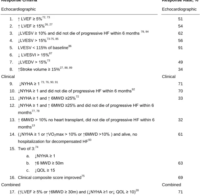
Table 2: Seventeen Different Response Criteria Identified From the Fornwalt (2010) review6
Response Criteria
Echocardiographic 1. ↑ LVEF ≥ 5%72, 73 2. ↑ LVEF ≥ 15%26, 27
3. ↓LVESV ≥ 10% and did not die of progressive HF within 6 months 78, 84
4. ↓LVESV > 15%73-75, 85 5. LVESV < 115% of baseline86 6. ↓ LVESVI > 15%87 7. ↓LVEDV > 15%73 8. ↑Stroke volume ≥ 15%27, 88, 89 Clinical 9. ↓NYHA ≥ 1 73, 76, 90, 91
10. ↓NYHA ≥ 1 and did not die of progressive HF within 6 months92 11. ↓NYHA ≥ 1 and ↑ 6MWD ≥25%72
12. ↓NYHA ≥ 1 and ↑ 6MWD ≥25% and did not die of progressive HF within 6 months77, 78
13. ↑ 6MWD > 10% no heart transplant, did not die of progressive HF within 6 months13
14. (↓NYHA ≥ 1 or ↑VO2max > 10% or ↑6MWD >10% ) and alive, no
hospitalization for decompensated HF93 15. Two of 3:74
a. ↓NYHA ≥ 1 b. ↑6 MWD ≥ 50m c. ↓QOL ≥ 15
16. Clinical composite score improved75 Combined
17. (↑LVEF ≥ 5% or ↑6MWD ≥ 30m) and (↓NYHA ≥1 or↓ QOL ≥ 10)28
Response Rate, % Echocardiographic 51 54 62 56 91 49 34 Clinical 71 70 33 32 61 63 69 Combined 71
↑ indicates increase; LVEF, left ventricular ejection fraction; ↓, decrease; HF, heart failure; LVESV, left ventricular end-systolic volume: LVESVI, LVESV indexed by body surface area; LVEDV, left ventricular end-diastolic volume; NYHA, New York Heart Association functional class; 6MWD, 6-minute walk distance; VO2max, oxygen consumption at peak exercise, and QOL, quality-of-life score.
If the authors did not specify whether death was considered a nonresponse, then it was assumed that deaths were excluded.
19
Fornwalt et al.6 collected seventeen different primary response criteria in the 26 most-cited publications on predicting response to CRT (table 2). Eight of these seventeen response criteria were based on echocardiography, eight were based on clinical measures, and one criterion was based on a combination of both echocardiographic and clinical measures. The percentage of patients defined as having a positive response to CRT ranged from 32% to 91% for the 15 response criteria.
The reasons for a lack of response to CRT are not well known7, 13, 14, 37.So far, better characterization of patients who will respond to CRT has been the main focus of ongoing research.
The identification of non responders to CRT may be also of interest. This therapy requires high costs and potential implantation related complications that may be avoided in patients who won’t have clinical benefit. Current inclusion criteria may not be accurate enough to differentiate patients who will or will not respond to CRT. Other pathophysiologic factors such as HF etiology, LV dimensions and function, mitral regurgitation, LV dyssynchrony, position of LV pacing lead, and extent/location of myocardial scar have also shown to influence CRT response7.
CRT was associated with increased total costs when compared with standard medical treatment. Over a mean follow-up of 29.6 months in CARE-HF94, the mean €4316 overcost was mainly attributable to the device itself, with an estimated cost of €5825.
The mean incremental cost-effectiveness ratio per life year gained was €29 40094 and $28 10095 with CRT-P and $46 700 with CRT-D95. These data suggest that the clinical benefits of CRT are economically viable and can be achieved at a reasonable cost in most European countries.
Long-term treatment with CRT-P appears cost-effective compared with medical therapy alone. From a life-time perspective, assuming a reasonable life expectancy when receiving effective treatment for HF, CRT–D may also be considered cost-effective when compared with CRT-P and medical therapy96.
The 2007 ESC/EHRA Guidelines for Cardiac Pacing46, the 2008 ESC Heart Failure Guidelines31, and the 2008 ACC/AHA/HRS Guidelines for Device Therapy97 provide class I A recommendation for CRT treatment with or without
20
an ICD function in patients with QRS width ≥ 120 ms, LVEF ≤ 35% and NYHA functional class III and IV. Those are the current inclusion criteria for CRT.
ECG recording should be taken to know the PR interval, QRS duration and morphology, and underlying rhythm to choose the most appropriate device. There is strong evidence that patients with prolonged QRS duration (≥120/ ≥130 ms) show worse prognosis but the impact of QRS duration to predict response to CRT is still unclear98.
Another important diagnostic tool is echocardiography evaluation for precise assessment of ventricular dimensions, presence of mitral regurgitation, the estimation of the LVEF (≤ 35%) and diagnosis of ventricular dyssynchrony. Lafitte et al.99 concluded that a multiparametric echocardiographic strategy based on the association of conventional criteria is a better indicator of CRT response than the existing single parametric approaches. Nevertheless, many currently used echocardiographic parameters failed to improve responder identification. There is no consensus about which echocardiographic parameters may best determine baseline dyssynchrony and which of these can predict response to CRT72, 85, 87, 89, 98, 100-108.
Besides ECG and echocardiographic parameters, cardiopulmonary exercise testing (CPET) is an important criterion for screening patients undergoing CRT18.
3.3 CARDIOPULMONARY EXERCISE TESTING
CPET and the 6-minute walk are the most common modalities for evaluating the functional capacity of patients with HF.
The 6-minute walk is usually used as an alternative to CPET, as it evaluates low-level or submaximal work and is more compatible with activities of daily living. This test and the NYHA classification may be helpful for assessing patient’s physical ability. Many clinical trials have used the 6-minute walk test to classify patients with HF into syndrome severity categories. A significant correlation between distance walked during 6 minutes and survival is noted. A total distance walked of less than about 300 meters in a study carried an annual mortality risk of 11%, in contrast to 4% among patients who could walk more than about 450 meters109.
21
CPET is used to evaluate maximal exercise capacity, for prognostic stratification, and for staging for possible cardiac transplantation. There is different exercise testing protocols in which the workload is progressively increased during the test, either on a bicycle or a treadmill. The selection of a particular protocol should be based on the experience of the testing physician, on the physical ability of the patient, and on the availability of the facility where the test is being performed. Exercise testing of patients with HF is supported by the American Heart Association for clinical and research application36.
The CPET has some advantage compared to the traditional exercise testing. Both of the tests are ECG monitoring however, the CPET used gas exchange analysis, which can provide directly the peak VO2, a measure of coupling
between central pulmonary gas exchange, cardiac output, and peripheral oxygen delivery to and use by skeletal muscle36.
Even so, this testing has some implications like time-consuming, expensive (it requires specialized equipment for gas exchange, coupled to ECG), and requires great skill in cardiopulmonary physiology.
In spite of these limitations, the measurements of carbon dioxide production and oxygen consumption during this test can provide numerous additional data that have both diagnostic and prognostic information, such as the ratio of volume of expired gas to carbon dioxide production (VE/VCO2), ventilatory
threshold, and respiratory exchange ratio.
HEART RATE RESPONSE
There is yet no consensus about the value of the resting heart rate (HR) relative to measure the risk to develop cardiovascular diseases and mortality.
Some authors defend that HR is not recognized as a factor for cardiovascular risk assessment or risk reduction in U.S. and European guidelines. A review of resting HR in cardiovascular disease, leaves some doubts that HR is a risk factor for cardiovascular mortality, independent of currently accepted risk factors and other potentially confounding demographic and physiological characteristics. It has been difficult to determine whether modulation of HR can beneficially alter risk; currently available interventions that lower HR, such as beta-blockers, certain calcium channel blockers, and
22
physical conditioning have multiple additional actions. Nonetheless, improved HR is important and potentially beneficial for patient care110.
More recently, there has been a study with patients with left ventricular dysfunction and a recent myocardium infarction or HF showed that resting HR was independently associated with increased risk of overall mortality over a 10 year follow-up period. The results suggest that the prognostic importance of resting HR is stronger in patients with myocardium infarction compared to patients with HF, especially in the short term111.
The immediate response of the cardiovascular system to exercise is an increase in HR due to a decrease in vagal tone. This increase is followed by an increase in sympathetic outflow to the heart and systemic blood vessels. During dynamic exercise, HR increases linearly with workload and VO2. Heart rate will
reach a steady state within minutes during low levels of exercise and at a constant work rate. As workload increases, the time necessary for the HR to stabilize will progressively lengthen. The HR response to exercise is influenced by several factors such as age, deconditioning, body position, type of exercise, and various states of health and therapy, including heart transplant29, 112. The HR peak is the highest value of the heart rate or pulse rate which can be attained and measured during incremental exercise.
HR recovery refers to the deceleration of the HR in early exercise recovery in association with vagal tone reactivation. It is the difference between HR at peak exercise and after one minute or other time defined of recovery. An abnormal value for the recovery of HR was defined as a reduction of 12 beats per minute (bpm) or less from the HR at peak exercise113, 114. The increase in HR that accompanies exercise is due in part to a reduction in vagal tone. HR recovery immediately after exercise is a function of vagal reactivation, a decrease of vagal activity is known to be a risk factor for death114.
Lipinski et al.115 and Tang et al.116 conclude that HR recovery on the first minute recovery is a significant predictor of mortality and may provide valuable prognostic information for patients with HF or LVSD. HR recovery should be evaluated along with VO2, age, HR peak, and other variables to predict mortality
and may also aid in determining which patients with HF and LVSD will require heart transplantation113, 115. Even after adjusting for other exercise derived
23
predictor variables and previously validated HF survival scores, post-exercise HR recovery remained an independent predictor of adverse clinical events116.
Moreover, a recent study concludes that CRT favorably alters the cardiac autonomic functions assessed by HR recovery indices117. This effect of CRT on cardiac autonomic functions was observed both in responders and in non responders. However, the degree of improvement in HR recovery indices is correlated with left ventricular reverse remodeling. In the Okutucu et al.117 study, the baseline HR recovery indices could not predict response to CRT, considering a responder as a decrease of ≥ 15% in LVESV at the 6-month follow-up was defined as a positive echocardiographic response.
OXYGEN UPTAKE
Maximum oxygen uptake (VO2max) or peak oxygen uptake (VO2peak) is the
maximum capacity of an individual's body to transport and use oxygen which can be attained and measured during an incremental exercise protocol for a specific exercise mode. It can, however, be affected by age, gender, muscle mass, aerobic conditioning and medication therapy. It is measured in liters per minute, but is often relativised for body mass (ml/kg/min) to allow better comparison between individuals of different body size.
Functional status and cardiac reserve of patients with CHF can be objectively characterized by determining exercise tolerance. Particularly important is the precise measurement of VO2peak consumption.
VO2peak is one of the most important independent predictors of mortality and
hospitalization for patients with HF19, 118. This functional variable represents functional effort capacity and is improved significantly by CRT119, 120, but is influenced by non-cardiac factors (age, motivation, anaemia and obesity). It has become probably the most important test to determine whether ambulatory patients are ill enough to list for cardiac transplantation36.
Mancini et al.19 analyzed if VO2peak can be used to identify ambulatory
patients in whom cardiac transplantation can be safely deferred. According to the study protocol, patients with a VO2peak > 14 ml/kg/min were denied
transplantation, whereas those with a VO2peak ≤ 14 ml/kg/min were offered
24
ml/kg/min have a far worse prognosis than those with VO2peak >14 ml/kg/min. It
is important to include in the follow-up of CHF patients the determination of VO2peak to establish more effectively the optimal therapeutic strategy23. Recent
recommendations for the International Society for Heart and Lung transplantation guidelines for CPET were VO2peak ≤ 12.0 ml/kg/min for patients
receiving beta-blockers and VO2peak ≤ 14.0 ml/kg/min for patients not receiving
such therapy121.
An accurate estimation of VO2peak might be underestimated and it is difficult
to obtain in patients with severe HF. It is difficult to assess whether a truly maximal test was performed and rarely is reach a true plateau of oxygen consumption with increasing workloads because of some limitations such as peripheral muscle fatigue, motivation or procedural difficulties (selection of an appropriate exercise protocol)98, 121.
HF patients rarely reach a plateau of VO2 with increasing workloads,
common determinants of a maximal exercise test have been respiratory exchange ratio (RER) > 1.1 and reaching an anaerobic threshold. The RER rises from a resting value of around 0.7 at rest during exercise. Healthy controls achieve a RER at peak exercise of between 1.10 and 1.20 or even higher, indicating that anaerobic metabolism is occurring. Such a RER is used as an indicator of maximal effort112. Patients with CHF are typically less able to exercise to a level with such a high RER, and some CHF patients are unable to reach a RER ≥ 1.0. Ingle et al.122 conclude that independent predictors of mortality were different in patients with a RER < 1.0 compared to those with a RER ≥ 1.0. In CHF patients with a RER < 1.0, traditional prognostic markers (VE/VCO2 slope, VO2peak) were not independently predictive of mortality.
However, a review of studies defining the criteria for VO2peak showed that 6 of
14 studies used an RER cutoff of 1.0 or 1.05, so these criteria may be too stringent. Decisions might also need to be made based on a submaximal test121.
In a study by Auricchio et al.12 patients with peak VO2 > 16 ml/kg/min did not
show significant cardiorespiratory improvements during CRT. In contrast, a study by Piepoli et al.123 showed that patients with a peak VO2 ≤ 7 ml/kg/min did
not benefit from CRT. A difference of approximately 2 ml/kg/min in VO2peak
25
In a recent study Berger et al.98 analyzed the impact of the cardiorespiratory functional reserve to predict the response to CRT. Submaximal cardiopulmonary treadmill exercise testing prior and 6 months after implantation of a CRT device was made. Responders to CRT, defined by a decrease in LVESV > 15% showed a significant lower cardiorespiratory reserve at baseline (prior CRT) as compared to the non responders. The conclusion of this study was that non responders to CRT showed a more preserved cardiorespiratory functional reserve as compared to responders despite similar NYHA classification.
Some authors have used percentage of predictive VO2peak rather than the
absolute value to stratify risk. In multivariate analysis, 50% or 55% of predicted peak oxygen uptake (when the respiratory exchange ratio is greater than 1.10) has generally been selected as the most significant predictor of cardiac death121. The proposed cutoff point of 50% has been confirmed by Stelken et al.20, who showed that in a cohort of 181 HF patients those with an oxygen uptake greater than 50% predicted value had a 94% possibility two year survival as compared to only 50% survival in patients with an oxygen uptake of below 50% predicted value.
Therefore, the percentage of predicted value in clinical reports must be interpreted in a patient specific context in view of other comorbidity conditions, for example, adjustments have to be made if the patient is taking a beta-blocker124.
SLOPE OF THE VENTILATORY RESPONSE
VE/VCO2 is calculated during a cardiopulmonary exercise test and is the
slope of the relationship between minute ventilation (VE) and oxygen uptake (VO2) during incremental exercise125. The ventilatory response evaluation is not
influenced by beta-blocker therapy and submaximal exercise. It is well recognized that VE/VCO2 slope is measurable at any point during exercise and
adds significant prognostic value in HF population126.
In patients with HF an increased ventilator response throughout exercise is observed. A VE/VCO2 slope value up to 35 was associated with a one-year
26
mortality rate of 30% in the Corra et al. study127. Francis et al.128 found a two-year mortality rate of 65% for patients with a value up to 55.
The VE/VCO2 slope seems to be a better predictor of outcome than VO2peak
with regard to submaximal effort128. The complementary prognostic value especially with the VO2peak is of great interest. Patients with VO2peak < 11
ml/kg/min and VE/VCO2 slope ≥ 34 are at particularly high risk for
transplantation or death. Thus, patients with well preserved exercise capacity and low VE/VCO2 slope are at low risk for transplantation or death. This
approach allows high-risk patients to be identified noninvasively and could provide guidance for intensified treatment (medical regimen, CRT, exercise training and heart transplantation) and monitoring129.
This parameter should be a routine component of exercise analysis in HF population. Consideration should be given to revising clinical guidelines to reflect the prognostic importance of the VE/VCO2 slope in addition to VO2peak129, 130
.
VENTILATORY ANAEROBIC THRESHOLD
In 1991, submaximal exercise parameters such as the ventilatory anaerobic threshold (VAT) were introduced to evaluate the cardiopulmonary functional reserve. In fact, most activities of daily living do not require maximal effort, so submaximal exercise parameters should be used like VAT.
There is a point during progressive exercise in which lactate accumulation caused a nonlinear increase in ventilation. The increase in blood lactate concentration during exercise is thought to cause a nonlinear increase in ventilation as a result of bicarbonate buffering of excess hydrogen ions from lactate in the blood and consequent production of carbon dioxide. The resulting hyperventilatory response has been commonly termed the VAT131.
VAT is assessed by ventilatory expired gas, defined by the exercise level at which VE begins to increase exponentially relative to the increase in VO2.
Nevertheless, VAT cannot be obtained in 25% to 30% of patients with HF because of severe deconditioning, early onset of acidosis, and the presence of an irregular breathing pattern132.
27
4 METHODOLOGY
4.1 INTRODUCTION
This chapter describes the methodological procedure of the study. In the first part, a description of the experimental concept such as the study design and the characterization of the sample are presented. In the second part, study variables are presented, including the independent and dependent variables and how they were evaluated. The statistical treatment of the data of interest to the present study was the last task of the procedure to be done.
4.2 STUDY DESIGN
The present study is an observational and analytical prospective cohort study. This study design requires a comparative analysis between data collected in CPET before and after the implantation of a CRT considering two types of criteria to define responders as well an echocardiography and clinical consultation to assess the ejection fraction and analyze the NYHA class.
Data were collected before and after the implantation of a CRT, under the same conditions, with the same procedures.
4.3 PARTICIPANTS
The study sample initially included 22 patients from both genders with CHF referred to Santa Marta Hospital, Lisbon. From the 22 patients that initially met the criteria for enrolment in the study, 7 were excluded for data analysis. Therefore, the total sample for this study consisted of 15 patients.
Patients were excluded for several reasons, such as: having traditional exercise testing done after CRT instead of CPET (n=2), missed some variables measured from CPET (n=2), missed CPET after CRT before data were analyzed (n=2) and CRT not working properly (n=1).
The study sample receiving CRT based on the following clinical criteria were considered for this study: patients presenting severe symptomatic heart failure despite optimal pharmacological therapy (NYHA functional Class III or IV),
28
LVEF < 35% and free of exercise-limiting comorbidities such as cerebrovascular disease, muskuloskeletal impairment, or peripheral vascular disease.
All patients were followed-up prospectively at Santa Marta Hospital. Clinical status (NYHA class) was assessed at baseline and approximately 5 months after CRT. The average time of the echocardiogram performed after CRT was 2 months ± 18 days. The CPET after CRT was 5 months ± 13 days.
RESPONSE CRITERIA
There are two possible criteria used to classify the CRT response according to the literature. The first is defined by an increase of 15% or more of LVEF, measured by conventional echocardiography from baseline to follow-up26, 27. The second was a combinination of two criteria, defined by clinical and echocardiography measures with an increase of 5% or more of LVEF and a decrease of 1 or more NYHA class, respectively28. These measures were chosen because of their percentage of response rate, the first with 54% and the second with 71% of response rate, as seen in table 2, page 18.
4.4 VARIABLES IN STUDY
4.4.1 DEPENDENT VARIABLES
In this study the following dependent variables were considered: in echocardiography, the ejection fraction; in the clinical category, the NYHA class; and in the CPET the, VO2 peak, predicted VO2, HR initial, HR peak, HR
recovery at first and third minutes, blood pressure at the start and maximum reached, RER, VAT and VE/VCO2 slope.
EQUIPMENTS AND PROTOCOLS OF ASSESSMENT
Echocardiography: resting cardiac echocardiogram was recorded in a lying position using a commercially available digital ultrasound scanner (Vivid 7, Vivid 3 and Vivid IE9, GE Vingmed Ultrasound, Horten, Norway). In this exam, LVEF
29
was measured which represents the volumetric fraction of blood pumped out of the left ventricle which ejects via the aortic valve into the systemic circulation. Although this value is one of the inclusion criteria for this study, their difference before and after CRT indicates one of the criteria to response.
NYHA class: the assessment was made according the table 1 on page 17. The classification was made by a cardiologist in the medical consultation before and after the implantation of CRT.
CPET: all subjects underwent maximal symptom-limited treadmill CPET (GE Marquette Series 2000 treadmill and Mortara instrument, Milwaukee, USA), using modified Bruce Protocol. The 12-lead electrocardiogram and HR were recorded continuously during the test and continued for six minutes of the recovery period. Blood pressure was measured at rest, during the last 30 seconds of each stage, at peak exercise and at the first, third and sixth minute of the recovery phase.
In no case did altered blood pressure, arrhythmia, chest pain or electrocardiographic changes lead to interruption of the test, in accordance with international standards133. All studies were, accordingly, interrupted by subjective fatigue or dyspnea preventing the patient from continuing the exercise. No medication was discontinued before the test.
Minute ventilation (VE in l/min) and oxygen uptake (VO2 in l/min/kg) were
acquired breath-by-breath, using a gas analyzer. Gas analysis was preceded by calibration of the equipment and began three minutes prior to exercise.
Patients were encouraged to perform exercise until the VCO2/VO2 ratio
(RER) was ≥ 1.10. But in this type of patients it is complicated achieve that ratio, however, ratios under 1.10 were considered to this study. Besides RER, other derived variables were calculated, including ventilatory equivalent for oxygen (VE/ VO2).
VO2peak was expressed as the highest VO2 attained during the final 30
seconds of exercise134. Predicted peak VO2 and the percentages of the
predicted values achieved were calculated by the system software. The VAT was determined using the V-slope method, and corrected, when necessary, using the VE/VO2 versus VE/VCO2 criterion and/or the end-tidal oxygen and
30
as the slope of the regression line relating VE to VCO2 during exercise, with
data obtained over the complete duration of exercise.
4.4.2 INDEPENDENT VARIABLES
The implantation of CRT was the independent variable; the most common being the CRT-D. According to the Guidelines for cardiac pacing and CRT46, implantation of a CRT device is more demanding than implantation of a conventional pacemaker or implantable cardioverter defibrillator. Thus, additional laboratory, operator, and technical support were considered.
In our study and according to the experts advise, patients with CRT should fulfill the following conditions: a) two or more cardiologists qualified for device implantation and management; (b) all physicians should possess knowledge and experience in haemodynamic monitoring and administration of cardiovascular support; (c) trained nurses and technical personnel; (d) pacing system analyzer and programmer of implanted device: electronic patient file is highly encouraged; (e) continuing medical education for physician, nurses, and technicians is mandatory.
Two operators were required, especially during extraction/insertion of guidewires, handling of wires, sheaths, and stylets. Ideally, two nurses were required. One nurse monitors patient status and manages all necessary impellent accesses, including the urine catheter and the intravenous administration of drugs. A second nurse provides implant assistance by monitoring some variables, handling over sterile material; and positioning the ECG screen.
Continuous anaesthesiological support is not obligatory, but quick anaesthesiological assistance must be available if a critical clinical situation develops.
4.5 STATISTICAL ANALYSIS
For statistical analysis it was used the Statistical Package for Social Science (SPSS 19.0 for Windows ®, SPSS Inc, Chicago, USA).
31
Efficacy of CRT was examined by comparing variables at baseline and after implantation of CRT. Differences between pre and post CRT were tested according to the variables categories, qualitative and quantitative, to evaluate the effect of this treatment.
For the quantitative variables, it was used the Student’s t-test for paired samples if the normal distribution is assumed or the Wilcoxon signed-rank test if normal distribution cannot be assumed. Regarding normality and homogeneity of variances data was tested using Shapiro-Wilk and Levene's tests, respectively. Pearson χ2
test was used for comparisons of qualitative variables. Results were expressed as means ± standard deviation.
The differences in quantitative variables (before CRT) between responders and non responders were analyzed using the Student’s t-test for independent samples or, when normality was not observed, the Mann-Whitney U test. Responders were defined by two parameters: an increase of 15% of the LVEF or an increase of 5% of the LVEF and a decrease of one level or more in the NYHA classification.
32
5 RESULTS AND DISCUSSION
INTRODUCTION
In this chapter, the results and discussion will be described, respectively, for each dependent variable. Initially, the results will be referred to the baseline, describing their characteristics, and then the comparison will be made between the group of responders and non responders according to the studied variables before CRT. Finally, the comparisons between the studied variables before and after CRT on exercise performance will be described.
PARTICIPANTS CHARACTERISTICS
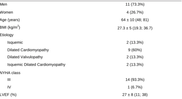
The clinical and demographic characteristics of the study population are summarized in table 3. Around 75% of the patients were medicated with beta-blockers, 60% with diuretics and 40% with angiotensin-converting enzyme inhibitors or/and angiotensin receptor blockers. A high percentage of patients had a CRT-D implanted (93%).
COMPARISON BETWEEN RESPONDERS AND NON RESPONDERS VARIABLES
Table 3: Clinical and demographic characteristics at baseline for the sample in the study (n=15)
Men 11 (73.3%) Women 4 (26.7%) Age (years) 64 ± 10 (48; 81) BMI (kg/m2) 27.3 ± 5 (19.3; 36.7) Etiology Isquemic 2 (13.3%) Dilated Cardiomyopathy Dilated Valvulopathy 9 (60%) 2 (13.3%)
Isquemic Dilated Cardiomyopathy 2 (13.3%)
NYHA class
III 14 (93.3%)
IV 1 (6.7%)
LVEF (%) 27 ± 8 (11; 38)
33
According to the different classification criteria to define responders to CRT (either an increase of 15% of the LVEF or an increase of 5% of the LVEF and a decrease of one level or more in the NYHA classification), the demographic characteristics, the patients’ medications, echotrocardiographic values and functional characteristics at baseline were listed in tables 4 and 5.
Table 4: Comparisons of baseline demographic, clinical, and functional characteristics in responders vs. non responders to CRT considering echocardiographic parameters
Responders (n=9) Non Responders (n=6) P-value
Age (years) 63 ± 9 66 ± 12 0.644
Men/women (n) 5/4 6/0 0.057
Heart failure etiology (n) Ischemic
Dilated Cardiomyopathy Dilated Valvulopathy
Isquemic Dilated Cardiomyopayhy
1 6 1 1 1 3 1 1 0.937 Medications (n) Angiotensin Diuretics Beta-Blockers 3 6 6 3 3 5 0.519 0.519 0.475 NYHA III/IV 3.11 ± 0.3 3 ± 0 0.435 LVEF (%) 27 ± 8.4 26.7 ± 8.9 0.942 Peak VO2 ≤ 14 ml/kg/min 4 3 0.833 VO2(ml/kg/min) Peak exercise Percentage of predicted VO2 (%) 14.7 ± 6.2 56.4 ± 17.8 17.7 ± 4.7 64 ± 6.3 0.348 0.399
VE/VCO2 slope (L/min) 30.5 ± 10.1 38.6 ± 4.4 0.200
Anaerobic threshold (n achieve) 4 5 0.132
Systolic blood pressure (mm Hg) Initial Peak 112 ± 19 143 ± 20 118 ± 18 140 ± 24 0.473 0.778 Diastolic blood pressure (mm Hg)
Initial Peak 60 ± 10 72 ± 10 68.33 ± 8 70 ± 9 0.115 0.711 Heart rate (bpm) Initial Peak 80 ± 12 125 ± 25 92 ± 13 132 ± 24 0.088 0.628 Heart rate recovery (bpm)
1st minute 3rd minute 14 ± 7 38 ± 17 18 ± 12 36 ± 19 0.437 0.813
34
Positive echocardiographic response to CRT was observed in 9 patients, corresponding to a 60% responder rate, as seen in table 4. No differences were observed between the two groups regarding demographic, clinical and functional characteristics, probably owing to the small study sample size.
As we can see in table 4, the baseline characteristics of the two groups were overall similar, including functional capacity and echocardiographic measurements.
The HR initial was lower in responders than non responders, the difference were 12 bpm, but did not reach statistical significance (p > 0.05). No significant differences were observed between patients with VO2peak ≤ 14 ml/kg/min.
No clear-cut value was found for separating responders and non responders.
Table 5: Comparisons of baseline demographic, clinical, and functional characteristics in responders
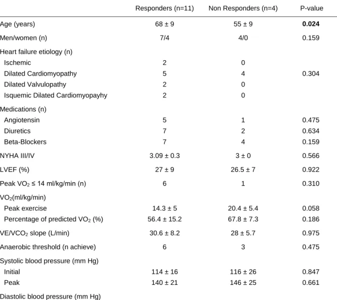
vs. non responders to CRT considering echocardiographic and clinical parameters (Combined)
Responders (n=11) Non Responders (n=4) P-value
Age (years) 68 ± 9 55 ± 9 0.024
Men/women (n) 7/4 4/0 0.159
Heart failure etiology (n) Ischemic
Dilated Cardiomyopathy Dilated Valvulopathy
Isquemic Dilated Cardiomyopayhy
2 5 2 2 0 4 0 0 0.304 Medications (n) Angiotensin Diuretics Beta-Blockers 5 7 7 1 2 4 0.475 0.634 0.159 NYHA III/IV 3.09 ± 0.3 3 ± 0 0.566 LVEF (%) 27 ± 9 26.5 ± 7 0.922 Peak VO2 ≤ 14 ml/kg/min (n) 6 1 0.310 VO2(ml/kg/min) Peak exercise Percentage of predicted VO2 (%) 14.3 ± 5 56.4 ± 15.2 20.4 ± 5.4 67.8 ± 7.3 0.058 0.186
VE/VCO2 slope (L/min) 30.6 ± 8.2 28 ± 5.7 0.975
Anaerobic threshold (n achieve) 6 3 0.475
Systolic blood pressure (mm Hg) Initial Peak 114 ± 16 140 ± 21 116 ± 26 146 ± 25 0.847 0.661 Diastolic blood pressure (mm Hg)
35
As we can see in table 5, in the combined parameters, positive responders to CRT corresponding to a 26.7% non responder rate. Overall, the baseline characteristics of the NYHA, LVEF and SBP (both at initial and peak of CPET) of the two groups were similar (table 5).
Among all variables examined, in our study, the only significant differences detected were the age, peak HR and HR recovery after 3 minutes from CPET performance.
The mean age was significantly higher approximately 13 years in responders compared to non responders (p = 0,024). Whether age negatively affects response to CRT is currently unknown; this is an important issue, because most patients with HF are of greater age136.
The peak HR before CRT was higher 39 bpm in non responders than in responders (p ≤ 0.05), whereas the minute 3 of HR recovery was lower 23 bpm in responders than in non responders before CRT (p = 0.014). In contrast to a previous finding, baseline HR recovery at minute 1 and 3 could not predict response to CRT, their values between responders and non responders were very similar117. In our study, this does not happen, there were different values at baseline. According to the literature, HR recovery immediately after exercise is a function of vagal reactivation, a decrease of vagal activity is known to be a risk factor for death114. Thus, the faster the recovery, in this case for non responders, there is an increase of vagal activity.
Responders, compared with non responders, were more likely to have dilated cardiomyopathy heart disease (33.3% vs. 26.7%, p > 0.05), and more diuretic and beta blocker medications (46.7% vs. 13.3% p > 0.05 and 46.7% vs. 26.7% p > 0.05, respectively). However, none of these variables were statistically significant. Initial Peak 60 ± 9 70 ± 10 70 ± 8 75 ± 6 0.108 0.338 Heart rate (bpm) Initial Peak 82 ± 10 118 ± 18 93 ± 19 157 ±13 0.145 0.002
Heart rate recovery (bpm) 1st minute 3rd minute 13 ± 9 31 ± 14 21 ± 8 54 ± 13 0.149 0.014
36
The group of non responders had a VO2peak and a percentage of predicted
VO2 higher than the group of responders although not reaching statistical
significance (20.4 ± 5.4 ml/kg/min vs. 14.3 ± 5 ml/kg/min p = 0.058 and 67.8 ± 7.3% vs. 56.4 ± 15.2%, p = 0.186). In a recent study, Arora et al.137 found a significant value for the VO2peak between responders and non responders. A
positive responder was considered if VO2peak ≥ 1 ml/kg/min. In the non
responder group, the value was higher than the group of responders (11.7 ± 2.4 ml/kg/min vs. 10.6 ± 2.3 ml/kg/min p < 0.05). The percentage of predicted VO2peak was not significantly different between groups but was higher in the non
responder’s group.
As mentioned previously, patients with a baseline VO2peak < 14 ml/kg/min
regularly benefit from CRT during the first year of treatment23. The German “Statement on cardiac resynchronization”25
already recommends a baseline VO2peak < 14 ml/kg/min as one criterion to indicate CRT. As we can observe in
table 5, the value of the VO2peak for the responder’s group was almost < 14
ml/kg/min, with a p = 0.058. Probably owing to the small study sample size, it was not possible to obtain statistically significance in the VO2peak, but there is a
trend towards a lower VO2peak.
The VE/VCO2 slope (30.6 ± 8.2 l/min vs. 28 ± 5.7 l/min) and the
achievement of the VAT (n = 6 vs. n = 3) were higher in the group of responders than non responders, but no significant differences were found.
In this study, it was possible to make a relation between the response criteria and their efficacy response rate according the Fornwalt review6 mentioned previously. In that review, the response rate for the response criteria parameter ≥ 15% improvement of LVEF was 54%; the response criteria for combined parameters was 71%, as we can see in table 2. Similar findings have also been demonstrated in this study, it was on the combined response criteria that were statistical significant values for the variables in study.