A
RTIGO DER
EVISÃOThe clinical importance of cardiopulmonary
exercise testing and aerobic training in patients
with heart failure
A importância clínica de testes de exercícios cardiopulmonares e treinamento
aeróbico em pacientes com insufi ciência cardíaca
Arena R1, Myers J2, Guazzi M3
Abstract
Introduction: The appropriate physiological response to an acute bout of progressive aerobic exercise requires proper functioning of the
pulmonary, cardiovascular and skeletal muscle systems. Unfortunately, these systems are all negatively impacted in patients with heart failure
(HF), resulting in signifi cantly diminished aerobic capacity compared with apparently healthy individuals. Cardiopulmonary exercise testing
(CPX) is a noninvasive assessment technique that provides valuable insight into the health and functioning of the physiological systems that
dictate an individual’s aerobic capacity. The values of several key variables obtained from CPX, such as peak oxygen consumption and
ventilatory effi ciency, are often found to be abnormal in patients with HF. In addition to the ability of CPX variables to acutely refl ect varying
degrees of pathophysiology, they also possess strong prognostic signifi cance, further bolstering their clinical value. Once thought to be
contraindicated in patients with HF, participation in a chronic aerobic exercise program is now an accepted lifestyle intervention. Following
several weeks/months of aerobic exercise training, an abundance of evidence now demonstrates an improvement in several pathophysiological
phenomena contributing to the abnormalities frequently observed during CPX in the HF population. These exercise-induced adaptations to
physiological function result in a signifi cant improvement in aerobic capacity and quality of life. Conclusions: Furthermore, there is initial
evidence to suggest that aerobic exercise training improves morbidity and mortality in patients with HF. This paper provides a review of the
literature highlighting the clinical signifi cance of aerobic exercise testing and training in this unique cardiac population.
Key words: ventilatory expired gas; cardiac output; skeletal muscle; survival.
Resumo
Introdução: A resposta fi siológica aguda ao exercício aeróbio progressivo demanda funcionamento adequado dos sistemas pulmonares,
cardiovasculares e músculo-esquelético. Infelizmente, todos estes sistemas estão negativamente afetados em pacientes com insufi ciência
cardíaca (IC), resultando numa redução signifi cativa da capacidade aeróbia comparada com indivíduos aparentemente saudáveis. O teste
de exercício cardiopulmonar (TCP) representa uma técnica não-invasiva de avaliação que fornece compreensão valiosa sobre a saúde e
funcionamento dos sistemas fi siológicos que ditam a capacidade aeróbia de um indivíduo. Os valores de várias variáveis-chave obtidas através
do TCP, como consumo pico de oxigênio e efi ciência ventilatória são encontrados frequentemente como anormais em pacientes com IC.
Além da capacidade das variáveis do TCP refl etir de maneira aguda os graus variáveis da fi siopatologia, também possuem forte signifi cância
prognóstica, aumentando ainda mais o seu valor clínico. A participação num programa de exercícios aeróbios crônicos, anteriormente era
contra-indicada em pacientes com IC. Agora é uma intervenção aceitável de estilo de vida. Após um período de treinamento com exercícios aeróbios,
durante várias semanas/meses, tem sido evidenciada uma melhora em vários fenômenos fi siopatológicos que contribuem às anormalidades
constatadas frequentemente durante TCP na população com IC. Conclusões:As adaptações fi siológicas induzidas por exercícios aeróbios
resultam em uma melhora signifi cativa de capacidade aeróbia e de qualidade de vida. Além disso, há evidências sugerindo que treinamento
com exercícios aeróbios melhora a morbidade e a mortalidade em pacientes com IC. Este artigo fornece uma revisão da literatura que destaca
a signifi cância clínica dos testes de exercícios aeróbios e treinamento nesta população cardíaca única.
Palavras-chave: gás expirado ventilatório; rendimento cardíaco; músculo esqueleto; sobrevivência.
Recebido: 15/01/2008 – Revisado: 17/01/2008 – Aceito: 04/02/2008
1 Departments of Internal Medicine, Physiology and Physical Therapy, Virginia Commonwealth University, Health Sciences Campus, Richmond, Virginia, United States 2 VA Palo Alto Health Care System, Cardiology Division, Stanford University, Palo Alto, California, United States
3 Cardiopulmonary Laboratory, Cardiology Division, University of Milan, San Paolo Hospital, Milan, Italy
Correspondence to: Ross Arena, PT, PhD, Associate Professor, Department of Physical Therapy, Box 980224, Virginia Commonwealth University, Health Sciences Campus, Richmond, VA, USA 23298-0224, e-mail: [email protected]
76
Introduction
Systems infl uencing the physiological response to
normal exercise
An individual’s capacity to perform aerobic exercise is de-pendent upon pulmonary, cardiovascular and skeletal muscle function. While proper physiological functioning of these three systems is important, cardiac output (Q), i.e. the product of heart rate and stroke volume, is the primary determinant of peak or
maximal oxygen consumption (VO2). Cardiac output is
ap-proximately i ve liters/minute at rest and increases to approxi-mately 20-25 and 30-35 liters/minute at maximal exercise in apparently healthy sedentary subjects and elite athletes, respec-tively. h e ability of skeletal muscle to increase oxygen extraction during aerobic exercise plays a lesser but still important role in determining aerobic capacity. In apparently healthy subjects,
the dif erence in oxygen (O2) concentration between arterial
and venous blood (a-vO2 dif ) increases from approximately
5 mlO2/100 ml at rest to 16 mlO2/100 ml at maximal exercise. h e Fick equation, dei ned as the product of Q and a-vO2 dif , is used to describe VO2. While pulmonary function is not included in the Fick equation, the ability to increase gas exchange (oxygen intake and carbon dioxide removal) is of paramount importance to aerobic exercise capacity. Minute ventilation (VE), the product of respiratory rate and tidal volume, normally increases 10-20 fold at maximal aerobic exercise compared with resting values. It should be noted that pulmonary function is not typically the primary limiter of aerobic capacity, either in apparently healthy individuals or among patients diagnosed with cardiovascular disease. Even when the pulmonary, cardiovascular and skeletal muscle systems are all functioning properly, maximal aerobic capacity remains a rather heterogeneous phenomenon, since it is also inl uenced by age, sex, genetic predisposition and exer-cise habits. Considering these factors, the approximate range for
maximal VO2 in the apparently healthy population is between
20-55 mlO2·kg-1·min-1(1).
Pathophysiological abnormalities associated
with diminished aerobic capacity in patients
with heart failure
Severely compromised cardiac function is a primary pathophysiological component in heart failure (HF), and pre-vious investigations have demonstrated a signii cant
relation-ship between cardiac output during exercise and peak VO2 in
this population2-5. It has furthermore been well established that patients with HF frequently present reduced capillary density6 and intrinsic skeletal muscle abnormalities, primarily in the
form of diminished aerobic (mitochondrial) function6-13. Given that aerobic capacity is reliant primarily on Q and secondarily on the a-vO2 dif , as dei ned by the Fick equation, the signii cant reduction in peak VO2 frequently observed in patients with HF should be of no surprise. On average, peak VO2 is approximately 50% lower in this patient population, compared with values observed in apparently healthy individuals matched according to age and sex. Moreover, peak VO2 is approximately 25% lower in patients with HF, compared with patients diagnosed with coronary artery disease14.
A relationship between pulmonary abnormalities and peak VO2 has also been demonstrated in patients with HF15-17. Both resting15 and maximal16 measures of pulmonary function (i.e. inspiratory capacity), as well as dif usion capacity17, have all demonstrated signii cant correlations with peak VO2. h e degree to which these pulmonary abnormalities contribute towards the diminished aerobic capacity observed in HF, after accounting for the contributions of cardiovascular and skeletal muscle dysfunction, is unknown.
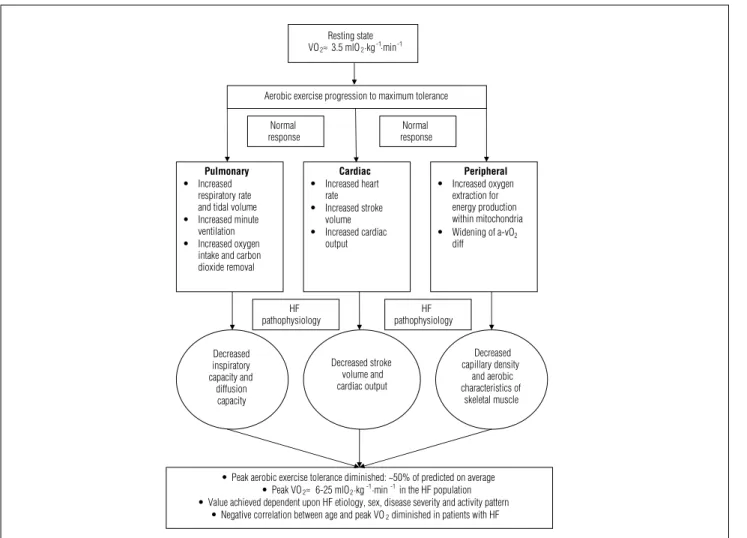
Figure 1 illustrates the systems involved in the physiologi-cal response to aerobic exercise and how HF af ects these systems.
The clinical applications of cardiopulmonary
exercise testing in patients with heart failure
Cardiopulmonary exercise testing (CPX) is a highly reliable18, well-accepted assessment technique in the HF population.
American19-21 and European22-24 associations have endorsed
its use. CPX is most often performed on a treadmill or lower-limb ergometer using highly conservative ramping protocols, which are appropriate given the severely diminished exercise tolerance often observed in this population25,26. h e addition of ventilatory expired gas analysis to the standard exercise
test enables measurement of VO2, carbon dioxide production
77
Resting state VO2 ≈ 3.5 mlO2·kg-1·min-1
Aerobic exercise progression to maximum tolerance
Pulmonary
t Increased
respiratory rate and tidal volume
t Increased minute ventilation
t Increased oxygen intake and carbon dioxide removal
Cardiac
t Increased heart rate
t Increased stroke volume
t Increased cardiac output
Peripheral
t Increased oxygen extraction for energy production within mitochondria
t Widening of a-vO2
diff Normal
response
Normal response
tPeak aerobic exercise tolerance diminished: ~50% of predicted on average
tPeak VO2 ≈ 6-25 mlO2·kg-1·min-1 in the HF population tValue achieved dependent upon HF etiology, sex, disease severity and activity pattern
tNegative correlation between age and peak VO2 diminished in patients with HF
Decreased inspiratory capacity and
diffusion capacity
Decreased stroke volume and cardiac output
Decreased capillary density
and aerobic characteristics of
skeletal muscle HF
pathophysiology
HF pathophysiology
Figure 1. Illustration of central and peripheral physiological adaptations from rest to maximal aerobic exercise and the impact of heart failure.
Table 1. Key considerations for cardiopulmonary exercise testing variables in patients with heart failure.
Variable Prognostic value Prognostic thresholds Response to interventions
VE/VCO2 Slope* Well established; >20 papers
Single best prognostic marker
</≥34 as dichotomous threshold; prognosis appears to progressively worsen as slope increases into 40s (VC-IV ≥45.0)
Signifi cant reduction with multiple interventions; should be assessed with interventional trials
Peak VO2*∑ Well established;
>20 papers
</≥10 mlO2·kg-1·min-1 in patients prescribed
a beta-blocker;
</≥14 mlO2·kg-1·min-1 in patients not
prescribed a beta-blocker
Signifi cant reduction with multiple interventions; should be assessed with interventional trials
EOV* Consistent results; 4 papers
Defi ned as oscillatory pattern in VE that occurs for ≥60% of the exercise test at an amplitude ≥15%
of average amplitude observed at rest
Not well investigated; signifi cant reduction in occurrence of EOV following milrinone treatment and respiratory muscle training
OUES Compelling results; 2 papers
Not well established at this time; lower = worse prognosis
Not well investigated; signifi cantly improved following exercise training
PETCO2 Rest and exercise
Compelling results; 2 papers
Not well established at this time; lower = worse prognosis
Response to interventions not investigated at this time
HRR Compelling results;
3 papers with small cohorts
Not well established at this time; lower = worse prognosis
Not well investigated; signifi cantly improved following exercise training
Abbreviations: EOV= Exercise oscillatory ventilation; OUES= Oxygen uptake efficiency slope; PETCO2= Partial pressure of end-tidal carbon dioxide; HRR= Heart rate recovery.
* Combination of high VE/VCO2 slope plus the presence of EOV and/or low peak VO2 may be highly indicative of poor prognosis; ∑ Use of peak VO
2 for prognostic purposes
78
Peak oxygen consumption
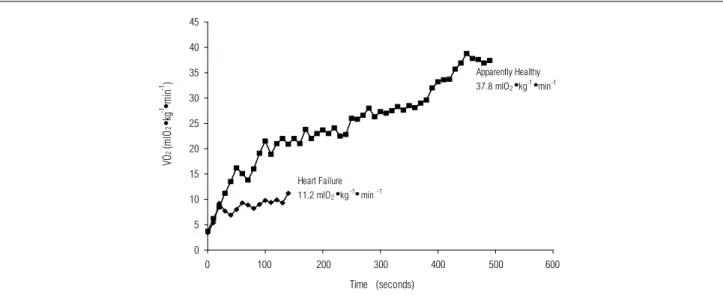
Oxygen consumption at peak exercise remains the most fre-quently assessed variable obtained from CPX in the HF population and is often signii cantly reduced, compared with normal pre-dicted values for a given age. It is usually referred to as “peak VO2” in patients with HF, since a plateau in oxygen uptake is uncommon. Although ventilatory expired gas systems provide absolute peak VO2 data (ml/min or l/min), it is most often reported clinically as a relative value (mlO2·kg-1·min-1). Figure 2 illustrates a comparison
of VO2 responses during symptom-limited CPX between an
ap-parently healthy individual and a patient diagnosed with HF. Both subjects were 55-year-old males. A plateau in VO2 is observed in the apparently healthy individual (VO2max) but is absent in the pa-tient with HF (peak VO2). Values of 37.8 and 11.2 mlO2·kg-1·min-1 place the apparently healthy individual and patient with HF in the 50th and below the 10th percentile for their age, respectively1.
Numerous investigations have reported relationships
be-tween peak VO2 and the pathophysiological abnormalities
associated with HF. Lower cardiac output during exercise2-5,
decreased alveolar-capillary membrane conductance28,
de-creased heart rate variability29, increased pulmonary vascular pressures30,31 and increased brain natriuretic peptide32-34 have all been signii cantly correlated with lower peak VO2 in patients with HF. Furthermore, several interventions have been shown to signii cantly improve peak VO2, including aerobic exercise training35, inspiratory muscle training36, left ventricular assistance device implantation37, cardiac resynchronization therapy38, ACE inhibition39 and sildenai l40. Beta-blockade, however, has consis-tently been shown to have no ef ect on peak VO241,42.
Given the ability of peak VO2 to rel ect varying degrees of disease severity, the consistently demonstrated prognostic value of this CPX variable should be of no surprise43-45. In fact, peak VO
2
remains the most frequently analyzed variable in clinical prac-tice with regard to prognostic assessment. A peak VO2 threshold of </≥ 14 mlO2·kg-1·min-1 was established for prognostic purposes by Mancini et al.43 in 1991 and is still used today. More recently, O’Neil et al.46 found that this threshold might be too high in pa-tients with HF who have been prescribed beta-blocking agents, a pharmacological class that improves survival but does not signii cantly improve peak VO2. Given these i ndings, a peak VO2 threshold of </≥ 10 mlO2·kg-1·min-1 may be more appropriate for present-day practice, particularly given the large percentage of patients with HF who are prescribed beta-blockers.
Although peak VO2 is clearly an important prognostic
variable, it does have limitations. h e central limitation is
dependence on maximal ef ort by the subject to attain a valid measurement. Mezzani et al.47 demonstrated that the prognos-tic value of peak VO2 ≤ 10 mlO2·kg-1·min-1 was signii cantly di-minished in subjects who attained peak respiratory exchange ratio (RER) < 1.15 (clinical signii cance of peak RER is discussed in the following section). For this reason, the prognostic veracity
of peak VO2 should be questioned among subjects who
volun-tarily terminate the exercise test and do demonstrate objective signs of maximal ef ort (i.e. high peak RER).
Peak respiratory exchange ratio
Achievement of at least 85% of age-predicted maximal heart rate is a classic indicator of maximal ef ort during the exercise test. h e maximal heart rate response to exercise, however, has wide variability (± 12 beats per minute) in the general population and this has a negative impact on the ability to use heart rate to accu-rately gauge subject ef ort. Furthermore, the use of beta-blocking agents, now commonplace in the HF population, dramatically and heterogeneously blunts the heart rate response at maximal
Figure 2. Oxygen consumption comparison during symptom-limited CPX: apparently healthy vs. heart failure. 0
5 10 15 20 25 30 35 40 45
0 100 200 300 400 500 600
Time (seconds)
VO
2
(m
lO
2
tkg
-1 t
mi
n
-1 )
Heart Failure
11.2 mlO2tkg-1tmin-1
Apparently Healthy
79 0
200 400 600 800 1000 1200
Time (seconds)
Ventilatory Threshold
VO
2
(ml /
min
)
0 100 200 300 400 500 600 700 800 900
0 15 30 45 60 75 90
105 120 135 150 165 180 195 210 225 240 255 260 275 290 305 320 335 350 365 380
0 15 30 45 60 75 90 105 120 135 150 165 180 195 210 225 240 255 260 275 290 305 320 335 350 365 380 Time (seconds)
yugyug
Ventilatory Threshold
VCO
2
(ml /
min
)
0.0 5.0 10.0 15.0 20.0 25.0 30.0 35.0 40.0 45.0
0 15 30 45 60 75 90
105 120 135 150 165 180 195 210 225 240 255 260 275 290 305 320 335 350 365 380 Time (seconds)
VE
(L/
min
)
Ventilatory Threshold exercise, thus negating the validity of age-predicted maximal heart
rate. h e RER, dei ned as the ratio between VCO2 and VO2, is the most accurate way to assess subject ef ort during CPX. As exercise progresses to higher intensities, lactic acid buf ering contributes towards VCO2, thereby increasing the numerator of this expression at a faster rate than the denominator. h is physiological response to exercise is consistent across all individuals, making peak RER a reliable method for determining subject ef ort. Peak RER ≥ 1.10 is an indication of excellent subject ef ort during CPX. As a mini-mal threshold, peak RER < 1.00 during CPX that is terminated at the subject’s request, with the absence of electrocardiographic and/or hemodynamic abnormalities (ST segment changes, ven-tricular arrhythmias, drop in systolic blood pressure, etc.), may be indicative of poor subject ef ort. Caution should therefore be ap-plied in using peak VO2 for prognostic purposes when coinciding with a low peak RER. Assessment of peak RER is also important during interventional trials, to ensure comparable subject ef ort from one test to the next. A signii cant increase in aerobic ca-pacity following a given intervention, with similar peak RER values, strongly supports the assertion that observed improvements are secondary to physiological adaptation.
Oxygen consumption at ventilatory threshold
Minute ventilation, VO2 and VCO2 all increase in a similar linear fashion during the initial stages of progressive exercise tests, because of increased aerobic metabolism. At a given sub-maximal level of exercise unique to each individual, anaerobic metabolism begins to increase. From this point to maximal exercise, there are two signii cant sources of CO2, consisting
of byproducts from metabolism and lactic acid buf ering. h is
causes a nonlinear rise in VCO2 in relation to VO248. Ventila-tion is driven by VCO2, thus causing a simultaneous nonlinear break in VE. h e ability to detect this break point through ven-tilatory expired gas (venven-tilatory threshold) enables noninvasive estimation of the anaerobic threshold. h e VO2, VCO2 and VE responses to progressive CPX are illustrated in Figure 3.
h e v-slope, ventilatory equivalents and end-tidal O2/CO2 methods have all been used to determine the ventilatory threshold. Techniques for these calculations are described
elsewhere49,50. Because of the signii cantly reduced aerobic
capacity and/or oscillations in exercise ventilation among patients with HF, accurate determination of the ventilatory
threshold is not always possible. When detectable, VO2 at
ventilatory threshold, like peak VO2, is often signii cantly re-duced in patients with HF. Although there is some evidence to indicate that VO2 at the ventilatory threshold is prognostically signii cant51, its analysis is at present more important as a core component of exercise prescription, with regard to the over-load principle (discussed in a subsequent section).
The minute ventilation – carbon dioxide
production relationship
Minute ventilation and VCO2 are tightly coupled during
exercise, since the former is driven by the metabolic and anaer-obic production of the latter. h e VE-VCO2 relationship is most often expressed as a slope value, calculated by linear regression
Figure 3. Detecting ventilatory threshold using oxygen consumption, carbon dioxide production and the minute ventilation response to exercise.
3A. Oxygen consumption
3B. Carbon dioxide production
80
(y = mx + b, b = slope). A VE/VCO2 slope < 30 is considered normal, while the range observed in HF is < 30 to > 70. Figure 4
illustrates normal and elevated VE/VCO2 slope responses to
progressive exercise tests on two patients diagnosed with HF.
h e pathophysiological mechanism behind an abnormally
elevated VE/VCO2 slope in HF patients appears to be
multi-factorial. Centrally, an elevated VE/VCO2 slope has been linked to ventilation-perfusion abnormalities (adequate ventilation and poor perfusion)52,53. Additionally, elevated VE/VCO
2 slopes have demonstrated signii cant correlations with abnormally increased chemo and ergoreceptor sensitivity54-56, both con-tributing towards exaggerated ventilatory response to exer-cise. Like peak VO2, the VE/VCO2 slope has been signii cantly
correlated with decreased cardiac output30,31,57, increased
pulmonary pressures30, decreased alveolar-capillary
mem-brane conductance58 and decreased heart rate variability32,33.
Also consistent with peak VO2, several interventions have
been shown to signii cantly improve the VE-VCO2
relation-ship, including aerobic exercise training35, inspiratory muscle training36, left ventricular assistance device implantation37, car-diac resynchronization therapy38, ACE inhibition39 and Sildena-i l40. In contrast to peak VO
2, beta-blockade has also been shown to signii cantly improve the VE-VCO2 relationship41,42.
Given the link between the VE-VCO2 relationship and
pathophysiology, considerable attention has been given to the prognostic value of this CPX variable. h e VE-VCO2 rela-tionship, again most often expressed as a slope, has consis-tently been shown to have high prognostic value in patients
with HF21,45,59-61. For prognostic purposes, the most frequently used dichotomous VE/VCO2 slope threshold is </≥ 3449,62. A four-level ventilatory classii cation (VC) scheme based upon the VE/VCO2 slope (VC-I: < 29.9, VC-II: 30.0-35.9, VC-III: 36.0-4.9, VC-IV: ≥ 45.0) may, however, better identify varying levels of risk of adverse events21. Irrespective of the VE/VCO
2 slope threshold that is used, this variable should be calculated using all the exercise data (beginning of exercise to peak exertion), as opposed to submaximal calculations. Using
all the exercise data to calculate the VE/VCO2 slope has
consistently been shown to provide stronger prognostic
information61,63. Furthermore, the VE-VCO
2 relationship
appears to be prognostically superior to peak VO262. One of the primary reasons for the consistent prognostic superiority of the VE-VCO2 relationship over peak VO2 is its independence from subject ef ort. Furthermore, diagnostic studies have
shown that, while both the VE-VCO2 relationship and peak
VO2 are correlated with the same pathophysiological markers
(reduced cardiac output, elevated neurohormonal markers, etc), the relationship with the former CPX variable and these pathophysiological markers is stronger62. h e body of evidence
in this area supports the use of the VE/VCO2 slope as the
primary variable assessed when CPX is performed for prog-nostic purposes in HF populations. It should be noted that,
while the VE/VCO2 slope has consistently been the strongest
prognostic marker in previous investigations, peak VO2 was
retained in multivariate regression analyses in approximately half of the studies comparing these CPX variables62. For this
Figure 4. VE/VCO2 slope comparison during symptom-limited CPX: normal vs. heart failure.
0 10 20 30 40 50 60 70 80 90
0 0.5 1 1.5 2 2.5 3
Normal Slope: 27.0
Elevated Slope: 48.0
VCO2 (L/min)
V
E (L/
m
in
81
reason, we recommend the analysis of both the VE/VCO2
slope and peak VO2 in clinical practice.
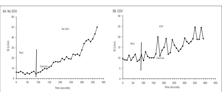
Exercise oscillatory ventilation
Minute ventilation generally increases linearly during pro-gressive exercise tests. In HF populations, however, a number of patients present a waxing/waning VE pattern than has been dei ned as exercise oscillatory ventilation (EOV). h e body of research investigating this phenomenon in patients with HF is not as robust as the work done in the areas of peak VO2 and
the VE-VCO2 relationship. h e analysis of EOV in HF does,
however, rather convincingly indicate that disease severity is signii cantly increased when this ventilatory abnormality is present64,65. Although there is at present no universal dei nition of EOV, an oscillatory VE pattern at rest that persists for ≥ 60% of the exercise test at an amplitude ≥ 15% of the average resting value has been proposed66,67. Figure 5 illustrates the VE pattern at rest and during a progressive exercise test in two patients diagnosed with HF: one with a normal pattern and the other with EOV.
Like elevated VE/VCO2 slopes, EOV has been linked to
increased chemosensitivity in patients with HF68. In addition, oscillations in cardiac function have been reported in patients
with EOV69. Using quantitative algebraic analysis of dynamic
cardiorespiratory physiology, Francis et al.70 concluded that the primary pathophysiological factors resulting in EOV are circulatory delay and an increased chemorel ex gain. While the
impact of interventions on EOV are limited, both milrinone64
and respiratory muscle training36 have been shown to reduce
the occurrence of EOV.
Like peak VO2 and the VE/VCO2 slope, the presence of EOV appears to be a signii cant predictor of adverse events66,67,71,72.
Furthermore, combined assessment of EOV and both peak VO267 and the VE/VCO
2 slope
72 appears to enhance prognostic
signii cance, thus warranting their inclusion when using CPX data to assess prognosis. h e combination of the independence of EOV from subject ef ort and its ability to rel ect cardiac pathophysiology may help to account for the strong prognostic value observed in previous investigations.
Other noteworthy cardiopulmonary exercise
testing variables
Several other CPX variables have been assessed for their
prognostic value in patients with HF. h e oxygen uptake
effi ciency slope (OUES), dei ned as the linear relationship
between VO2 and the logarithmic transformation of VE73,74, the partial pressure of end-tidal carbon-dioxide produc-tion at rest75 and during exercise76, and heart rate recovery
(HRR)77-79 have all demonstrated prognostic value among
patients with HF. Furthermore, both the OUES80 and
HRR81 have been shown to signii cantly increase (improve)
following an aerobic exercise training program among pa-tients with HF. h e additive prognostic value of these variables
to peak VO2, the VE/VCO2 slope and EOV is unclear at this
time. Future investigations are needed in order to determine whether one or more of the variables mentioned should be added to multivariate modeling. Lastly, although not central to the prognostic assessment of patients with HF, monitoring of the hemodynamic and electrocardiographic response to CPX should be performed, particularly to identify potentially life-threatening situations that warrant test termination. A fall in systolic blood pressure during exercise compared with baseline measurements is a test termination criterion20,26 that potentially rel ects worsening left ventricular performance
0 10 20 30 40 50 60
0 50 100 150 200 250 300 350 400
Time (seconds)
VE
(
L/
m
in
)
Exercise Rest
No EOV
0 5 10 15 20 25 30
0 50 100 150 200 250 300 350 400 450
Time (seconds)
VE
(L
/m
in
)
Exercise Rest
EOV
5A. No EOV 5B. EOV
82
and may be a particularly ominous prognostic marker82-85.
Likewise, electrocardiographic evidence of ischemia and/or ventricular arrhythmia is a potentially serious indicator of worsening cardiac function during exercise and may also warrant termination of the CPX26.
Aerobic exercise training considerations
in patients with heart failure
General principles of aerobic exercise training
h e overload, specii city and reversibility principles are key considerations in developing an ef ective aerobic exercise
program. h e overload principle relates to the fact that the
training stimulus must be greater than what the physiologi-cal systems (i.e. cardiovascular and skeletal muscle) are
ac-customed to, for a positive adaptation to occur. h e mode,
intensity, duration and frequency of aerobic exercise are con-sidered in combination, in order to safely use the overload principle for a given training program. Among patients with HF, overload can typically be achieved at a lower training level, particularly during the initial phases of the exercise program,
compared with apparently healthy subjects. h e specii city
principle states that physiological improvements are unique to the mode of exercise performed. For example, walking performance will be optimized with a training program primarily focusing on treadmill training as opposed to lower-limb ergometry or swimming. However, the positive health-related adaptations observed in patients with HF who participate in aerobic exercise training (discussed in a subse-quent section) are achieved with any type of exercise using large muscle groups on a continuous basis (walking/running, lower-limb ergometry, elliptical devices, etc). For the over-whelming majority of patients with HF, the specii city prin-ciple is less important than the fact that moderate aerobic activity of any type has numerous health benei ts. h e type of exercise should therefore be driven by individual preference and the availability of necessary equipment. Lastly, the re-versibility principle states that positive training adaptations are not maintained if an individual returns to a sedentary behavior pattern. Life-long participation in the prescribed aerobic exercise program should therefore be a primary goal.
Specifi c recommendations
for aerobic exercise prescription
Once contraindicated, aerobic exercise training is now a well-accepted lifestyle intervention for patients with
compensated HF. h e general frequency, duration and intensity recommendations for aerobic exercise in this population are 3-5 days/weeks, 30-60 minutes and 50-80% of maximal aerobic capacity, respectively7,14. Walking (treadmill, track or other mea-sured course), lower-limb cycle ergometry (mobile or station-ary) or elliptical units enable physical stressing of larger muscle groups and are therefore acceptable types of exercise. Patients with HF should be guided to progress in frequency, duration and intensity towards the upper end of these aerobic exercise recommendations (i.e. 5 days per week, ~60 minutes per ses-sion, 70-80% of maximal aerobic capacity) over several weeks/ months. While all patients should strive to ultimately achieve these recommendations, it should be recognized that some level of physical activity is always preferable to a sedentary lifestyle.
While continuous aerobic exercise is the ultimate goal, some debilitated patients with HF will not be able to sustain an exercise session for the entire time period at a given intensity, particularly during the initial stages of the training program. In these instances, interval training, i.e. periods consisting of 1-2 minutes of exercise at the desired intensity followed by a lower intensity recovery period, should be used. Progres-sion for patients performing interval training entails a gradual increase in the training duration at a given exercise intensity (1-2 to 2-4 to 4-6 minutes, etc.) before it becomes necessary to start the lower intensity recovery period. h e goal is to guide these patients to progress to continuous bouts of aerobic activity (i.e. 30-60 minutes) over several weeks/months of training.
83 lower end of this range (50%) and gradually progress to ~80% of
the baseline peak VO2 over several weeks or months of aerobic exercise training. h e heart rate associated with this peak VO2 range can be used to monitor compliance with the prescribed exercise intensity during individual training sessions. Because of the potential day-to-day variability associated with heart failure medical management and/or stability, setting an indi-vidual exercise session at ± 5% of the specii c target intensity is recommended14. For example, for a patient with a target exer-cise heart rate of 120 beats per minute, a ± 5% range would be 114-126 beats per minute. Alternatively, a perceived exertion level of 12-14 (on the Borg scale from 6 to 20) may be used to set the exercise intensity for patients who rate their exertion appropriately during the baseline exercise test.
h e level of supervision, particularly at the initial stages of the exercise program, is an important consideration for this high-risk patient population. It is no longer considered neces-sary to recommend that all patients with HF undergo super-vised exercise training with continuous electrocardiographic
monitoring. h is advanced level of supervision should,
how-ever, be strongly considered for patients with a history of car-diac arrhythmias, documented coronary artery disease that has not been surgically addressed or a low ejection fraction (≤ 25%), or whose characteristics resemble those of patients
who suf ered sudden cardiac death14. Furthermore,
irrespec-tive of past medical history, patients who demonstrate an abnormal hemodynamic (hypertensive/hypotensive) response and/or electrocardiographic (ischemia/ventricular arrhyth-mias) abnormalities during the baseline exercise test should undergo supervised exercise training for some period of time.
h e duration and number of supervised exercise sessions
is at the discretion of the health professional responsible for the training program. As a general guideline, patients should demonstrate an ability to appropriately self-monitor the exer-cise session and not have any abnormal physiological responses for several weeks before progressing to unmonitored exercise.
Documented benefi ts of aerobic exercise training
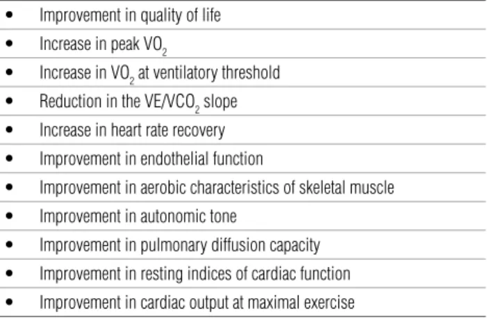
h ere is now a rather impressive body of research
demonstrating numerous health-related benei ts associated with aerobic exercise training among patients with HF7,35,62,81,86,87.
h e benei ts that have been documented are listed in Table
2. Furthermore, the adverse event rate with exercise training appears to be low7.
While one large trial examining the impact of aerobic exercise training on survival and hospitalization among patients with HF is ongoing88, no i ndings have been published to date. A meta-analysis on this topic, pooling together a number of smaller exercise trials (combined n= 801), demonstrated a signii cant
increase in survival and signii cant reduction in hospitalization in the exercise training group, compared with controls. h ese re-sults need to be coni rmed by future prospective investigations. Lastly, the work cited in this section was exclusively performed on patients diagnosed with systolic HF. h e initial evidence indi-cates that the improvements in peak VO2 and quality of life fol-lowing exercise training are similar in patients with systolic and diastolic HF89. Despite these initial i ndings, caution should be applied in extrapolating the documented benei ts of exercising training listed in Table 2 to the diastolic HF population.
Complementary interventions also shown to
improve aerobic capacity
Several other interventions within allied health profession-als’ scope of practice have been shown to improve peak VO2 and should be considered as potential complements to the aerobic exercise training program on an individual basis. Unlike in ap-parently healthy populations, resistance training programs have
been shown to signii cantly improve peak VO2 among patients
with HF90. In addition, resistance training improves bone mineral density, muscle mass and muscle force production to a greater extent than aerobic exercise programs do. In general, resistance training programs for patients with HF should focus on higher numbers of repetitions (≈1-3 sets of 10-12 repetitions) at a lower
load (≈50% of one-repetition maximum). Additional general
recommendations include a training frequency of 1-3 days per week, targeting large muscle groups with 4-9 training stations. Cable or hydraulic resistance systems may be preferable to free weights, from a patient-safety perspective. Subjects with a greater level of HF severity (New York Heart Class I-II vs. Class II-III) should be set tasks at the lower range of these recommendations90. As pre-viously mentioned, subjects with HF may present varying levels of inspiratory capacity impairment that seems to be correlated with peak VO215,16. Inspiratory muscle training may improve respiratory muscle function and peak VO236. h is treatment alternative should
Table 2. Benefi ts of aerobic exercise training in patients with heart failure.
Improvement in quality of life •
Increase in peak VO
• 2
Increase in VO
• 2 at ventilatory threshold
Reduction in the VE/VCO
• 2 slope
Increase in heart rate recovery •
Improvement in endothelial function •
Improvement in aerobic characteristics of skeletal muscle •
Improvement in autonomic tone •
Improvement in pulmonary diffusion capacity •
Improvement in resting indices of cardiac function •
84
be considered when an HF patient presents an inspiratory capacity that is below the normative values predicted for the age and sex. Lastly, chronic electrical myostimulation has been shown to sig-nii cantly improve muscle force production91, VO
2 at ventilatory threshold92 and peak VO
2
92,93 in patients with HF. h ese programs
typically consist of myostimulation to lower extremity muscle groups (bilateral quadriceps plus hamstring or calf muscles), for one to several hours most days of the week for several weeks. Implementation of a myostimulation program may be particularly advantageous for severely debilitated patients who initially are un-able to perform continuous aerobic exercise sessions.
Summary
There is now a robust body of evidence demonstrating the clinical value of both CPX and aerobic exercise training
for systolic HF populations. Cardiopulmonary exercise testing provides valuable prognostic information, is valu-able in assessing the response to numerous interventions and is important in developing individualized exercise pre-scriptions. Participation in an aerobic exercise program is a safe means for improving functional capacity, quality of life and numerous physiological measurements. There is also promising evidence to indicate that aerobic exercise training improves morbidity and mortality in systolic HF populations. These findings need to be reproduced in pa-tients with diastolic HF before concrete CPX and aerobic exercise training recommendations are made for this sub-group. Allied health professionals who are responsible for assessing and treating patients with HF should be aware of the importance of CPX, aerobic exercise training and complementary interventions and, when appropriate, ad-vocate their implementation.
References
1. Armstrong L, Balady G, Berry M et al. Health-Related Physical Testing and Interpretation. In: Whaley MH, Brubaker PH, Otto R, editors. ACSM’s Guidelines for exercise testing and prescription. 7th ed. Philadelphia: Lippincott Williams and Wilkins; 2007. p. 55-92.
2. Tanabe Y, Nakagawa I, Ito E, Suzuki K. Hemodynamic basis of the reduced oxygen uptake relative to work rate during incremental exercise in patients with chronic heart failure. Int J of Cardiol. 2002;83:57-62.
3. Matsumoto A, Itoh H, Eto Y, Kobayashi T, Kato M, Omata M, et al. End-tidal CO2 pressure decreases during exercise in cardiac patients: association with severity of heart failure and cardiac output reserve. J Am Coll Cardiol. 2000;36:242-9.
4. Metra M, Faggiano P, D’Aloia A, Nodari S, Gualeni A, Raccagni D, et al. Use of cardiopulmonary exercise testing with hemodynamic monitoring in the prognostic assessment of ambulatory patients with chronic heart failure. J Am Coll Cardiol. 1999;33:943-50.
5. Myers J, Gujja P, Neelagaru S, Burkhoff D. Cardiac output and cardiopulmonary responses to exercise in heart failure: Application of a new bio-reactance device. J Card Fail. 2007;13:629-36.
6. Duscha BD, Kraus WE, Keteyian SJ, Sullivan MJ, Green HJ, Schachat FH, et al. Capillary density of skeletal muscle: a contributing mechanism for exercise intolerance in class II-III chronic heart failure independent of other peripheral alterations. J Am Coll Cardiol. 1999;33:1956-63.
7. Pina IL, Apstein CS, Balady GJ, Belardinelli R, Chaitman BR, Duscha BD, et al. Exercise and heart failure: A statement from the american heart association committee on exercise, Rehabilitation, and Prevention. Circulation. 2003;107:1210-25.
8. Hambrecht R, Fiehn E, Yu J, Niebauer J, Weigl C, Hilbrich L, et al. Effects of endurance training on mitochondrial ultrastructure and fi ber type distribution in skeletal muscle of patients with stable chronic heart failure. J Am Coll Cardiol. 1997;29:1067-73.
9. Bekedam MA, van Beek-Harmsen BJ, Boonstra A, van MW, Visser FC, van der Laarse WJ. Maximum rate of oxygen consumption related to succinate dehydrogenase activity in skeletal muscle fi bres of chronic heart failure patients and controls. Clin Physiol Funct Imaging. 2003; 23:337-43.
10. Witte KK, Clark AL. Why does chronic heart failure cause breathlessness and fatigue? Prog Cardiovasc Dis. 2007;49:366-84.
11. Mettauer B, Zoll J, Garnier A, Ventura-Clapier R. Heart failure: a model of cardiac and skeletal muscle energetic failure. Pfl ugers Archiv. 2006;452:653-66.
12. Sullivan MJ, Knight JD, Higginbotham MB, Cobb FR. Relation between central and peripheral hemodynamics during exercise in patients with chronic heart failure. Muscle blood fl ow is reduced with maintenance of arterial perfusion pressure. Circulation. 1989;80:769-81.
13. Myers J, Froelicher VF. Hemodynamic determinants of exercise capacity in chronic heart failure. Ann Intern Med. 1991;115:377-86.
14. Myers J. Principles of exercise prescription for patients with chronic heart failure. Heart Fail Rev. 2008;13:61-8.
85 16. Papazachou O, Anastasiou-Nana M, Sakellariou D, Tassiou A, Dimopoulos
S, Venetsanakos J, et al. Pulmonary function at peak exercise in patients with chronic heart failure. Int J Cardiol. 2007;118:28-35.
17. Agostoni P, Bussotti M, Cattadori G, Margutti E, Contini M, Muratori M, et al. Gas diffusion and alveolar-capillary unit in chronic heart failure. Eur Heart J. 2006;27:2538-43.
18. Meyer K, Westbrook S, Schwaibold M, Hajric R, Peters K, Roskamm H. Short-term reproducibility of cardiopulmonary measurements during exercise testing in patients with severe chronic heart failure. Am Heart J. 1997;134:20-6.
19. Gibbons RJ, Balady GJ, Beasley JW, Bricker JT, Duvernoy WF, Froelicher VF, et al. ACC/AHA Guidelines for exercise testing. A report of the American College of Cardiology/American Heart Association Task Force on Practice Guidelines (Committee on Exercise Testing). J Am Coll Cardiol. 1997; 30:260-311.
20. Gibbons RJ, Balady GJ, Timothy BJ, Chaitman BR, Fletcher GF, Froelicher VF, et al. ACC/AHA 2002 guideline update for exercise testing: summary article. A report of the American College of Cardiology/American Heart Association Task Force on Practice Guidelines (Committee to Update the 1997 Exercise Testing Guidelines). J Am Coll Cardiol. 2002; 40:1531-40.
21. Arena R, Myers J, Abella J, Peberdy MA, Bensimhon D, Chase P, et al. Development of a ventilatory classifi cation system in patients with heart failure. Circulation. 2007;115:2410-7.
22. Piepoli MF, Corra U, Agostoni PG, Belardinelli R, Cohen-Solal A, Hambrecht R, et al. Statement on cardiopulmonary exercise testing in chronic heart failure due to left ventricular dysfunction: recommendations for performance and interpretation. Part I: defi nition of cardiopulmonary exercise testing parameters for appropriate use in chronic heart failure. Eur J Cardiovasc Prev Rehabil. 2006;13:150-64.
23. Piepoli MF, Corra U, Agostoni PG, Belardinelli R, Cohen-Solal A, Hambrecht R, et al. Statement on cardiopulmonary exercise testing in chronic heart failure due to left ventricular dysfunction: recommendations for performance and interpretation Part II: How to perform cardiopulmonary exercise testing in chronic heart failure. Eur J Cardiovasc Prev Rehabil. 2006;13:300-11.
24. Piepoli MF, Corra U, Agostoni PG, Belardinelli R, Cohen-Solal A, Hambrecht R, et al. Statement on cardiopulmonary exercise testing in chronic heart failure due to left ventricular dysfunction: recommendations for performance and interpretation Part III: Interpretation of cardiopulmonary exercise testing in chronic heart failure and future applications. Eur J Cardiovasc Prev Rehabil. 2006;13:485-94.
25. Arena R, Humphrey R, Peberdy MA, Madigan M. Predicting peak oxygen consumption during a conservative ramping protocol: implications for the heart failure population. J Cardiopulm Rehabil. 2003; 23:183-9.
26. Fletcher GF, Balady GJ, Amsterdam EA, Chaitman B, Eckel R, Fleg J, et al. Exercise standards for testing and training: a statement for healthcare professionals from the American Heart Association. Circulation. 2001;104:1694-740.
27. Guazzi M, Myers J, Arena R. Cardiopulmonary exercise testing in the clinical and prognostic assessment of diastolic heart failure. J Am Coll Cardiol. 2005;46:1883-90.
28. Guazzi M, Pontone G, Brambilla R, Agostoni P, Reina G. Alveolar-capillary membrane gas conductance: a novel prognostic indicator in chronic heart failure. Eur Heart J. 2002;23:467-76.
29. Ponikowski P, Chua TP, Piepoli M, Banasiak W, Anker SD, Szelemej R, et al. Ventilatory response to exercise correlates with impaired heart rate variability in patients with chronic congestive heart failure. Am J Cardiol. 1998;82:338-44.
30. Reindl I, Wernecke KD, Opitz C, Wensel R, Konig D, Dengler T, et al. Impaired ventilatory effi ciency in chronic heart failure: possible role of pulmonary vasoconstriction. Am Heart J. 1998;136:778-85.
31. Myers J, Dziekan G, Goebbels U, Dubach P. Infl uence of high-intensity exercise training on the ventilatory response to exercise in patients with reduced ventricular function. Med Sci Sports Exerc.1999; 31:929-37.
32. Kruger S, Graf Ju, Kunz D, Stickel T, Hanrath P, Janssens U. brain natriuretic peptide levels predict functional capacity in patients with chronic heart failure. J Am Coll Cardiol. 2002;40:718-22.
33. Passino C, Poletti R, Bramanti F, Prontera C, Clerico A, Emdin M. Neuro-hormonal activation predicts ventilatory response to exercise and functional capacity in patients with heart failure. Eur J Heart Fail. 2006;8:46-53.
34. Scardovi AB, De MR, Coletta C, Aspromonte N, Perna S, Infusino T, et al. Brain natriuretic peptide is a reliable indicator of ventilatory abnormalities during cardiopulmonary exercise test in heart failure patients. Med Sci Monit. 2006;12:CR191-5.
35. Guazzi M, Reina G, Tumminello G, Guazzi MD. Improvement of alveolar-capillary membrane diffusing capacity with exercise training in chronic heart failure. J Appl Physiol. 2004;97:1866-73.
36. Dall’Ago P, Chiappa GR, Guths H, Stein R, Ribeiro JP. Inspiratory muscle training in patients with heart failure and inspiratory muscle weakness: a randomized trial. J Am Coll Cardiol. 2006;47:757-63.
37. de Jonge N, Kirkels H, Lahpor JR, Klopping C, Hulzebos EJ, de la Riviere AB, et al. Exercise performance in patients with end-stage heart failure after implantation of a left ventricular assist device and after heart transplantation: an outlook for permanent assisting? J Am Coll Cardiol. 2001;37:1794-9.
38. Auricchio A, Stellbrink C, Sack S, Block M, Vogt Ju, Bakker P, et al. Long-term clinical effect of hemodynamically optimized cardiac resynchronization therapy in patients with heart failure and ventricular conduction delay. J Am Coll Cardiol. 2002;39:2026-33.
39. Guazzi M, Marenzi G, Alimento M, Contini M, Agostoni P. Improvement of alveolar-capillary membrane diffusing capacity with enalapril in chronic heart failure and counteracting effect of aspirin. Circulation. 1997; 95:1930-6.
40. Guazzi M, Samaja M, Arena R, Vicenzi M, Guazzi MD. Long-term use of Sildenafi l in the therapeutic management of heart failure. J Am Coll Cardiol. 2007;50:2136-44.
41. Agostoni P, Guazzi M, Bussotti M, De Vita S, Palermo P. Carvedilol reduces the inappropriate increase of ventilation during exercise in heart failure patients. Chest. 2002;122:2062-7.
86
43. Mancini DM, Eisen H, Kussmaul W, Mull R, Edmunds LH Jr., Wilson JR. Value of peak exercise oxygen consumption for optimal timing of cardiac transplantation in ambulatory patients with heart failure. Circulation. 1991;83:778-86.
44. Myers J, Gullestad L, Vagelos R, Do D, Bellin D, Ross H, et al. Cardiopulmonary exercise testing and prognosis in severe heart failure: 14 mL/kg/min revisited. Am Heart J. 2000;139:78-84.
45. Arena R, Myers J, Aslam SS, Varughese EB, Peberdy MA. Peak VO2 and VE/VCO2 slope in patients with heart failure: a prognostic comparison. Am Heart J. 2004;147:354-60.
46. O’Neill JO, Young JB, Pothier CE, Lauer MS. Peak oxygen consumption as a predictor of death in patients with heart failure receiving {beta}-blockers. Circulation. 2005;111:2313-8.
47. Mezzani A, Corra U, Bosimini E, Giordano A, Giannuzzi P. Contribution of peak respiratory exchange ratio to peak VO2 prognostic reliability in patients with chronic heart failure and severely reduced exercise capacity. Am Heart J. 2003;145:1102-7.
48. Wilson JR, Ferraro N, Weber KT. Respiratory gas analysis during exercise as a noninvasive measure of lactate concentration in chronic congestive heart failure. Am J Cardiol. 1983;51:1639-43.
49. Arena R, Myers J, Williams MA, Gulati M, Kligfi eld P, Balady GJ, et al. Assessment of functional capacity in clinical and research settings: a scientifi c statement from the American Heart Association Committee on Exercise, Rehabilitation, and Prevention of the Council on Clinical Cardiology and the Council on Cardiovascular Nursing. Circulation. 2007;116:329-43.
50. Myers J. Information from ventilatory gas exchange data. In: Washburn R, editor. Essentials of cardiopulmonary exercise testing. Champaign: Human Kinetics; 1996. p. 83-108.
51. Gitt AK, Wasserman K, Kilkowski C, Kleemann T, Kilkowski A, Bangert M, et al. Exercise anaerobic threshold and ventilatory effi ciency identify heart failure patients for high risk of early death. Circulation. 2002;106:3079-84.
52. Uren NG, Davies SW, Agnew JE, Irwin AG, Jordan SL, Hilson AJ, et al. Reduction of mismatch of global ventilation and perfusion on exercise is related to exercise capacity in chronic heart failure. Br Heart J. 1993;70:241-6.
53. Wada O, Asanoi H, Miyagi K, Ishizaka S, Kameyama T, Seto H, et al. Importance of abnormal lung perfusion in excessive exercise ventilation in chronic heart failure. Am Heart J. 1993;125:790-8.
54. Ponikowski P, Francis DP, Piepoli MF, Davies LC, Chua TP, Davos CH, et al. Enhanced ventilatory response to exercise in patients with chronic heart failure and preserved exercise tolerance: marker of abnormal cardiorespiratory refl ex control and predictor of poor prognosis. Circulation. 2001;103:967-72.
55. Chua TP, Clark AL, Amadi AA. The relationship between chemosensitivity and the ventilatory response to exercise in chronic heart failure. J Am Coll Cardiol. 1996; 27:650-7.
56. Piepoli M, Clark AL, Volterrani M. Contribution of Muscle Affarents to the Hemodynamic, Autonomic, and Ventilatory Responses to Exercise in Patients with Chronic Heart Failure. Circulation. 1996; 93:940-52.
57. Sullivan MJ, Higginbotham MB, Cobb FR. Increased exercise ventilation in patients with chronic heart failure: intact ventilatory control despite hemodynamic and pulmonary abnormalities. Circulation. 1988; 77:552-9.
58. Guazzi M, Reina G, Tumminello G, Guazzi MD. Alveolar-capillary membrane conductance is the best pulmonary function correlate of exercise ventilation efficiency in heart failure patients. Eur J Heart Fail. 2005; 7:1017-22.
59. Francis DP, Shamim W, Davies LC, Piepoli MF, Ponikowski P, Anker SD, et al. Cardiopulmonary exercise testing for prognosis in chronic heart failure: continuous and independent prognostic value from VE/VCO(2) slope and peak VO(2). Eur Heart J. 2000; 21:154-61.
60. Kleber FX, Vietzke G, Wernecke KD, Bauer U, Opitz C, Wensel R, et al. Impairment of ventilatory effi ciency in heart failure: prognostic impact. Circulation. 2000;101:2803-9.
61. Bard RL, Gillespie BW, Clarke NS, Egan TG, Nicklas JM. Determining the best ventilatory effi ciency measure to predict mortality in patients with heart failure. Heart Lung Transplant. 2006; 25:589-95.
62. Arena R, Myers J, Guazzi M. The clinical and research applications of aerobic capacity and ventilatory effi ciency in heart failure: an evidence-based review. Heart Fail Rev. In press 2007.
63. Arena R, Myers J, Aslam S, Varughese EB, Peberdy MA. Technical considerations related to the minute ventilation/carbon dioxide output slope in patients with heart failure. Chest. 2003;124:720-7.
64. Ribeiro JP, Knutzen A, Rocco MB, Hartley LH, Colucci WS. Periodic breathing during exercise in severe heart failure. Reversal with milrinone or cardiac transplantation. Chest. 1987;92:555-6.
65. Feld H, Priest S. A cyclic breathing pattern in patients with poor left ventricular function and compensated heart failure: a mild form of Cheyne-Stokes respiration? J Am Coll Cardiol. 1993; 21:971-4.
66. Corra U, Pistono M, Mezzani A, Braghiroli A, Giordano A, Lanfranchi P, et al. Sleep and exertional periodic breathing in chronic heart failure: prognostic importance and interdependence. Circulation. 2006;113:44-50.
67. Corra U, Giordano A, Bosimini E, Mezzani A, Piepoli M, Coats AJ, et al. Oscillatory ventilation during exercise in patients with chronic heart failure: clinical correlates and prognostic implications. Chest. 2002; 121:1572-80.
68. Lahiri S, Hsiao C, Zhang R, Mokashi A, Nishino T. Peripheral chemoreceptors in respiratory oscillations. J Appl Physiol. 1985; 58:1901-8.
69. Yajima T, Koike A, Sugimoto K, Miyahara Y, Marumo F, Hiroe M. Mechanism of periodic breathing in patients with cardiovascular disease. Chest. 1994;106:142-6.
70. Francis DP, Willson K, Davies LC, Coats AJ, Piepoli M. Quantitative general theory for periodic breathing in chronic heart failure and its clinical implications. Circulation. 2000;102:2214-21.
71. Guazzi M, Raimondo R, Vicenzi M, Arena R, Proserpio C, Sarzi BS, et al. Exercise oscillatory ventilation may predict sudden cardiac death in heart failure patients. J Am Coll Cardiol. 2007;50:299-308.
87 73. Arena R, Myers J, Hsu L, Peberdy MA, Pinkstaff S, Bensimhon D, et al. The
minute ventilation/carbon dioxide production slope is prognostically superior to the oxygen uptake effi ciency slope. J Card Fail. 2007;13:462-9.
74. Davies LC, Wensel R, Georgiadou P, Cicoira M, Coats AJ, Piepoli MF, et al. Enhanced prognostic value from cardiopulmonary exercise testing in chronic heart failure by non-linear analysis: oxygen uptake effi ciency slope. Eur Heart J. 2006;27:684-90.
75. Arena R, Peberdy MA, Myers J, Guazzi M, Tevald M. Prognostic value of resting end-tidal carbon dioxide in patients with heart failure. Int J Cardiol. 2006;109:351-8.
76. Arena R, Guazzi M, Myers J. Prognostic value of end-tidal carbon dioxide during exercise testing in heart failure. Int J Cardiol. 2007; 117:103-8.
77. Arena R, Guazzi M, Myers J, Peberdy MA. Prognostic value of heart rate recovery in patients with heart failure. Am Heart J. 2006;151:851.
78. Lipinski MJ, Vetrovec GW, Gorelik D, Froelicher VF. The importance of heart rate recovery in patients with heart failure or left ventricular systolic dysfunction. J Card Fail. 2005;11:624-30.
79. Sheppard RJ, Racine N, Roof A, Ducharme A, Blanchet M, White M. Heart rate recovery - a potential marker of clinical outcomes in heart failure patients receiving beta-blocker therapy. Can J Cardiol. 2007; 23:1135-8.
80. Van Laethem C, Van De Veire N, Backer GD, Bihija S, Seghers T, Cambier D, et al. Response of the oxygen uptake effi ciency slope to exercise training in patients with chronic heart failure. Eur J Heart Fail. 2007;9:625-9.
81. Myers J, Hadley D, Oswald U, Bruner K, Kottman W, Hsu L, et al. Effects of exercise training on heart rate recovery in patients with chronic heart failure. Am Heart J. 2007;153:1056-63.
82. Froelicher V, Morrow K, Brown M, Atwood E, Morris C. Prediction of atherosclerotic cardiovascular death in men using a prognostic score. Am J Cardiol. 1994; 73:133-8.
83. Morris CK, Morrow K, Froelicher VF, Hideg A, Hunter D, Kawaguchi T, et al. Prediction of cardiovascular death by means of clinical and exercise test variables in patients selected for cardiac catheterization. Am Heart J. 1993;125:1717-26.
84. Dubach P, Froelicher VF, Klein J, Oakes D, Grover-McKay M, Friis R. Exercise-induced hypotension in a male population. Criteria, causes, and prognosis. Circulation. 1988; 78:1380-7.
85. Morrow K, Morris CK, Froelicher VF, Hideg A, Hunter D, Johnson E, et al. Prediction of cardiovascular death in men undergoing noninvasive evaluation for coronary artery disease. Ann Intern Med. 1993;118:689-95.
86. Mezzani A, Corra U, Giannuzzi P. Central adaptations to exercise training in patients with chronic heart failure. Heart Fail Rev. 2008;13:13-20.
87. Haykowsky MJ, Liang Y, Pechter D, Jones LW, McAlister FA, Clark AM. A Meta-Analysis of the effect of exercise training on left ventricular remodeling in heart failure patients: The benefi t depends on the type of training performed. J Am Coll Cardiol. 2007;49:2329-36.
88. Whellan DJ, O’Connor CM, Lee KL, Keteyian SJ, Cooper LS, Ellis SJ, et al. Heart failure and a controlled trial investigating outcomes of exercise training (HF-ACTION): Design and rationale. Am Heart J. 2007;153:201-11.
89. Smart N, Haluska B, Jeffriess L, Marwick TH. Exercise training in systolic and diastolic dysfunction: Effects on cardiac function, functional capacity, and quality of life. Am Heart J. 2007;153:530-6.
90. Braith R, Beck D. Resistance exercise: training adaptations and developing a safe exercise prescription. Heart Fail Rev. 2008;13:69-79.
91. Quittan M, Wiesinger GF, Sturm B, Puig S, Mayr W, Sochor A, et al. Improvement of thigh muscles by neuromuscular electrical stimulation in patients with refractory heart failure: a single-blind, randomized, controlled trial. Am J Phys Med Rehabil. 2001; 80:206-14.
92. Nuhr MJ, Pette D, Berger R, Quittan M, Crevenna R, Huelsman M, et al. Beneficial effects of chronic low-frequency stimulation of thigh muscles in patients with advanced chronic heart failure. Eur Heart J. 2004;25:136-43.