Vol. 51, n. 3 : pp.539-545, May-June 2008
ISSN 1516-8913 Printed in Brazil BRAZILIAN ARCHIVES OF BIOLOGY AND TECHNOLOGY
A N I N T E R N A T I O N A L J O U R N A L
Optical and Ultrastructural Study of the Pollen Grain
Development in Hermaphrodite Papaya Tree (
Carica papaya
L
.
)
Lídia Márcia Silva Santos1, Telma Nair Santana Pereira1*, Margarete Magalhães de Souza2, Pedro Correa Damasceno Junior1, Fabiane Rabelo da Costa1, Beatriz Ferreira Ribeiro1, Noil Gomes de Freitas1 and Messias Gonzaga Pereira1
1
Universidade Estadual do Norte Fluminense Darcy Ribeiro (UENF); Centro de Ciências e Tecnologias Agropecuárias; Laboratório de Melhoramento Genético Vegetal; Av. Alberto Lamego, 2000; Parque Califórnia; [email protected]; 28013-60; Campos dos Goytacazes - RJ - Brazil. 2Departamento de Ciências Biológicas; Universidade Estadual Santa Cruz (UESC); Km 16 Rodovia Ilhéus-Itabuna; 45.650-000; Ilhéus - BA - Brazil
ABSTRACT
The objective of this study was to describe the pollen grain development in hermaphrodite papaya tree. The flower buds were collected at different stages of the development and the anthers were treated chemically for observation under optical and electronic transmission microscopes. The pollen grain development followed the normal pattern described for the Angiosperms. The pollen grain development was described from meiocyte to the mature pollen grain. In the microsporogenesis, the microspore mother cells or the meiocytes underwent meiosis giving rise to the tetrads that were enclosed by the calose. Later, the tetrads were released by the dissolution of the calose by calase activity and microspores underwent mitosis. Microgametogenesis was characterized by asymmetrical mitotic division of each microspore giving rise to bi-nucleate pollen grains. The structures similar to the plastids were found in the cytoplasm and close to the nucleus of the generative cell. Gradual degeneration was observed in the tapetum during the male gamete development.
Key words: Carica papaya, microsporogenesis, microgametogenesis, pollen grain, ultrastructure
*
Author for correspondence
INTRODUCTION
The papaya tree is a polygamous species presenting individuals with male, female and hermaphrodite flowers. The male flower is grouped in panicle-type inflorescences, with a long, straight corolla tube and five free petals. It has ten functional stamens, five upper and five lower, fused to the petals. It may present a very rudimentary and generally sterile female organ (Ronse Decraene and Smets, 1999). The female flower is observed in a cluster of two or three
and yellow colored anthers. The female organ consists of an elongated ovary and papillae stigma. Several floral alterations, such as five-stamened pentandria, carpelloidy and female sterility are observed in hermaphrodite papaya trees, probably due to the environmental influence during flower development (Storey, 1958).
Several studies have been carried out on the papaya tree, but there are few on male and female gamete development. This type of study can generate data of practical use on the formation of the pollen grains and their relationship with the reproduction mechanism of the crop. Considering the lack of data on the reproductive biology of the papaya, it is necessary to establish the pattern of gamete development as well as to generate knowledge that helps to clarify other aspects of the reproductive biology of the crop. The objective of this work was to study the anatomic modifications that occurred during the pollen grain development in the hermaphrodite flowers of the papaya.
MATERIALS AND METHODS
The flower buds at different stages of the development were collected at random from the hermaphrodite plants of the Solo variety. The anthers were fixed in 2.5% glutaraldehyde and 4% paraformaldehyde in 0.1 M sodium cacodylate buffer solution, pH 7.2, for two hours at the room temperature. After fixing, the flower buds were washed three times in 0.1M cacodylate buffer for 30 minutes each time, and post fixed with 1% osmium tetroxide in 0.1M sodium cacodylate buffer for one hour. The anthers were dehydrated in ethanol series 50, 70, 90, and 100% (twice) for a period of one hour at each step. They were blocked in Spurr resin at an ethanol/resin proportion of 3:1, 2:1, 1:1, 1:2, 1:3, and pure Spurr resin (Spurr, 1969); at each step the material remained in the solution for eight hours at room temperature. After infiltration, the anthers were included in the special silicon molds, covered by the Petri dishes and submitted to 70°C for 12 h. For the observation under optical microscope (OLYMPUS BX60), the anthers included in the resin were cut in semi fine sections (0.3 - 0.5µm) with an ultra microtome (LEICA RM2145). The cuts were collected on the slides and stained with the methylene azur II blue for five minutes on a heated plate and counterstained in 2.5% basic fuchsine solution for 30 seconds at room
temperature (Humphrey and Pittman, 1974). After staining, the cuts were set with the entelan and cover sliped.
For observation under the transmission electronic microscope (ZEISS 900, A 60KV), ultra fine cuts were made (50 to 60 nm) with a diamond knife of the same blocks used for the optical microscope observations. The cuts were collected on the copper grids of 200 mesh, stained with the uranyl acetate for 20 minutes, washed in the distilled water, and then contrasted with the lead citrate for five minutes (Venable and Coggeshall, 1965), washed once again in the distilled water and dried on the filter paper.
RESULTS AND DISCUSSION
presented large but fewer vacuoles (Fig. 1, e), probably resulting from the fusion of the several small vacuoles as described by Horner and Palmer (1995). Each microspore nucleus underwent asymmetrical mitosis, giving rise to bi-cellular pollen grains with the vegetative and generative cell (Fig. 1, f), although Frankel and Galun (1977) reported tri-cellular pollen grains for the
Caricaceae family. Bi-cellular pollen grains are
found in at least 192 and tri-cellular pollen grains in at least 150 Angiosperm families (Esau, 1974). In the bi-cellular pollen grain, the generative cell undergoes two mitotic divisions during the pollen grain germination and the pollen tube growth occurs, thus generating the two-gamete nuclei that participate in the double fertilization. The papaya mature pollen grain presented a rounded shape with three germination pores or the apertures (Fig. 1, f).
During the pollen grain development, the tapetum cells showed signs of the degeneration, with altered cytoplasmic contents and cell morphology. These cells seemed to exude some material similar to polenkitt. Gabarayeva (1995) stated that polenkitt conferred variety to the pattern of exine projections, spines and grooves and functioned as a barrier, preventing pathogen entry in the pollen grain, and also was resistant to biodegradation. Ultrastructurally, the MMC presented a dense cytoplasm containing many small vessels, a conspicuous centralized nucleus, with dispersed chromatin and a well-developed nucleolus (Fig. 2, a). The MMCs were isolated from the others by a layer of relatively thin calose, secreted by the tapetum cells. At a more advanced stage, the layer of calose became thicker; the cytoplasm remained dense with a centralized nucleus and an even more developed nucleolus (Fig. 2, b). McCormick (1993) suggested that there was communication between the meiocytes through the plasmodesmas, establishing synchrony in the first meiotic division. In this study, some cells were very close to one another, but no cytoplasmic bridges were observed. The tapetum cells around the meiocyte were, at times, binucleate with a vacuolated cytoplasm, a large quantity of dictiosomes and endoplasmatic reticulum (Fig. 2, c). Frankel and Galun (1977) stated that there was interdependency among the meiocytes and they depended on the cytoplasmic connections with the tapetum cells. These connections decreased as the calose thickened and non-synchronization of the stages of the second meiocyte division could arise,
suggesting that the nucleous was independent of the meiocyte. The tapetum and the other anther cell layers were preserved until the intermediate microspore stage as was reported in soybean by Horner and Palmer, (1995). At this stage, the tapetum cell layers that were still quite thick, presented large vacuoles in their cytoplasm; these cells were mononucleate or binucleate, with one or two nucleoli. These cytoplasmatic characteristics were probably related with the stocking necessary substances and essential proteins synthesis that were possibly used for the nutrition and perfect development of the future pollen grains (Polowick and Sawhney, 1993).
The tetrad presented a thick calose sheath (Fig. 2, d) that increased the distance between the cells; at this time, microspore wall formation was initiated. Gabarayeva (1995) studying Anaxagorea brevipes
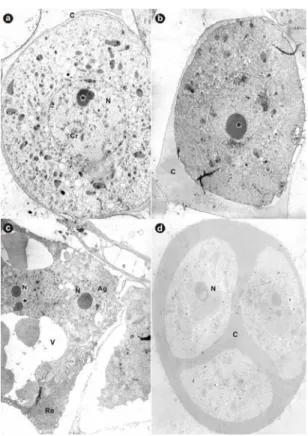
Figure 2 - Transmission electron microscope photographs of pollen grain development in papaya.
a) Meiocyte showing the central nucleus, nucleolus (*), and dispersed chromatin, all enclosed into the callose (x 3.000). b) Meiocyte in advanced development stage showing a prominent nucleolus (*) and thicker callose (x 4500). c) Tapetum cell showing the nucleus and nucleolus (*) (x 4500). d) Detail of tetrad showing the three nuclei and callose around them (x 4500). C: calose; Cr: cromatina; N: nucleus; V: vacuole; Ag: apparatus Golgi; Re: endoplasmatic reticulum;
In addition to the vacuoles, structures similar to the plastids, probably amyloplasts and leucoplasts, and lipid droplets were found widely distributed throughout the cytoplasm (Fig. 3, d-f). The vegetative cell had a smaller nucleus than the generative cell, with diffused genetic material. A developed endoplasmatic reticulum was observed close to its nucleus, and next to this, bodies similar to the lipids (Fig. 3, e). Polowick and Sawhney (1993) and Rodríguez-Garcia et al. (2003) believed that the appearance of lipid bodies in the pollen grain could be related to the energy production for the pollen grain germination or for the pollen tube wall synthesis.
The generative cell presented very little cytoplasm, a relatively large nucleus with a large nucleolus. In the present study, structures similar to the plastids were observed very close to the nucleus of the generative cell (Fig. 3, f). The nature of papaya pollen grain was lipidic as observed by
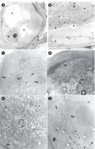
Figure 3 - Ultra structural microphotographs of late phase of pollen grain development.
a)Microspore showing the nucleus located close to the cell wall, vacuoles and the exine and intine (x 3000). b) Detail do the microspore showing the aperture pollen grain (x 3000). C)Detail of binuclear mature pollen grain showing the vegetative nucleus and the generative nucleus (x 4400). d) Detail of mature pollen grain showing the reticular endoplasmitic net, vacuoles and plastid (x 12000). e) Detail of mature pollen grain showing lipid droplets close to the endoplasmic reticulum (x 4400). f) Generative cell involved by structures plastid like showing the prominent nucleolus (x 12000). V: vacuole; N: nucleus; E: exine; I: intine; A: aperture; T: tapetum; P: plastid; Re: endoplasmatic reticulum; Nv: vegetative nucleus; Ng: generative nucleus
ACKNOWLEDGEMENTS
We thank Márcia Adriana S.C. Dutra and Arthur Rodrigues, CBB/UENF Photographic Laboratory technicians, for help in developing and amplifying the photographs for this article. This study was supported by FAPERJ and CNPq.
RESUMO
Foram descritos os estádios de desenvolvimento do grão de pólen desde meiócitos ou células mães de micrósporos até o grão de pólen binucleado. Na microesporogênese, as células esporogênicas sofreram meiose dando origem as tétrades, com posterior liberação dos micrósporos da tétrade. A
microgametogênese caracterizou-se, principalmente, pela divisão mitótica assimétrica
de cada micrósporo originando grãos de pólen binucleados. Estruturas semelhantes a plastídios foram encontradas no citoplasma da célula generativa e agregados próximos ao núcleo da célula generativa. Observou-se degeneração gradativa das células do tapete no decorrer da formação do gameta.
REFERENCES
Esau, K. (1974), Anatomia das plantas com sementes. Tradução de Berta Lange de Morretes. São Paulo: Ed.
Edgard Blucher LTDA.
Damasceno Júnior, P. C. (2004), Estudo reprodutivo em mamoeiro (Carica papaya L.). Doctorate Thesis,
Universidade Estadual do Norte Fluminense Darcy Ribeiro, Campos dos oytacazes, RJ, Brasil.
Frankel, R. and Galun, E. (1977), Pollination Mechanisms, Reproduction and Plant Breeding. Springer-Verlag, Berlin Heidelberg New York. Gabarayeva N. I. (1995), Pollen wall and tapetum
development in Anaxagorea brevipes (Annonaceae): sporoderm substructure, cytoskeleton, sporollenin precursor particles, and the endexine problem. Review of Palaeobotany and Palynology, 85:123-152. Horner, H.T. and Palmer, R.G. (1995), Mechanisms of
genic male sterility. Crop Science,35, 1527-1535. Humphrey, C. D. and Pittmann, F. E. (1974), A simple
methylene blue-azure II basic fuchsin stain for epoxi-embedded tissue sections. Stain Technology, 49, 9-14.
Maheshwari, P. (1950), An introduction to the embryology of angiosperms. New York: McGraw-Hill.
McCormick, S. (1993), Male gametophyte development. The Plant Cell, 5, 1265-1275.
Noher de Halac I., Cismondi, I. A., Rodríguez-Garcia, M. I. and Famá, G. (2003), Distribution of pectins in the pollen apertures of Oenothera hookeri. Velansster/+ster. Biocell, 27, 11- 18.
Polowick P. L. and Sawhney v. K. (1993), An structural study of pollen development in tomato (Lycopersicon esculentum Mill.) .II. Pollen maturatin. Canadian Journal of Botany, 71, 1048-1055.
Rodríguez-García, M.I., M’rani-Alaoui, M., Fernández, M. (2003), Behavior of storage lipids during development and germination of olive (Olea europaea L.) pollen. Protoplasma, 221, 237- 244.
Ronse Decraene, L.P. and Smets, E.F. (1999), The floral development and anatomy of Carica papaya (Caricaceae). Canadian Journal of Botany, 77, 582-598
Souza, M. M. and Pereira, T. N. S. (2000), Development of pollen grain in yellow passion- fruit (Passiflora edulis f.flavicarpa; Passifloraceae). Genetics and Molecular Biology, 23, 469-473.
Spurr A. R. (1969), A low viscosity epoxy resin embedding medium for electron microscopy. Journal of Ultrastructure Research, 26, 31- 43.
Storey, W. B. (1958), Modifications of the sex expression in papaya. Horticultural Advances, 2, 49 – 60.
Storey, W. B. (1969), Papaya. In: Ferwerda, F. P. and Wit, F. (eds.). Outlines of Perennial Crop Breeding in the Tropics. H. Vienman and Zonen N. V. Wageningen. pp. 389-407.
Venable, J. H. and Coggeshall, R. (1965), A simplified lead citrate stain for use in electron microscopy. Journal of Cell Biology, 25: 407.