Carlos André de Oliveira Faria
Robotic Implantation of Intracerebral
Electrodes for Deep Brain Stimulation
Car los Andr é de Oliv eir a F ar ia 2 R obo tic Im plant ation of Intracer ebral Electrodes f
or Deep Brain Stimulation
Universidade do Minho
Tese de Mestrado
Ciclo de Estudos Integrados Conducentes ao
Grau de Mestre em Engenharia Biomédica
Trabalho efetuado sob a orientação de
Professora Doutora Estela Bicho
Neurocirurgião Doutor Manuel Rito
Carlos André de Oliveira Faria
Robotic Implantation of Intracerebral
Electrodes for Deep Brain Stimulation
Universidade do Minho
Acknowledgements
I want to express a very special thanks to my scientific advisor Prof. Estela Bicho, for the opportunity to work in this fascinating project, for the extent knowledge, experience and guidance provided. Her unwavering support and enthusiasm were deeply appreciated in both good and difficult moments and a determinant factor for the final outcome of this work.
I would like to thank my co-advisor Dr. Manuel Rito for proposing the idea of the dissertation, for the motivation, support and availability. His knowledge and experience were fundamental.
I would also like to express my gratitude to all my lab colleagues, Eliana Silva, Emanuel Sousa, Flora Ferreira, Hung Vu, Luís Louro, Miguel Sousa, Rui Silva, Tiago Malheiro and Toni Machado. To them i am sincerely thankful for all they taught me, for their warm welcoming and for their friendship.
I want to thank Coimbra University Hospitals Neurosurgery Service for the avail-ability and welcoming environment, and for the opportunity to visit a neurosurgery operating room and attend a real surgery.
I want to thank Prof. Wolfram Erlhagen for the opportunity to work on the project Pest-C/MAT-UI0013/2011 with the Centre of Mathematics of the University of Minho with the grant support provided by "Fundação para a Ciência e a Tecnolo-gia" with the reference UMINHO/BIC/8/2012.
I would like to finish my acknowledgments by expressing my deepest gratitude to my parents and my brother for the unconditional support in all moments of my life, because without them it would be impossible to have reached this far.
Abstract
The objective of this dissertation is to develop an initial approach of a robotic system to play an assistive role in Deep Brain Stimulation (DBS) stereotactic neurosurgery. The robot is expected to position and manipulate several surgi-cal instrumentation in a passive or semi-active role according to pre-operative directives and to medical team instructions. The current impact of neurological disorders sensitive to DBS, the underlying knowledge of neurostimulation and neu-roanatomy, and practical insight about DBS surgery is studied to understand the ultimate goal of our project. We elaborated a state of the art search on neuro-surgery robots to get the picture of what was done and what could be improved. Upon determining the optimal robotic system characteristics for DBS surgery, we conducted a search on industrial robotic manipulators to select the best candi-dates. The geometric and differential kinematic equations are developed for each robotic manipulator. To test the kinematic equations and the control application in a virtual operating room environment, we used the CoopDynSim simulator. Be-ing this simulator oriented to mobile robots, we introduced the serial manipulator concept and implemented the selected robots with all specifications. We designed a control application to manoeuvre the robot and devised an initial interface towards positioning/manipulation of instrumentation along surgical trajectories, while em-phasizing safety procedures. Although it was impossible to assess the robot’s precision in simulation, we studied how and where to place the manipulator to avoid collisions with surrounding equipment without restricting its flexibility. Keywords: Deep Brain Stimulation; Robotic Neurosurgery; Non-redundant and Redundant Manipulator Kinematics; 3D Robotics Simulator
Resumo
O objectivo desta dissertação é o desenvolvimento de uma abordagem inicial a um sistema robótico para desempenhar um papel de assistência em neurocirurgia estereotáxica de Estimulação Cerebral Profunda (DBS). O robô deve posicionar e manipular variados instrumentos cirúrgicos de uma forma passiva ou semi-ativa de acordo com diretivas pré-operativas ou com as instruções da equipa médica. O im-pacto atual dos distúrbios neurológicos sensíveis a DBS, o conhecimento subjacente de neuro-estimulação e neuro-anatomia, e conhecimento prático sobre a cirurgia de DBS são estudados para concluir sobre o objectivo final do nosso projeto. Nós ela-borámos uma pesquisa sobre o estado da arte em robots neurocirúrgicos para per-ceber o que tem sido feito e o que pode ser melhorado. Após determinar o conjunto óptimo de características de um sistema robótico para cirurgia de DBS, nós procu-ramos manipuladores robóticos industriais para escolher os melhores candidatos. As cinemáticas geométricas e diferenciais são desenvolvidas para cada manipula-dor robótico. Para testar as equações cinemáticas e a aplicação de controlo num ambiente virtual de uma sala de operações, nós usamos o simulador CoopDynSim. Sendo este manipulador orientado a robôs móveis, nós introduzimos o conceito de manipuladores em série e implementamos os robôs selecionados com todas as especificações. Nós projetamos uma aplicação de controlo para manobrar os robôs e desenvolvemos uma interface inicial no sentido do posicionamento/manipulação de instrumentação ao longo de trajetórias cirúrgicas, enfatizando os procedimen-tos de segurança. Embora não tenha sido possível avaliar a precisão do robô em simulação, nós estudamos como e onde posicionar o manipulador de forma a evitar colisões com o equipamento circundante sem restringir a sua flexibilidade.
Contents
I Introduction
1
0 Aim and Outline of the dissertation 3
1 Neurological Disorders and Deep Brain Stimulation 7
1.1 Neurological Disorders Impact in Public Health . . . 7
1.1.1 General Burden . . . 8
1.1.2 Parkinson’s Disease . . . 10
1.1.3 Epilepsy . . . 11
1.1.4 Essential Tremor . . . 12
1.1.5 Dystonia, Psychiatric and other Neurological disorders . . . 12
1.2 Deep Brain Stimulation Principles . . . 13
1.2.1 Patient Selection . . . 15
1.2.2 Basal ganglia anatomy and circuitry . . . 19
1.2.3 Neurostimulation . . . 22
1.3 Deep Brain Stimulation Surgery . . . 25
1.3.1 Preoperative Management and Target Selection . . . 26
1.3.2 Intraoperative . . . 29
1.3.3 Which tasks can be accomplished by a robotic manipulator? 33 2 Robotic Systems for Deep Brain Stimulation Neurosurgery 37 2.1 Motivation . . . 37
2.2 Robotic Systems for Neurosurgery . . . 38
2.3 State of the Art Robotic Systems . . . 43
2.3.2 NeuroMate . . . 46 2.3.3 Pathfinder . . . 48 2.3.4 Robocast . . . 50 2.3.5 Rosa . . . 51 2.3.6 Evolution 1 . . . 52 2.3.7 Minerva . . . 54 2.3.8 NeuroArm . . . 55
2.3.9 Other robotic systems for stereotactic procedures . . . 57
2.4 Current Challenges and Directions . . . 58
II Developed Solution
63
3 Industrial Robot System Search 65 3.1 Robot features . . . 663.1.1 Mechanism type . . . 66
3.1.2 Degrees of Freedom . . . 67
3.1.3 Rigidity . . . 67
3.1.4 Workspace . . . 68
3.1.5 Force and Position Control . . . 69
3.1.6 Precision and Repeatability . . . 70
3.2 Available robotic systems . . . 71
4 Manipulator Kinematics 77 4.1 Kinematics Problem . . . 77
4.2 Spatial Descriptions and Transformations . . . 79
4.3 Geometric Kinematics . . . 83
4.3.1 Geometric Direct Kinematics . . . 85
4.3.2 Geometric Inverse Kinematics . . . 87
4.4 Differential Kinematics . . . 95
4.4.1 Differential Direct Kinematics . . . 95
4.5 Selected Robots Specifications . . . 99
4.5.1 ABB IRB 120 . . . 99
4.5.2 Motoman MH5 . . . 103
4.5.3 Schunk LightWeightArm II . . . 106
5 Simulator 111 5.1 CoopDynSim 3D robotics simulator . . . 113
5.2 Adaptation of CoopDynSim . . . 119
5.2.1 Objects and World . . . 119
5.2.2 Manipulator Robots . . . 125 5.3 Controller Interface . . . 141 5.3.1 Utilities . . . 143 5.3.2 Kinematics . . . 144 5.3.3 Client Robot . . . 150 5.3.4 Client Communication . . . 151 5.3.5 Client DBS . . . 152 5.3.6 User Interface . . . 155 5.3.7 Safety . . . 160
III Conclusions
169
6 Results 171 6.1 Fitting in the Operating Room . . . 1716.1.1 ABB IRB 120 results . . . 176
6.1.2 Motoman MH5 results . . . 178
6.1.3 Schunk LWA II results . . . 180
6.1.4 Comparative analysis . . . 181
6.2 Trajectory execution . . . 183
7 First Conclusions 187 7.1 Summary and Discussion . . . 187
Nomenclature
BDI Beck Depression Inventory CNS Central Nervous System CT Computed Tomography
DALY Disability Adjusted Life Year DBS Deep Brain Stimulation
DOF Degrees of Freedom ET Essential Tremor
FDA Food and Drug Administration GABA Gamma-AminoButyric Acid GBD Global Burden of Disease GPe Globus Pallidus Externus GPi Globus Pallidus Internus HRI Human Robot Interaction Hz Hertz
IPG Implanted Pulse Generator MDRS Mattis Dementia Rating Scale
MMES Mini Mental Status Examination MRI Magnetic Resonance Imaging PD Parkinson’s Disease
PPN Pedunculopontine Nucleus
RMIS Robotic-assisted Minimally Invasive Surgery SNc Substantia Nigra (pars compacta)
SNr Substantia Nigra (pars reticulata) STN Subthalamic Nucleus
UI User Interface
UPDRS III Unified Parkinson’s Disease Rating Scale, Part III(Motor Subscale) VMI Ventral Intermediate Nucleus of the Thalamus
WHO World Health Organization YLD Years Lived with Disability YLL Years of Life Lost
List of Figures
1.1 Disability adjusted life-year. . . 9
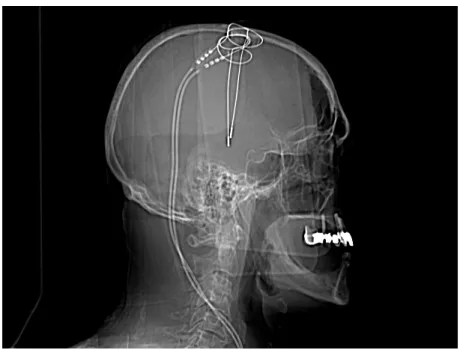
1.2 Postoperative X-Ray scan after the attended DBS surgery that took place at the Coimbra University Hospitals. . . 14
1.3 Schematic of the basal ganglia anatomy. . . 20
1.4 Diagram of the basal ganglia pathways. . . 21
1.5 Preoperative MRI scans previous to the attended surgery at the Coimbra University Hospitals. . . 27
1.6 Setting phantom coordinates. . . 30
1.7 Marking the entry position. . . 30
1.8 Surgical steps. . . 31
1.9 Electrode placement, signal registration and stimulation. . . 32
1.10 Calibration of stimulation parameters. . . 32
2.1 Neuromate. . . 47
2.2 Pathfinder . . . 48
2.3 Rosa Robot. . . 52
2.4 Universal Robots Systems, Evolution1. . . 53
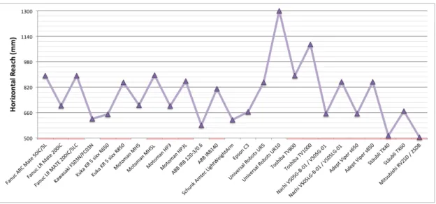
2.5 NeuroArm concept . . . 56 2.6 BrainTumorRobot concept . . . 58 3.1 Manipulator weight . . . 72 3.2 Controller weight . . . 73 3.3 Payload . . . 73 3.4 Horizontal reach . . . 74
3.5 Repeatability . . . 74
4.1 Joint types . . . 84
4.2 Anthropormorphic arm. . . 88
4.3 Plane projection formed by links 2 and 3. . . 89
4.4 Spherical wrist. . . 91
4.5 Elbow redudancy. . . 92
4.6 Elbow angle ✓4. . . 93
4.8 ABB IRB 120 manipulator at starting position with the frames assigned to each link, according to Denavit-Hartenberg convention. 101 4.10 Motoman MH5 manipulator at home position with the frames as-signed to each link, according to Denavit-Hartenberg convention. . . 105
4.12 Schunk Light Weight Arm at starting position with the frames as-signed to each link, according to Denavit-Hartenberg convention. . . 108
5.1 CoopDynSim interface window (operating room environment). . . . 112
5.2 CoopDynSim interface window (mobile robotic simulator). . . 114
5.3 Diagram of CoopDynSim basic architecture. . . 115
5.4 Object’s properties in the simulated world. . . 116
5.5 Middleware abstraction layer modularity. . . 118
5.6 Communication message protocol. . . 118
5.7 Operating Table 3D Model with Stereotactic Frame in Solidworks. . 121
5.8 Utility Cabinet 3D Model in Solidworks. . . 121
5.9 3D model conversion to OpenGL readable cpp files. . . 122
5.10 Simulator architecture on object classes and dependencies. . . 123
5.11 Surgical lights graphical and physical representations. . . 124
5.12 Abb IRB 120, Motoman MH5 and Schunk LWA II assembled in their home position. . . 126
5.15 Robot end-effectors. . . 135
5.16 Probe end-effector guided by the MH5 robotic arm towards a sur-gical target. . . 138
5.19 Euler Angles Z-X’-Z” convention. . . 154
5.21 Control Application UI, Developer panel. . . 156
5.22 Control Application UI, User panel. . . 158
6.1 Generic trajectories to be reached by the manipulator. . . 172
6.2 Generic trajectories representation. . . 173
6.3 End-effector positioned at X mm from the entry point collinear to the desired trajectory. . . 174
6.5 ABB IRB 120 trajectory reaching results. . . 176
6.6 Motoman MH5 trajectory reaching results. . . 179
List of Tables
1.1 Summary of generally accepted criteria of deep brain stimulation
candidacy for treatment of Parkinson’s disease. . . 17
1.2 Summary of generally accepted criteria of deep brain stimulation candidacy for treatment of Tourette syndrome. . . 19
2.1 Advantages and disadvantages of human and robots capabilities. . . 40
2.2 Main neurosurgical robotic projects able to perform DBS and prin-cipal features (adapted from [1]). . . 44
4.1 ABB IRB 120 manipulator limits. . . 101
4.2 ABB IRB 120 arm segments length. . . 102
4.3 Denavit-Hartenberg parameters for the ABB IRB 120 manipulator. 102 4.4 Motoman MH5 manipulator limits. . . 104
4.5 Motoman MH5 arm segments length. . . 105
4.6 Denavit-Hartenberg parameters for the Motoman MH5 arm. . . 106
4.7 Schunk Light Weight Arm manipulator limits. . . 107
4.8 Schunk Light Weight Arm segments length. . . 108
4.9 Denavit-Hartenberg parameters for the Schunk Light Weight Arm. . 109
Part I
Chapter 0
Aim and Outline of the dissertation
The aim of this dissertation is to contribute to the development of a robotic system able to facilitate the implantation of intracortical electrodes for Deep Brain Stim-ulation (DBS), for symptomatic treatment of neurological disorders. The robotic system should be able to hold and manipulate instrumentation according to the planned trajectories and assist other neurosurgeon needs. More important, the aimed solution should be pragmatic, easy to integrate in a healthcare institution, with low budget acquisition and maintenance costs. We will focus in implementing an initial solution in simulation, to test the control application, the implemented algorithms, the robotic systems selected and the feasibility of the overall project. Due to time and resources limitations real robots implementation will not be cov-ered in the dissertation.
Deep Brain Stimulation is a surgical treatment based in applying controlled electrical pulses directly to specific regions of the basal ganglia, and it has proven to be rather successful in mitigating symptoms of neurological disorders (e.g. Parkinson’s disease, epilepsy, dystonia, among others) when other conventional drug therapies or resection neurosurgeries fail [2] [3]. The number of cases as-signed to DBS has been steadily increasing, however due to the time demanding and exhausting characteristics of the procedure, they can not be performed at the necessary pace [4]. Medical teams involved in DBS neurosurgery believe that the procedure could be made less physically and cognitively demanding if robots
could take over some tasks, but naturally always under the ultimate control of neurosurgeons. Skull drilling, implantation of multiple electrodes for monitoring and stimulating brain structures are some examples of tasks that could be accom-plished by a robotic system. Additionally, the precision, steadiness and tirelessness so characteristic of robotic systems are a major contribute for improving the final outcome of the treatment.
The presented dissertation is organized in three major parts that subdivide in chapters and are presented as follows.
Part I, includes the introductory chapters:
In Chapter 1, we present the impact of the DBS sensitive diseases in current society, some principles about neurostimulation and neuroanatomy. We describe a typical DBS surgery and identify the potential benefits of using a robotic system. In Chapter 2 is formalized the motivation of the dissertation and provided some general concepts about robotic neurosurgery. It is also elaborated a state of the art review on the robots in neurosurgery that can be potentially adapted to DBS. In the end, we present the current trends and challenges for neurosurgery robotically assisted.
Part II, describes the steps to reach an initial solution:
In Chapter 3 it is presented a search in industrial robotic systems that can be used in our project. It starts by explaining the robotic features and its impact in light of our aimed system. We defined an optimal robot profile and compared the products of the most renowned industrial robots industries to find the best candidate.
Chapter 4 describes the algorithms followed to develop the 6 DOF and 7 DOF geometric and differential kinematics. It is also described some of the selected robots specifications that have direct implication on the developed kinematic equa-tions.
In Chapter 5, we present the work developed in upgrading the CoopDynSim robotics simulator, the creation of the operating room environment and the
imple-mentation of the selected robotic serial manipulators. We also present the designed controller application and all the embedded features, taking into account the ex-pected DBS assistive behavior.
Part III, shows some first conclusions about the ongoing work:
Chapter 6, it is presented some results regarding the size and flexibility impli-cations of each robotic system considering the intraoperative environment and the placement of the robot within the operating room. It is also depict the robot’s trajectory execution.
Chapter 7 has a brief overview of the developed solution, discusses some of the findings and options taken along the dissertation, and suggests some objectives to be answered in future work.
Chapter 1
Neurological Disorders and Deep
Brain Stimulation
This chapter reviews the epidemiology of neurological disorders sensitive to DBS treatment and attends theoretical principles regarding DBS treatment and surgery. It also discusses the utility of a robotic manipulator as an assistive tool within the operating room. This review is based in the following textbooks [5] [6] [7] [2] [8].
1.1 Neurological Disorders Impact in Public Health
Public health has always been a main concern in any complex society and a growing effort has been put towards health promotion and disease prevention. Health promotion is a process that enables people to have a better control over their health and to further enhance it. It aims to raise the global consciousness about health risks, establish healthy policies and provide environments to sustain good health practices. The World Health Organization structured a healthy public philosophy as a conjunction of health education, community development and interventions. On the other hand, disease prevention stands for a policy of actively fighting the disease in its various stages:• Primary prevention, consists in quantifying health parameters to avoid the appearance of a disease;
• Secondary prevention, stands for a correct diagnosis and administration of the standard treatment while taking into account the risks involved;
• Tertiary prevention, includes rehabilitation, palliative care or treatment to restrain complications and miniaturize the effect of the disease.
While recognizing the importance of each strategy, in this dissertation we will focus the DBS treatment which is part of the tertiary prevention.
Neurological disorders are one of the major public health threats, causing mo-tor and cognitive impairment that directly restricts an ordinary life style. Such disabilities imply direct and indirect costs to the society, so it is of great interest to prevent or manage its consequences. Several studies and projects have been commenced to evaluate the impact of neurological diseases, more specifically the epidemiology and burden to the community. Among the other epidemiology stud-ies relative to specific neurological disabilitstud-ies, it was also regarded a broader essay known as the Global Burden of Disease leaded by WHO, the World Bank and the Harvard School of Public Health. The primary objective of this section is to envis-age the panorama of neurological diseases and the impact they exert in the society, without acknowledging the causes. Furthermore, from all neurological disorders we will narrow our scope to the ones sensitive to DBS therapy [9] such as Parkinson’s Disease, Dystonia, Essential Tremor, Epilepsy, Neuropathic Pain and Psychiatric Disorders.
1.1.1 General Burden
The Global Burden of Disease study, presents not only the raw numbers of in-cidence and prevalence of each disease, but also quantifies the impact it has in peoples lives in terms of DALY, YLD and YLL, (figure 1.1). The neurological disorders consequences go beyond the statistical data, and to assess the real reper-cussions one needs to consider the impairment caused to each afflicted individual. Quoting data presented in GBD, neurological disorders contributed to 92 mil-lion DALYs in 2005, overcoming Tuberculosis, HIV/AIDS, Malignant neoplasms and Ischaemic heart, Respiratory and Digestive diseases. Epilepsy and Parkinson’s
Figure 1.1: Disability adjusted life-year1.
disease alone constitute almost 9 million DALYs which represents more than 0.6% of the total DALYs for all the diseases covered in the GBD essay. One of the most worrying facts, is the detected growing tendency of both disease DALYs, which is expected to ascend 6% by 2030.
Another interesting information regarding the motto of this dissertation, is to quantity how DBS sensitive neurological disorders affect each stratum of society. GBD presents in their table 2.5 [5], the impact of each neurological disease in countries of high, upper middle, lower middle and low income categories according to the World Bank. The Epilepsy and Parkinson’s disease, the only ones sensi-tive to DBS treatment among the studied have the highest DALYs per 100 000 population in low income countries. Almost half of the total DALYs caused by these disorders are associated to low and lower middle income categories, or in other words, population with hardly any access to excellence healthcare services, or rather expensive treatments like DBS or involving robotic surgery.
The large impact that these neurological disorders have in today’s society fur-ther reinforce the idea that efficient solutions like DBS are welcome to brief the problem. Along with the DBS treatment the numbers displayed in the subsec-tions below justify the need of an auxiliary robotic system that would assist the neurosurgical team to achieve better outcomes in less time, with better working
1subsection 1.1.1 Free licensed media from Wikimedia Commons, with the authorship
of Planemad http://commons.wikimedia.org/wiki/File:DALY_disability_affected_life_ year_infographic.png
conditions and therefore allow more DBS procedures to be performed in the same time interval. An affordable robotic equipment would enable more healthcare in-stitutions, even in low income countries, to acquire and exploit all the advantages of an assistive robotic system in DBS surgeries.
We will hereby list the more important diseases responsive to DBS treatment, introducing its definition, enumerating the symptoms associated and providing an overall perspective of its significance in todays society.
1.1.2 Parkinson’s Disease
Parkinson’s disease is a chronic and progressive neurodegenerative disorder of insid-ious onset, recognized by the bradykinesia, rest tremor and posture disturbances. It can be later on, associated to other motor and non-motor symptoms like postural instability, falls, freezing gait, speech and swallowing difficulties, among others [5]. It holds the second position among the most common neurodegenerative disor-ders, next only to Alzheimer’s disease, and is expected to cause increasing econom-ical and social burden due to the populations aging. Parkinson’s disease causes are largely unknown and furthermore its subtle progression and symptoms are also similar to other motor disorders. Thus a neurologist can at best provide a probable diagnosis, which may later be definitely confirmed post-mortem.
Adding to the variability inherent to an epidemiology study from sampling the population, there is yet room for subjective methodologies relative to the diagnosis criteria. To reduce the impact of these inconsistencies, we gathered information from large sample studies. A broad study carried on in 2006, states that the prevalence of PD in industrialized countries is estimated to be 0.3% and reaches 4% in elder population [10]. The same study reports an incidence rate of 8-18 per 100 000 person-years, however this range englobes the population from all ages and the onset of PD is rarely noticed before 50 years old. As a result of being a chronic disease, the prevalence is much higher than its incidence.
The disease’s course and outcome varies from patient to patient, and the symp-toms which are usually mild and unilateral at the first stages, if left untreated after
several years, can lead to significant motor deterioration with loss of independence. The symptoms limit an ordinary lifestyle, emotionally, socially and economically as it usually implies an early retirement, the stigma of a chronic and incurable condition and an additional burden to families or communities.
1.1.3 Epilepsy
Epilepsy is a chronic neurological disorder prevalent in both genres and contrary to PD, common at all ages. The WHO defined it as a disorder of the brain char-acterized by an enduring predisposition to generate epileptic seizures, and by the neurobiological, cognitive, psychological and social consequences of this condition. The definition of epilepsy requires the occurrence of at least one epileptic seizure. Theoretically any individual with a functioning brain can have a seizure, the factor that triggers it is related to a threshold which can be set by genetic variations or premature birth. Epileptic seizures can manifest in various forms depending on the underling cause and lead to different prognosis, which ultimately condition the treatment approach. The epilepsy can be characterized by the age of the be-ginning of symptoms. When the seizures begin at childhood they normally remit spontaneously, while when they begin in adolescence they are often lifelong but frequently sensitive to antiepileptic drugs.
Epilepsy affects people of all ages, so the YLDs related to this disease are in average more per individual than PD, and thus the need to measure the different impact it has in society. According to Sander et al. [11], the incidence of Epilepsy ranges 40-70 per 100 000 person year, and 100-190 per 100 000 person year in poorer resource countries. So not only are the people in this countries more prone to develop Epilepsy, they have also less access to quality healthcare. In the study presented by the WHO, it states that the incidence among the children is higher and more variable ranging from 25 to 840 cases per 100 000 population per year. Even knowing from community based studies that 70 to 80% of the people afflicted will achieve remission, the disability and stigma caused to the others makes it one of the most dramatic psychological and social impairments since an early age.
The burden of Epilepsy extends beyond the estimated 50 million peopled af-fected worldwide, to roughly 200 million family members or friends who are ac-countable for them and therefore indirectly affected too [5].
1.1.4 Essential Tremor
Essential tremor is known as the most prevalent tremor disorder, and one of the most common neurological disorders. Despite this alarming fact, the etiology and pathophysiology are still not well understood according to neurologists [12]. This movement disorder is a chronic, progressive and degenerative brain disease, dis-tinguishable by a 4-12 Hz kinematic tremor that occurs during volitional motions unlike the tremor latent in PD, which is predominantly basal. Aside from the tremor, ET afflicted patient’s often experience ataxic gait and balance problems. Moreover in a psychological level it is usually diagnosed anxiety, depressive symp-toms and social phobia [13].
Once again for ET, it is not an easy task to present a definite study that describes the real impact of the disease in terms of prevalence and incidence in population due to the flexibility of diagnose criteria. Quoting Louis, ED. [14], it is estimated that 30 to 50% of supposed cases of ET are misdiagnosed as parkin-sonian or other forms of tremor, which means that the epidemiology data may be underestimated. Nonetheless, ET is expected to have a prevalence range from 0.4 to 3.9%, and an incidence rate of 77-326.3 cases per 100 000 person years as stated by recent studies.
1.1.5 Dystonia, Psychiatric and other Neurological
disor-ders
Besides the previously mentioned nearological disorders, also Dystonia [15], Neu-ropathic pain [16], and Psychiatric disorders [3] like Obsessive Compulsive Disor-der [17] and Gilles de la Tourette [18] symptom’s can be lessened by means of Deep Brain Stimulation. Despite displaying lower incidence and prevalence rates, they are not in any way easier to bear than the previous ones. Patient’s afflicted face
unsurmountable and incapacitating problems that go from the inability to carry on a normal life, to the non-acceptance of disease related symptoms by society, which ultimately results in depression, obsessive-compulsive behaviors, stress and in extreme cases self-injurious behaviors or suicide.
Dystonia is a syndrome of sustained muscle contractions frequently causing twisting and repetitive movements, or abnormal posture, which may be classified according to the age of beginning of symptoms, body distribution and cause. It has a prevalence of 2-50 cases per million population year for early onset, that is before 20 years old, and 30 to 7320 per million of population year for the late onset, after 20 years old [15].
Gilles de la Tourette is condition whose symptoms include repeated and quick movements or sounds performed without the control of the patient, that last longer than a year. These motor and vocal disorders are referred as tics. Tourette syn-drome is also characterized by the relative young age of its onset that ranges from 2 to 21 years with a mean of 7 years old [18]. It has therefore a tremendous nega-tive impact in any afflicted individual. Despite the expected remission [19] a study by Pappert et al. showed that from a set of cases followed since the onset of the disease, around 90% of the patients still presented tics in adulthood [20].
1.2 Deep Brain Stimulation Principles
Deep brain stimulation is a technique used in functional neurosurgery, which consists in applying controlled electrical pulses directly to deep brain structures through implanted electrodes linked to a neuropacemaker [21] [2]. It has proven successful in mitigating symptoms of neurological disorders (e.g. Parkinson’s dis-ease, Epilepsy, Essential Tremor, Dystonia, other Neurological and Psychiatric disorders), where other conventional drug therapies or resection neurosurgeries fail [3] [9]. Despite the previous works on brain stimulation through electric signals, only in the 1990s did Benabid and his colleagues brought together the definitive electrode implantation associated to a neuropacemaker device which introduced the long-term chronic DBS [22]. The growing acceptance and success of this
tech-nique is in large measure due to its non-destructive and reversible characteristics, with unknown side effects to date [23]. Even though the theory of localized electri-cal stimulation is known for decades, it has only recently been employed in patients as a consequence of recent developments in imaging technology and clinical instru-mentation [24].
The major impediment for the adoption of DBS surgical treatment is the re-quired knowledge of both electrophysiology and neural principles with applied electrical signals, which stands as a challenge to already overworked healthcare professionals. As healthcare professionals are more familiarized with pharmaco-logic concepts, the choice of treatment is often biased to drug therapy. Moreover, the DBS method does not seem to be as attractive as genetics or stem cell studies for treatment of Parkinson or Epilepsy disorders, if we compare the number and impact of papers submitted per year for each solution. Specialists also tend to amplify risks involved in a standard DBS procedure and disregard the drawbacks latent in today’s drug based treatments. However the most relevant fact is the fact that DBS symptomatic treatment clinical results far exceed those from stem cell treatment [23].
Figure 1.2: Postoperative X-Ray scan after the attended DBS surgery that took place at the Coimbra University Hospitals.
Literature brings some guiding and approaches to obtain the best results from DBS therapy, and addresses topics like number and configuration of electrodes, type of stimulation used in terms of amplitude and shape of the signal, and the type of electrical field generated by electrodes. DBS is focused in symptomatic management, but also provides an unique chance to study brain function and dysfunction theories [23] [9] [2].
One of the main drawbacks of this therapy is the absence of an absolute knowl-edge about the electrical stimulation mechanisms of action on deep brain struc-tures. The treatment works like a black box in which, the neurosurgeons place stimulation electrodes in planned coordinates to cover a volume around the ex-pected optimal target. Then it is applied controlled electrical inputs and assessed the patient’s symptomatic variation in a qualitative scale by neurologists. Even the slightest change in the input parameters, like electrode position or signal prop-erties, has a significant impact in the symptomatic response. Consequently, it is imperative to guarantee the correct positioning of each electrode, carefully se-lect and program the input stimulus to obtain the optimal symptomatic response, which will be in part referred in our work.
The success of DBS therapy is strongly dependent on three factors: i) a careful selection of patients, ii) the correct placement of the leads into the sensorimotor regions of the target nuclei, and iii) the optimal choice of electrical parameters for stimulation. In this subsection we will address the candidate selection step and discuss the major guidelines to choose the electrical parameters. Electrode placement methodologies will be addressed in the Deep Brain Stimulation Surgery section (cf. section 1.3).
1.2.1 Patient Selection
Patient selection is the first and one of the most crucial steps of DBS as not all candidates are apt to receive the treatment. The final decision whether to apply the treatment is usually deliberated based on a multidisciplinary group composed of neurologists, neurosurgeons and neuropsychologists [25]. They must always weight
the risks of both the surgery and treatment in light of the potential benefits. Not all the patients afflicted by DBS sensitive neurological disorders are eligible for this treatment. Therefore, it is particularly important to attend to patient selection also as a mean to quantify the impact that a treatment like DBS can have in society. However and as it will be shown, despite the relative high percentage of people who would benefit from this treatment, a large number is left apart [26]. DBS treatment is currently considered a case of success, fact that lead to a widespread interest and as a result, large and increasing numbers of patients are regularly assigned to DBS surgery. The large number of patients adding to the limited resources to provide this treatment, the limited experienced neurosurgeons and movement disorders neurology teams, dictate the need to define a criteria to restrict the number of patients with access to DBS. The selection method should consider the potential surgery morbidity and mortality, all costs associated to DBS, the time and effort demanded from the patient, caregivers and all the medical team assigned [26].
Neurologists have understood to a certain point the advantages and limita-tions of DBS and are able to predict different treatment improvements based on the symptoms. DBS tends to be an effective solution for symptoms like tremor, bradykinesia, rigidity, dyskinesia and motor fluctuations. On the other hand, the therapy response is not convincing enough for symptoms such as dysautonomia, cognitive dysfunction, dysphagia, micrographia, hypophonic speech and gait or balance problems [27].
One of the first steps when evaluating the eligibility of a PD patient is to con-firm the idiomatic PD diagnosis as it is the more sensitive to DBS than other parkinsonian syndromes. Being this condition confirmed, the neurologist evalu-ates the patient’s feedback to Levodopa2. Despite the fact that DBS is often a
substitute to conventional pharmacological treatments when they become ineffec-tive, neurologists expect a minimum feedback from drugs as a sign of potential improvement.
2subsection 1.2.1 One of the most common drugs used to treat PD and dopamine-responsive
Neurologists do not recommend DBS to patients above a certain age limit. This decision is backed up by higher risks of intraoperative cardiopulmonary events and to a direct relation between age and incidence of cognitive changes after surgery. Patients who present preoperative cognitive decline tend to get worse postoper-atively as concluded from empirical data. However it is hard to specify a rigid threshold for a chronological age from which to deny DBS treatment, as it does not necessarily correlate with the biological age.
The table 1.1 taken from [27] presents some practical and explicit criteria to decide the eligibility of a PD patient for DBS treatment [25].
Table 1.1: Summary of generally accepted criteria of deep brain stimulation candidacy for treatment of Parkinson’s disease.
Inclusion Criteria Exclusion Criteria
Diagnosis of idiopathic PD Serious surgical comorbidities Disabling or troubling motor
symp-toms, including motor fluctuations or dyskinesia, despite optimized pharma-cological treatment
Uncontrolled psychiatric illness, in-cluding anxiety and mood disorder (BDI3 > 15)
Robust motor response (other than tremor) to levodopa (> 30% improve-ment of UDPRS III score4)
Dementia (MMSE5 24, MDRS6
130)
Clear understanding of risks and real-istic expectations from surgery
Preoperative MRI with extensive white matter changes or severe cerebral atro-phy
Also for Epilepsy, the cost and complexity associated to DBS surpasses the anti epileptic drug treatment. Regardless of this fact, brain stimulation is often
3subsection 1.2.1 Multiple choice self-report inventory to assess the severity of depression 4subsection 1.2.1 Scale to assess motor signs in patients with PD
5subsection 1.2.1 Brief questionnaire to screen for cognitive impairment and/or dementia 6subsection 1.2.1 Scale that measures cognitive functioning dementia
used as a suplementar method for patients affected with localized epilepsy and low responsive/intolerable to drugs or that lack the precondition to undergo surgery. One of the disadvantages of DBS is that, patients treated this way have the number of seizures reduced but rarely become seizure-free, unlike in resection surgery. A particular interesting advantage of DBS, is that its toxicity does not overlap the medical therapy one, and until date there are no known side-effects [28].
Dystonia is one of the most difficult to evaluate movement disorders since it is a clinical syndrome instead of a disease, which results from several causes. The patient selection criteria for Dystonia follows the standard preventive and aware-ness guidelines common to other disorders when entrusted to DBS. Neurologists believe that when the patient is afflicted by focal dystonia, one can be successfully treated with botulinum toxin injections in most cases. However in hemidystonia, segmental or generalized dystonia as a result of the extent of muscles involved, the medical treatment is often inefficient, which leaves stereotactic surgery as the most indicated solution [29].
In 2006, Mink et al. [30] elaborated a viewpoint considering the current outcome expectancy of DBS when treating Tourette syndrome patients, and presented some guidelines to help select the best candidates for the treatment and consequently assure the best results. Tourette syndrome is often associated to neurological or psychiatric comorbid symptoms, that have a direct impact in the disorder ex-pression, in the surgery outcome and in the recovery [30]. Regarding the patient selection problem, the authors defined both inclusion and exclusion criteria, table 1.2.
To conclude the topic of patient selection it is essential for neurologists to elucidate the patient about all the procedure since the preoperative until treatment period, explaining the risks as well as the realistic expectations for the DBS. By submitting to this therapy the patient should understand that it consists in a palliative care and therefore will not have any effect in both curing or delaying the progression of the disease, instead it is only focused in suppressing the symptoms to provide a better quality of life. Patients should be aware of the duration of the treatment and the necessity of tuning the parameters of DBS to reach an
Table 1.2: Summary of generally accepted criteria of deep brain stimulation candidacy for treatment of Tourette syndrome.
Inclusion Criteria Exclusion Criteria
> 25 years old7 Movement disorders resulting from other conditions
Chronic and severe tic disorder, with major functional impairment (YGTSS > 358)
Severe medical, neurological or psy-chiatric disorders that may affect the surgery outcome or the recovery Failed conventional medical therapy
(lacking efficiency or unbearable side-effects)
Sensitive to other non-invasive treat-ments
Patient fit for surgery, with treated co-morbidity conditions
Patient unwilling to undergo surgery, or not totally aware of its implications.
optimal symptomatic response as it is not a straightforward solution. Furthermore, candidates to DBS should not expect a functional improvement greater than the best peak of effect of medication, for most symptoms except tremor.
1.2.2 Basal ganglia anatomy and circuitry
Being the deep cortical structures like the basal ganglia, the aim for electrode placement and stimulation, we want to provide some insight regarding the basic anatomy and circuitry of this area.
Deep inside the brain gray matter is located the basal ganglia which along with the diencephalon and the cerebral cortex constitute the prosencephalon or forebrain. This anatomical region is connected to the cortex and thalamus and consists of four main nuclei: the striatum, the globus pallidus, the subthalamic
7subsection 1.2.1 Stable degree of severity with low chances for remission.
8subsection 1.2.1 Yale Global Tic Severity Scale that goes from 0 to 50, and provides score
nucleus and the substantial nigra. In figure 1.3 we present a diagram layout of a brain coronal section with the principal elements that form the basal ganglia subtitled [31]. The basal ganglia system, is according to current standards sub-divided into three functional territories comprising sensorimotor, associative and limbic which process motor, cognitive and emotional or motivational information, respectively. For that reason this anatomical region is the main target of DBS electrode implants.
Figure 1.3: Schematic of the basal ganglia anatomy. [31]
The interaction between structures functions as a "center-surround" mecha-nism to execute desired actions or inhibit unwanted movements. The caudate and putamen are considered the input nuclei of the basal ganglia for sensorimotor in-formation, and together form the striatum which receives excitatory information mostly from the cerebral cortex. The information then flows from the striatum to GPi and SNr, which are thought to be the main output, through anatomi-cal and neurochemianatomi-cal distinct projections. From there it goes to the thalamus, and afterwards to the frontal cortex or dorsolateral prefrontal cortex, depending on whether the desired output is pallidal or nigral respectively. This
corticocor-tical loop passes through the well-known direct pathway basal ganglia circuitry. However to correctly modulate its functioning, it is important to refer the indirect pathway which involves the GPe and then the STN. After reaching the subthalamic nucleus, the information then proceeds to the GPi and SNr, ending in the same structure as in the direct pathway. The striatum, the GP and the STN receive a dopaminergic input from the SNc, which controls and balances the activity of the direct and indirect pathways. Moreover, the STN also receives excitatory inputs from both motor and premotor cortexes and applies a strong excitatory stimuli on its target nuclei. Figure 1.4 summarily explains the relations between nuclei and how the information is handled [8] [32].
Figure 1.4: Diagram of the basal ganglia pathways [33].
As we can see in figure 1.4, the dopamine has a differential effect on different subpopulations of striatal neurons, playing an excitatory role in the direct pathway and inhibitory role in the indirect pathway. This control decreases the inhibitory
effect of the system and allows the execution of a desired movement/behaviour. In the Parkinsonian state the lack of dopamine leads to a disinhibition of the GPi and SNr and an increase inhibition of the thalamocortical projections. Such events have as final effect the appearance of a resting tremor consequence of the non-suppression of involuntary movement and bradykinesia or akinesia due to the stronger inhibition felt at the thalamocortical projections, which are common symptoms of PD.
According to a largely cited review by Perlmutter et al. [24], the neurosur-geons/neurologists team select different neurological targets depending on the dis-order to be treated. For instance, when treating essential tremor, the VIM stim-ulation presents an average tremor reduction over 80% in most patients, however stimulating the VIM region to reduce PD symptoms only affects limb tremor hav-ing no effect in other symptoms. Still in PD, GPi stimulation can reduce most of the motor disorders as well as brief medication related side-effects but it does not free the patient from drug treatment which is a major drawback. So regard-ing DBS to treat PD, the most common target is the STN, which can reproduce similar symptom reduction effects while also reducing the patient dependency of dopaminergic medication. However, the final target selection, follows a bit more complex analysis than the one here described.
Since Dystonia manifestations are similar to PD and other tremor disorders and in front of the success of GP and thalamus stereotaxic ablative procedures, it was a matter of time before trying DBS on this very same structures. The result of this trials, defined GP as the primary target to lessen Dystonia symptoms with an improvement ranging from 30 to 50%, having the thalamus as a secondary target. In a relatively recent application of DBS to treat Tourette syndrome, the target selected was the centromedian-parafascicular complex of the thalamus.
1.2.3 Neurostimulation
Being the Neurostimulation the ultimate goal of our project, we decided to briefly describe the fundamental theoretical basis behind the brain stimulation of deep
structures.
The fact that DBS is relatively recent, the lack of complete knowledge about the interaction between electrical pulses and brain activity plus the physiological variability among the patients makes the programming process of DBS variables an extensive and complex equation. To understand the effect of all electrode configu-rations and stimulation parameters, it would be necessary to conduct a systematic and thorough assessments [23]. The choice of mono over multipolar configurations, the polarity and distribution of microelectrodes, even characteristics of stimulation like amplitude, pulse width and rate of signals need to be adjusted while consid-ering the inherent variability of each patient. An iterative approach would take countless hours to determine the optimal selection of variables.
Despite the variety of combinations of DBS parameters for stimulation that may rise to 12964 possible combinations of pulse width, frequency, and voltage plus 65 electrode configuration combinations, the majority of patients responded well to a known and specific set of configurations [34]. To the other cases however, it is necessary to spend more time studying the patient and calibrating variables to reach an optimal response.
One can brief the problem of DBS programming into two subquestions. The first that indicates how to adjust the properties of the generated electric field, which allows the selective stimulation of different neural elements within the field. This method explores DBS’s electrophysiological principles and is based in the influence of each variable such as pulse width, electric current or voltage of the stimulus and electrode configuration. The management of this parameters has a key influence in the improvement of DBS symptomatic control. The second takes into account parameters like size, shape and anatomical positioning of the electric field. It reduces or even eliminates side-effects and/or other possible complications. Improved DBS efficiency
Dostrivsky et al. [35] believed that high frequency stimulation of GPi, STN and VIM, the most common method used to suppress neurological disorder’s symptoms had an inhibitory effect that lead to a decrease of the simulated structure output,
by analogy to the outcome of ablative/resection methods. On the other hand, low frequencies were thought to worsen symptoms and signs of PD, as it would excite the target. However, Dostrivsky explanation left key issues untouched and has recently been refuted due to clear evidence from studies like [36] and [37], which confirm that both low and high frequency DBS stimulation can excite and increase the output of the target structure [38].
Low frequencies have already proven their usefulness when compared to high frequencies, by providing a better symptomatic control over dystonia, speech or gait problems. High frequencies present finer symptomatic control in motor dis-orders, ET and typical signs of PD. The exact mechanism of how the frequency modulates the neural activity is yet to be uncovered.
In terms of signal modulation, there seems to be clear advantages pointing out to current stimulation. When the signal is voltage modulated, the injected current varies in inverse proportion to the local impedance obeying the Ohm’s Law. However, the impedance is difficult to calculate due to the subjective nature of each patient and to the properties of the brain volume around the target. Consequently there exists a risk of the current overcoming the safety limits due to an unforeseen abrupt drop of impedance9. If current modulation is chosen, the signal current is
controlled and its waveform is preserver [39].
Reduction of Side-effects
As previously referred in subsection 1.2.2, a stimulus can produce different re-sponses depending on the stimulation site. This variations can be ascribed to the singular neuronal circuitry related to each structure, to the variations of neurons shape and properties among each region. The variation of symptomatic response is so marked that even inside the same neural structure, the stimulation can re-produce very distant effects as a result of the non homogenous shape of a nuclei.
There are authors like Montgomery et al. [37], that consider DBS success to be a trade-off between efficient stimulation of desired targets and the ability to avoid
the spreading of electrical current to nearby neural structures. By applying greater intensities of current or stimulating larger areas it is possible to recruit more action potentials in a greater number of neural elements. Aside from the increased efficacy on briefing the symptoms, more unwanted structures are also being stimulated which leads to more side-effects. This fact supports the categorical importance of a precise electrode placement and also helps neurologists/neurosurgeons teams to troubleshoot the current physiological position of the electrode based on the symptoms and side-effects.
If the stimulation side-effects still have a significant impact even upon reaching optimal stimulation coordinates, the medical team may change the combinations of current, voltage, signal shape, area of effect and even the distribution of the electrical field that can be modulated through the use of various contacts of the quadripolar electrode.
Other policies to restrict the activation of undesired structures exploit the prin-ciples of how neural elements react to the different characteristics of the stimulus. For instance, axons with a larger diameter have lower thresholds than smaller ones. Additionally, axons have lower thresholds when compared to dendrites, which in their turn are easier to excite than cell bodies. Knowing this beforehand allows the adjustment of the applied stimuli to exclusively affect more sensitive targets like larger diameter axons or axonal terminals.
1.3 Deep Brain Stimulation Surgery
Deep brain stimulation affects only local brain structures and circuitry, instead of the whole body as in medication. However, the amplitude of the stimulation sig-nal decreases with the distance from the electrode, so the correct placement of the lead is crucial for the success of the therapy. After addressing the basic anatomy and circuitry of the Basal Ganglia and unveiling some of the principles behind neurostimulation, we will now talk about the general DBS surgical procedure. As we aim to develop a robotic oriented solution to aid in the intraoperative DBS pro-cess it is of key importance to fully comprehend each surgery step. Understanding
how the surgery unfolds and perceiving any pre or intraoperative requirements, environment or procedure constraints is key information to set how the robotic system will operate.
In order to acquire practical insight about the surgery, to grasp when and how can a robotic manipulator be of use, why would it improve both working conditions and the final outcome, we assisted a DBS surgery in the Service of Neurosurgery, Coimbra University Hospitals in Portugal. It will be henceforth described a bilateral DBS surgery conducted in a patient affected by Parkinson’s disease. The surgical steps performed in a DBS procedure are independent of the neurological disorder that affects the patient as the only difference is the location of the target to be stimulated10. More information about DBS surgical technique
can be found in ( [9] chapter 1, [40], [24] and [25]).
One can briefly divide this procedure in sequential substeps that include: initial image guided anatomical targeting, skin incision and burr hole drill and attach-ment of the head reference system, intraoperative microelectrode recording and micro/macroelectrode stimulating and finally macroelectrode quadripolar stimu-lation upon reaching the optimal coordinates. This quadripolar electrodes are then connected to a neurostimulator device called IPG or implantable pulse generator, which goes under the skin [8].
1.3.1 Preoperative Management and Target Selection
After deciding the eligibility of the patient to DBS treatment, the next step is to plan the surgical procedure regarding his symptoms, tolerance to surgery, medical team preferences and available equipment. Furthermore, the medical team must also decide whether to apply unilateral or bilateral stimulation according to the pretended control over the patient’s symptoms and medication reduction. Neuro-surgeons can either chose to implant bilateral DBS leads and IPGs in the same day; or implant bilateral leads one day and insert both IPGs the day after; or
10section 1.3 The surgical technique described, does not exclude the fact that other surgery
do an unilateral insertion of both devices simultaneously or in separate interven-tions. Surgeons usually master the use of their institution’s medical equipment, and comply with the center operating policies.
About a week previous to the surgical placement of the electrodes, the patient undergoes a MRI scan to determine the targets to stimulate, figure 1.5. Although other exams like CT or ventriculography are also eligible for the same effect, MRI is the most advantageous because CT lacks the contrast and definition when analyz-ing soft tissues with close densities, and ventriculography may provoke hemorrhage, confusion and headache [41].
(a) T1-weighted preoperative MRI (GPi pointed by black arrow).
(b) T2-weighted preoperative MRI. (STN pointed by white arrow).
Figure 1.5: Preoperative MRI scans previous to the attended surgery at the Coimbra University Hospitals.
In the surgery day, the reference system of the stereotaximeter is attached to the patient’s head11. To assure the patient safety and prevent infections his head
is shaved and prepared with betadine12. Then the patient is sedated and the
11subsection 1.3.1 The reference system of the stereotaximeter used in the CHUC
neuro-surgery service had a ring shape. In order to simplify its notation we will henceforth call it stereotactic ring.
12subsection 1.3.1 Common topical antiseptic used in hospitals to prepare patient’s skin
pins of the stereotactic ring are inserted with caution to avoid the supraorbital nerve. When placing the ring there must be a special care so that it does not contact with the nose or the occipital bone/neck as it may cause skin erosion. The stereotactic ring should also not block the patient medic line of sight and thus allow the assessment of the ocular movement during the surgery. The pins should firmly secure the ring to keep it from being displaced during each step of the procedure, but not overly tightened, which may cause twists in the structure and affect the system’s accuracy.
With the stereotactic ring correctly placed the patient undergoes a CT scan. Computed tomography scans are more suited at this point, as it is compatible with the ferromagnetic material of the ring, and unlike MRI, CT is less affected by distortional artifacts caused by inhomogeneities in the magnetic field. In the end, an imaging planning software merges the high contrast MRI scans where the neurosurgeons segmented the targets, with the spatially precise CT scans and superimposes the information of each exam to achieve a three dimensional repre-sentation as similar as possible to the patient’s brain. This information is used to aid the neurosurgeon to pick the target and the entry coordinates, so that the final electrode trajectory does not cross any functional or vascular structures.
The final target coordinates to place the leads, can either be achieved using the direct or indirect targeting method. Direct targeting is done by visualizing STN and GPi in imaging scans like T2-weighted MRI or inversion recovery im-ages. Being the STN one of the most common targets for treating PD’s symptoms, together with GPi, both regions are generally identified recurring to T2-weighted MRI scans due to the marked contrast between the white matter and both nu-cleus. Some image processing techniques like windowing can facilitate this task and improve the overall accuracy. Axial and coronal are the most important perspec-tives when it comes to defining the spatial coordinates of the target. The indirect method involves registering and overlaying brain anatomical atlases according to the patient’s scans. From here the target position is inferred or calculated based on established formulas that return the target coordinates based in the distance to well identified landmarks. Indirect targeting while using atlases is both
depen-dent on the accuracy of the given atlas and the topographical correlation between the atlas and the patient’s neuroanatomy. The final coordinates may not be the spot where the stimulation will produce the optimal symptomatic relief, but it is still regarded as an initial target. In the surgery we attended, the neurosurgeons team extrapolated the initial coordinates based in known distances from of well identified landmarks.
1.3.2 Intraoperative
A stereotactic frame is mounted in the stereotactic ring, which orients the electrode cannulae along the trajectory provided by the image planning software. Since the skull has approximately a spherical form, the use of the arc stereotaxic frame set with spherical coordinates is advantageous because it allows the selected trajectory to be concentric with the hole rims in both skull outer and inner limits. The settings for the stereotactic frame are directly provided by the imaging planning software.
After setting the stereotactic frame spherical coordinates using mechanical screws, the frame will orient the driver along the defined electrode insertion tra-jectory. Then, the driver itself is responsible for lowering the electrode’s depth. Each input coordinate in the stereotactic frame is confirmed via a millimetric scale engraved between moving components, thus its precision is directly dependent on the human visual precision.
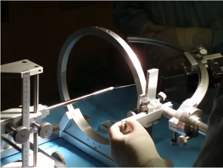
Inside the operating room, while the patient is being prepared, the target coordinates are simulated by means of a phantom device. This phantom has similar sockets to the stereotactic ring fixated on the patient’s skull. For each electrode or target coordinate, the x, y and z cartesian components have to be introduced in the phantom and other 3 spherical coordinates this time are set in the frame, so that in the end, the probe’s tip and the phantom’s tip are coincident, guaranteeing this way that the electrode reaches the target position with a defined orientation, figure 1.6.
Figure 1.6: Setting phantom coordinates.
stereotactic ring on the patient’s head and once again it is set along the desired trajectory. A thin straight metal rod placed in the driver, marks the entry position in the patient’s scalp, figure 1.7.
Figure 1.7: Marking the entry position.
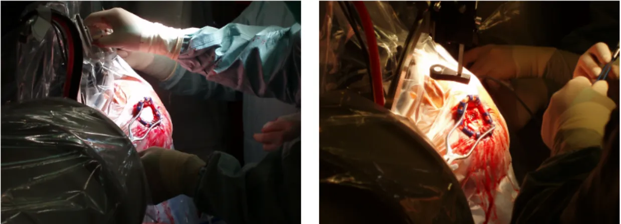
Marked the entry position, the stereotactic arc is set aside to make the scalp incision and to drill the hole in the skull to access the brain, figure 1.8.
(a) Making the scalp incision. (b) Hole drilling in the skull. Figure 1.8: Surgical steps.
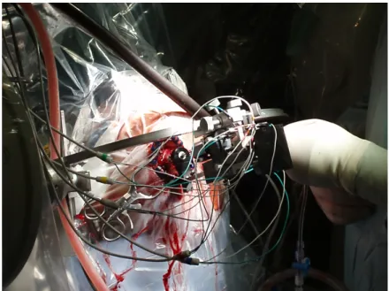
cannulae collinear to the defined trajectory. Firstly, a set of microelectrodes re-sponsible for recording cortical electrical signals are positioned 15mm above the target coordinates defined at the image planning software, along the trajectory and the patient’s sedation lowered. The medical team used 5 electrodes cross posi-tioned, set apart 1mm from each other to cover a wide area and thus increase the probability to find the optimal stimulation site, figure within the nucleus (fig. 1.9). The electrodes are lowered millimeter by millimeter in an iterative process until reaching 5mm from the target. Upon reaching this point the electrodes advance now half a millimeter between iterations, and by each step the signal is recorded. At each iteration the neurophysiological readings are saved, compared and ana-lyzed to assess the most proximal locations to the sensorimotor region, or in other words, the coordinates where the electrodes retrieve an electrophysiological record closer to the expected.
At the coordinates where the electrodes recorded the best signals, these record-ing microelectrodes are replaced by stimulation macro/microelectrodes. For each of this sub-solutions, the electrical current and depth of leads are changed individ-ually in iterative steps. At each step the neurologist team qualitatively evaluates the patient’s symptoms as responses to the stimulus applied, and once again they save each in order to find the optimal electrode trajectory, depth and current, figure 1.10.
Figure 1.9: Electrode placement, signal registration and stimulation.
Figure 1.10: Calibration of stimulation parameters.
Depending in which nuclei we are stimulating there are different current limits and levels of responsiveness, in other words, the GPi in comparison to the STN needs higher current amplitudes to achieve the required therapeutic effect. This becomes an important fact because if the patient complains about side-effects when stimulation parameters should not provoke any, it may indicate a bad electrode positioning. Another important fact to be aware of when introducing multiple
elec-trodes is that it may cause edemas or bleedings which will change the interstitial impedance allow the signal to spread to other structures and cause undesirable effects. This kind of problem is one of the reasons that leads some neurosurgi-cal teams to avoid using recording microelectrodes and implant the stimulation electrode alone [42].
Upon finding the ideal stimulation signal properties and position, the electrodes are replaced by a definitive quadripolar macroelectrode, that should later on be connected to an Implanted Pulse Generator (IPG). If a bilateral brain stimulation is needed, all intraoperative process must be repeated for the other side 13.
1.3.3 Which tasks can be accomplished by a robotic
manip-ulator?
After doing some research about robotic systems oriented to stereotactic keyhole surgery, more specifically DBS surgery, the information found almost exclusively describes the features of the system and displays qualitative data about its effi-ciency. Notwithstanding the factual importance of such details, which shall be discussed in chapter 2, there is quite scarce information about the practical and clinical advantages of including such technology within the operating room. How does it improve the work conditions for neurosurgeons, for neurologists and to other staff? What tasks could be achieved by the robotic system? What are the benefits for the patient?
As stated previously, a typical DBS surgery lasts several hours through which the surgical team must remain utterly focused while performing each step to avoid any failure. Due to its characteristics, robotic systems could provide a substantial contribute in consistency, steadiness and precision and therefore improve at the same time, the working conditions for neurosurgeons and the surgical outcome for the patient [43]. After watching DBS surgery and upon a meeting between neurosurgeons and robotic experts, we concluded that a simple robotic system could upgrade the standard procedure in various aspects:
1. Enable coordinates and trajectory information retrieved from the image plan-ning software to be managed by a communication protocol that would link this program to a robotic controller software. The trajectories information would be handled through software instead of having the medical team manu-ally register and insert it both in stereotactic frame and the phantom screws. 2. Avoid the slow process of, mounting and setting the phantom, frame and driver coordinates to test the position and trajectory generated by the image planning software. Dismount the frame, attach it to the patient stereotactic ring, input coordinates again (just to mark the entry position); disassemble, make incision and drill burr hole, and assemble again to finally introduce each set of electrodes. This procedure repeated for each target.
A robotic manipulator would position itself according to the desired tra-jectory in a few seconds and would also open the possibility for frameless stereotactic procedures with guaranteed precision.
3. Avoid stereotactic frame and driver mechanical slacks or loose parts, which may require preoperative calibration. In figure 1.6, it is possible to see that after setting each coordinate for both the phantom and the frame, there is a slight mismatch in their tips meaning that when placing the electrodes in the patient’s brain, they will not reach the target location but a nearby one, which may lead to decrease in the treatment efficiency and increase of side-effects.
4. Enable robotic manipulator to handle multiple end-effectors and surgical instrumentation to execute consistent skull drilling based on force feedback, swift positioning and driving of various sets of electrodes with improved precision. The manipulator could swiftly and precisely constrain drilling and insertion tasks to a predefined trajectory, instead of executing them based in a marked entry position.
5. Enable medical teams to easily take control over the task of advancing the depth of electrodes while evaluating the patient’s symptoms by simple
inter-acting with the robot controller interface, therefore aiding neurosurgeons on that task.
6. Enable an online monitoring of the instrumentation tips absolute coordinates based on their physical dimensions and on the manipulator position relative to the base referential, using geometric direct kinematics. However, this feature can only be used if the instrumentation is moved along the trajectory by the manipulator.
7. Providing assistance to younger and less experienced neurosurgeons as an assistive and training platform and enabling senior highly experienced neu-rosurgeons, who might lost some dexterity, to continue performing surgeries later in their careers. [44].
8. Reduce the risk of data loss or human errors.
Cooperation between neurosurgeons and robots opens a new horizon of possible improvements to quality healthcare and is therefore recognized as the final step in the transition of surgery from the Industrial to Information Age [45].
Chapter 2
Robotic Systems for Deep Brain
Stimulation Neurosurgery
In this chapter we will briefly talk about neurosurgical robots, point out their ad-vantages and current problems when compared to human standard performances, with a focus in DBS neurosurgery. We will also review the available robotic sys-tems oriented or possibly adaptable to DBS procedures, designating the category they fall in and their main features.
2.1 Motivation
As mentioned in the last chapter, neurological disorders have a tremendous impact in today’s society, and burden millions of patients, families and caretakers. Accord-ing to patient selection rules, not all patients affected by the previous enumerated neurological disorders are eligible for DBS, but a significant part is [4] [27]. On the other hand, DBS has proven to be a top notch treatment that despite having no effect in disease progression, it shows very good results as a palliative care by reducing the symptoms and allowing the patient and family to carry on a normal life. Alongside the growing awareness for such procedure, also the number of actual prescribed DBS surgeries is increasing [46]. Furthermore, DBS neurosurgeries tend to be rather long procedures as stimulation parameters are yet to be programmed
![Figure 1.4: Diagram of the basal ganglia pathways [33].](https://thumb-eu.123doks.com/thumbv2/123dok_br/17802989.840961/37.893.197.730.467.926/figure-diagram-basal-ganglia-pathways.webp)