Fausto Daniel dos Santos Queda
Licenciado em Bioquímica
Studies towards modified chitooligosaccharides: a
new approach to NAG-NAM moiety
Dissertação para obtenção do Grau de Mestre em
Bioorgânica
Orientador: Maria Manuel Marques, Investigadora
Auxiliar, FCT-UNL
Júri:
Presidente: Prof. Doutora Jorge Parola Arguente(s): Doutor Sérgio Raposo Filipe
Vogais: Doutora Maria Manuel Marques Doutor Sérgio Raposo Filipe
Fausto Daniel dos Santos Queda, Copyright
Agradecimentos
Resolvi escrever esta secção como forma de exprimir a minha gratidão para todos aqueles que me acompanharam e ajudaram durante esta etapa que culmina uma série provas e desafios algo longo dos últimos anos.
Antes de mais gostaria de agradecer à minha orientadora Prof. Maria Manuel Marques por me ter orientado e por ter acreditado nas minhas capacidades para a realização deste trabalho que se revelou um desafio desde o inicio, sem a sua orientação seria impossível ultrapassar todos os contratempos.
Gostaria de agradecer às pessoas que trabalharam comigo, nomeadamente ao pessoal do laboratório 202, FCT-UNL, Alexandra Loupas, Cátia Santos, Luísa Carvalho, Luísa Maria, Marina Pires, Tomé Silva e Vasco Bonifácio, laboratório Bacterial cell surfaces and parhogenesis, ITQB-UNL Gonçalo Covas e Filipa Vaz pelo apoio, pela paciência e pelas horas em convívio, sem esquecer a Cecília Bonifácio e a Ana Teresa que juntamente com a Rede Nacional de RMN foram importantes para a realização deste projecto, bem como o financiamento PTDC/SAU-IMU/111806/2009 e PTDC/QEQ-QOR/2132/2012
Gostaria agora de agradecer à minha namorada Nídia Almeida, pelo apoio nesta
jornada, que foi e tem sido uma peça fundamental para que as coisas decorressem da forma
mais harmoniosa possível.
Quero também deixar um especial obrigado ao André Dias, João Cascão e Tiago Costa,
pelos jantares de orientação e pelos momentos de diversão. À Helena Coelho e Mariana
Romão por todo o apoio antes e durante a escrita.
Aos meus amigos de longa data Cristina Montenegro, Filipe Sério, José Ribeiro Mariana
Mesquita, Samuel Figueiredo, Rita Nóbrega, Rui Oliveira, que embora uns mais distantes do
que outros também eles peças fundamentais durante esta jornada.
Não podia deixar de agradecer aos quatro amigos que compõem a banda da qual faço
parte, The Big Odds, Bruno Salgado, Fernando Dias, Gustavo Marques e Pedro Fonseca,
que se não fossem eles a tirar-me do trabalho em alguns momentos teria sido muito mais
difícil.
Quero também agradecer à minha mãe Alexandrina Santos por todo o apoio e paciência
ao longo destes anos nesta etapa que agora termina. Ao meu pai Fernando Queda, que
infelizmente não conseguiu ver o terminar desta caminhada, tendo sido vencido nesta recta
final, mas que de certo, estaria muito orgulhoso por me ver completar este ciclo.
O meu obrigado a todos os outros que não menciono mas que foram eles também
Resumo
O peptidoglicano (PGN) bacteriano tem sido associado a infecções bacterianas e à resistência antibióticos. Desta forma, este trabalho surge no enquadramento do problema com qual a comunidade científica se tem vindo a deparar aquando da obtenção de fragmentos de peptidoglicano puros e em quantidade que permita a realização de estudos biológicos. A síntese de fragmentos de PGN é complexa e envolve inúmeros passos de síntese e o seu isolamento e purificação de fontes naturais é difícil.
Este trabalho consistiu em investigar uma estratégia que permitisse transformar um produto considerando um desperdício da indústria alimentar, quitosano, num produto valioso, mureina, um polímero de unidades alternadas de NAG-NAM que se encontram interligadas por pontes peptídicas. Para isso explorou-se uma aproximação quimioenzimática. Numa primeira estratégia procedeu-se à protecção regioselectiva dos grupos hidroxilo da posição O-6 com dois grupos, TBDPS e TPS, com consequentemente introdução da unidade lactil na posição O-3, apenas contando com o impedimento estéreo que estes grupos proporcionavam à entrada do grupo lactil.
Numa segunda estratégia, foi utilizado uma pinça molecular, “molecular clamp”, com vista a forçar uma entrada da unidade lactil de uma forma alternada, recorrendo a uma di-esterificação, usando um ácido di-carboxílico nas posições O-6 alternadas.
As amostras obtidas por estas duas vias foram submetidas a uma hidrólise enzimática. As enzimas utilizadas foram a lisozima e mutanolizina. As amostras digeridas foram ainda avaliadas em relação à afinidade para a enzima mCherry-PGRP-SA. Os produtos obtidos foram avaliados quanto à sua quantidade relativa de NAG e NAM, por uma cromatografia de troca iónica.
Os resultados obtidos demonstraram que a via “molecular clamp” é a estratégia mais promissora permitindo a conversão de quitosano em unidade de NAG-NAM (1:1,18) numa composição semelhante à do PGN natural extraído de S. aureus.
Abstract
The bacterial peptidoglycan (PGN) has been directly associated with bacterial infections and antibiotic resistance. This work deals with the scientific community problem of obtaining peptidoglycan in a pure and reliable amounts for biological studies. The synthesis of PGN fragments. The synthesis of PGN fragments involves multi-step approaches and the isolation and purification from natural sources is usually difficult.
This work consisted on the investigation of a strategy that allows conversion of a food industry disposal, chitosan, in the valuable murein, a polymer of alternated NAG-NAM units that are cross-linked via short peptide bridges. Thus a chemoenzymatic approach was explored.
In a first strategy a regioselective O-6-hydroxyl group protection was performed using two different groups, TBDPS and TPS, with subsequent introduction of the lactyl moiety at O-3 position relying on the steric hindrance of the groups at O-6.
On a second approach, a molecular clamp strategy was applied to block the access to some of the O-3 positions in alternate units. This approach involved a di-esterification at the -O6 positions of two alternate units, using a di-carboxylic acid, resulting in an alternated lactyl insertion in the following step.
After the completion of the chemical modification of chitosan, the samples obtained were digested with two lytic enzymes, Lysozyme and Mutanolyzin. The digested samples were evaluated against its binding to the enzyme mCherry-PGRP-SA. Moreover the content in NAG:NAM of the samples produced was quantified by ionic chromatography.
to the results obtained in this study revealed that the molecular clamp strategy is a promising route to convert chitosan into NAG-NAM oligosaccharides in a composition, NAG:NAM ratio (1:1.18) similar to the PGN isolated from S. aureus.
Key words: peptidoglycan, chitosan, NAG-NAM, chemoenzymatic, molecular clamp,
Figure index
Figure Page
Figure 1.1 Drosophila melanogaster model of Toll and Imd pathway activation,
adapted from Lemaitre et al (5) 2
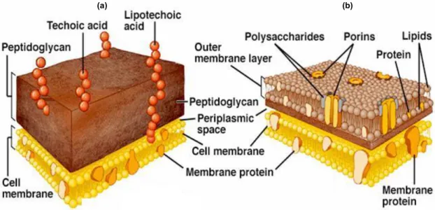
Figure 1.2: Representation of PGN; (a) Gram-positive; (b) Gram-negative;
adapted from Royet et al (71b) 3
Figure 1.3 Bacterial cell wall representation; (a) Gram-positive bacteria cell wall;
(b) Gram-negative bacteria cell wall adapted from 4
Figure 1.5: Schematic representation of glycosylation reaction; (a) via NAG
donor; (b) via N-disubstituted donors 5
Figure 1.4 Schematic NAG-NAM retro-synthesis approaches 6
Figure 1.6 Retrosynthetic view of a dimeric PGN 7
Figure 1.7 Schematic NAG-NAM coupling to the peptide
D-Ala-L-Lys-D-Gln-L-Ala, then coupled with five glycines and finally cleavageof the resin 7
Figure 1.8: Synthesis of glucosamine Donor (A) and Acceptor (B) 8
Figure 1.9 Synthesis of disaccharide NAG-NAM 9
Figure 1.10 Molecular skeleton of chitin (R=acetyl) and chitosan (R=H>50%) 9
Figure 1.11- Antiparallel chain arrangment in α-chitin.(1); parallel arrangement in
β-chitin. adapted from colocar aqui autores reference 33(e) (2) ou colocar A e B 11
Figure 1.12 Scheme of amine moiety protection with phthalic anhydride 12
Figure 1.13 Schematic water-influence in the solvent composition on the
chitosan and phthalic anhydride, reaction product 12
Figure 1.14 Synhesis of a regioselective amphiprotic chitosan; (a)N -phtaloylation; (b)position 6 bromination; (c) position 6 azidation; (d) Husigen
cycloadition; (e) phtaloyl group removal
13
Figure 1.15 Synthetic scheme of N-(4-carboxybutyroyl) chitosan derivatives using different mol ratios of glutaric anhydride 14
Figure 1.16 Synthesis of N,N,N-trimethyl O-carboxymethyl chitosan 16
Figure 1.17 O-3 and O-6 N-acetyl/phtaloyl chitosan simultaneous protection; (a)
Silylation; (b) Acetylation; (c) Tosylation 16
Figure 1.18 Selective O-6 Tosylation then FGI; (b)O-ethyl benzoate chitin; (c)
6-deoxy-diethyl malonate chitin; (d) 6-(deoxydiethyl) phosphitechitin 17
Figure 1.19 O-6-N-phtaloyl chitosan selective protection; (a) Trytilation; (b)
Silylation (TBDMS); (c) Silylation (TBDPS) 18
Figure 1.20 Synhesis of a regioselective amphiprotic chitosan; (a) N -phtaloylation; (b)position 6 bromination; (c) position 6 azidation; (d) Husigen
cycloadition; (e) phtaloyl group removal
19 Figure 1.21 Selective protection/deprotection of chitosan; (a)N-phtaloylation; (b)
O-6 tritylation; (c) O-3 benzylation; (d) O-6 trityl group removal; (e) N-phtaloyl
group removal; (f) O-3 benzyl group removal.
20
Figure 1.22 Conversion of the azide moiety into amine group 21
Figure 2.1 PGN’s sugar backbone retrosynthetic plan 23
Figure 2.2 5 hours chemical chitosan hydrolysis 23
Figure 2.3 Mechanism of chitosan acid assisted hydrolysis 23
Figure 2.4 TLC of 5 hours dialysis. (G) – I.5; (I) - inside of dialysis membrane; (F) - outside of dialysis membrane. Eluent 70% H2O 20% isopropanol 10% aqueous ammonia, revelled with 10%
sulphuric acid in ethanol
24
Figure 2.5 MALDI-TOF spectra from II.8 25
Figure 2.6 N-modification synthesis strategy 26
Figure 2.7 MALDI-TOF spectra from II.10 27
Figure 2.8 Synthesis plan of PGN sugar backbone or murein; (A) asymmetric
protection route; (B) silylation route 28
Figure 2.9 Generic strategy to the silylation route 29
Figure 2.10 Silylation of N-phthaloyl chitosan 30
Figure 2.11 Polymeric chitosan amine moiety protection 30
Figure 2.12 Schematic representation of lactyl moiety insertion 31
Figure 2.14 IR spectra of polymeric chitosan modifications, black – (I.30); red –
(I.32); dark green – (I.47); blue – (II.18); light green – (II.24) 33
Figure 2.15 NMR CP/MAS spectra green I.32; black I.47; 33
Figure 2.16 Linkage between two odd units 34
Figure 2.17 Dicarboxylic acids shynthetized during this work 34
Figure 2.18 Example of a molecular clamp reaction scheme 34
Figure 2.19 NMR CP/MAS green I.32; black II.33 35
Error! Reference source not found. 35
Figure 2.21 Silylation of the remaining O6 free positions 36
Figure 2.22 Muramic acid moiety insertion of O6 molecular clamp and O
6-TBDMS N-phthaloyl chitosan 37
Figure 2.23 Sequence of deprotection and N-acetylation reactions 37
Figure 2.24 IR spectra of polymeric chitosan modifications, black (I.32); red
(II.34); green (II.42); light blue (II.44); blue (II.48) 38
Figure 2.25 Comparison of lysozyme hydrolysis profiles, 206 nm 40
Figure 2.26 Hydrolysis profile of II.48 treated with mutanolyzin; (green); (black)
negative control 41
Figure 2.27 HPLC-UV/MS chromatogram of sample II.48; (red) II.48 +
mutanolyzin; (green) negative controlFigure 2.28 II.48 ESI+ MS, (8.18 min) 42
Figure 2.28 II.48 ESI+ MS, (8.18 min) 42
Figure 2.29 8% SDS-PAGE gel electrophoresis of the mCherry-PGRP-SA
pull-down assay in the presence of native PGN 43
Figure 2.30 mCherry-PRGP-SA percentage of binding of the different samples 44
Figure 2.32 8% SDS-PAGE gel electrophoresis of the mCherry-PGRP-SA
pull-down assay without native PGN 45
Figure 2.31 mCherry-PGRP-SA intensity in the PGRP-SA pull-down assay
without native PGN 45
Figure 4.1 Schematic representation of hydrolysis reactions 48
Figure 4.2 Schematic representation of N-protection reactions 50
Figure 4.3 Schematic representation of molecular clamp synthesis 51
Figure 4.4 Schematic representation of molecular clamp insertion 56
Figure 4.5 Schematic representation of the protection of the remaining O6 free positions 58
Figure 4.6 Schematic representation of muramic acid moiety insertion 59
Figure 4.7 Schematic representation of protecting group removal and and N-acetylation reaction 60
Figure 4.8 Schematic representation of silylation of N-protected chitosan 62
Figure 4.9 Schematic representation of lactyl moiety insertion 63
Figure 4.10 Schematic representation of all deprotection reactions an N-acetylation 65
Figure 4.11 HPLC-UV eluent profile 67
Table index
Table Page
Table 2-1 Summary of the hydrolysis’ work
23
Table 2-2 Summary of first enzymatic, lysozyme, digestion 40
Table 2-3 Summary of the second enzymatic study (92 hours)
41 Table 4-1 Summary of the hydrolysis’ work
52
Table 4-2 HPLC UV protocol eluent composition 67
Table 4-3 Concentration gradient used during the HPLC-UV program 67
Table 4-4 Concentration gradient used during the HPLC-MS program 68
Table 4-5 SDS gel composition
69 Table 4-6 Loading Buffer
69
Table 4-7 Electrophoresis Buffer 69
Table 4-8 Concentration gradient used during the anionic exchange
List of abbreviations
[M+] Molecular Ion
13
C NMR Carbon Nuclear Magnetic Ressonance
1
H NMR Proton Nuclear Magnetic Ressonance
Ac Acetyl
Ar Aromatic
alloc Allyloxycarbonyl
Bn Benzyl
CDI 1,1'-Carbonyldiimidazole
CHPTMAC 3-Chloro-2-Hydroxy-Propyl Trimethyl Ammonium Chloride
d Doublet
DA Deacetylation Degree
DAP Diaminopimelic
DCC N,N'-Dicyclohexylcarbodiimide
DCM Dichloromethane
DIC Carbodiimide
DMF Dimethylforamide
DMSO Dimethylsulfoxide
DP Polimerisation Degree
DS Degree Of Substitution
EC Effective Concentration
equiv. Equivalent
Et Ethyl
Fmoc Fluorenylmethyloxycarbonyl
g Gram
GNBP Gram-Negative Binding Protein
GPC Gel Premeation Chromatography
h Hour
HPLC High Performance Liquid Chromatografy
Hz Hertz
Im Imidazol
J Coupling Constant
m Multiplet
MALDI Matrix-Assisted Laser Desorption/Ionization
Me Methyl
MHz Megahertz
min Minute
mL Milliliter
mmol Millimole
Mn Number Average Molecular Weight
MS Molecular Sieves
MS Mass Spectroscopy
Mw Weight Average Molecular Weight
MW Molecular Weight
N.D. Nothing Detected
NAG N-Acetyl Glucosamine
NAM N-Acetyl Muramic Acid
NMR Nuclear Magnetic Ressonance
PDI Polydispersity Index
PGN Peptidoglycan
PGRP Peptidoglycan Recognition Protein
Ph Phenyl
Phth Phthaloyl
ppm Parts Per Million
q Quartet
rt Room Temperature
s Singlet
SN2 Substitution Nucleophilic Bi-Molecular
T Temperature
t Triplet
TA Teichoic Acid
TBAF Tetra-N-Butylammonium Fluoride
TBDPS Tert-Butyldiphenylsilyl
TCA Trichloroacetyl
TCP Trichlorophthalimide
TEA Triethylamine
TFA Trifluoroacetic Acid
THF Tetrahydrofuran
TLC Thin Layer Chromatography
TMC N,N,N-Trimethyl Chitosan
TMHTMAPC
N,N,N-Trimethyl O-(2-Hydroxy-3-Trimethylammonium Propyl)
TMS Trimethylsilyl
TPS Triphenylsilyl
Tr Trityl
Troc Trichloroethoxycarbonyl
Ts Tosyl
UV Ultraviolet
1
INTRODUCTION
1
1.1
B
ACTERIALI
NFECTIONS1
1.2
D
ROSOPHILA MELANOGASTER INNATE IMMUNE RESPONSE1
1.3
B
ACTERIALC
ELL-W
ALL3
1.3.1
S
YNTHETIC APPROACHES TOPGN
5
1.4
C
HITIN ANDC
HITOSAN9
1.4.1
C
HEMICAL MODIFICATION OF CHITIN AND CHITOSAN10
1.4.1.1
N-modifications
11
1.4.1.2
O-modification
16
2
RESULTS AND DISCUSSION
21
2.1
P
RELIMINARY STUDIES22
2.1.1
C
HITOSAN HYDROLYSIS23
2.1.2
N
-
MODIFICATION OF THE OLIGOMERS PRODUCED26
2.2
P
OLYMERIC CHITOSAN CHEMICAL MODIFICATION28
2.2.1
O
-6
S
ILYLATION ROUTE29
2.2.2
M
OLECULAR CLAMP ROUTE34
2.3
E
NZYMATIC STUDIES OF THE FINAL PRODUCTS39
2.3.1
E
NZYMATIC DIGESTION39
2.3.2
M
C
HERRY-PGRP
PULL-
DOWN ASSAYS43
2.3.3
M
ONOSACCHARIDE COMPOSITION ANALYSIS46
3
CONCLUSIONS AND FINAL REMARKS
49
4
EXPERIMENTAL PROCEDURE
51
4.1
G
ENERALM
ETHODS51
4.2
G
ENERAL PROCEDURES FOR CHITOSAN MODIFICATION52
4.2.1
C
HITOSAN HYDROLYSIS52
4.2.1.1
General procedure for chemical hydrolysis
52
4.2.2
C
HITOSANN
-
PROTECTION53
4.2.3
C
HITOSANO
6-
PROTECTION54
4.2.3.1
Molecular Clamp route
54
4.2.3.2
Silylation route
62
4.2.4
M
URAMICA
CID MOIETY INSERTION66
4.2.5
N
-
ACETYLATION66
4.3
G
ENERAL PROCEDURES TO EVALUATE THE FINAL PRODUCT66
4.3.1
E
NZYMATIC DIGESTION66
4.3.1.1
HPLC-UV protocol
67
4.3.1.2
HPLC-MS protocol
67
4.3.2
M
C
HERRY-PGRP
PULL-
DOWN ASSAY68
4.3.2.1
Electrophoresis
69
4.3.3
M
ONOSACCHARIDE COMPOSITION ANALYSIS69
5
BIBLIOGRAPHY
71
1 Introduction
1.1 Bacterial Infections
There is a huge biodiversity of bacteria, in fact bacteria are the most abundant of all organism with an approximate population of 5x1030 individuals. These bacteria have a large number of shapes and sizes and can be found everywhere even if in radioactive waste or at the Mariana Trench.1
Although the majority of bacteria are harmless, there are a few, called pathogenic bacteria that are responsible for the bacterial infections. In matter of fact the two most deadliest diseases caused by bacterial infections are tuberculosis, caused by the Mycobacterium tuberculosis, which kills almost two million people every year and pneumonia, caused by Streptococcus pneumoniae, Gram-positive, and Pseudomonas aeruginosa, Gram-negative bacteria, killing almost four million people every year.
The innate immune system is the first line of host defence against infection by pathogens. Infection by pathogenic microbes trigger several host-pathogen complex interactions that are mediated by pattern recognition receptors (PRR) that are highly conserved in evolution. Thus, during the early stages of infections, the innate immune responses play crucial roles in host defense and exert an intense influence on the resulting generation of adaptative immune responses. The understanding of the PRR that mediate the innate responses and their subsequent effect after receptor ligation will open room for the discovery and/or improvement of vaccines.2
In order to understand these immunologic responses, insects are the most usual model organism used in these investigations, since there are many mechanisms and pathways of their immune system that have been conversed during the species evolution. Drosophila melanogaster, or commonly fruit fly is one particular insect used in these kind of studies, since its genome was sequenced in the year of 2000, and Drosophila melanogaster has a high rate of reproduction, leading to the emergence of new generations in a short period of time.3
1.2
Drosophila melanogaster
innate immune response
Although the Drosophila melanogaster genome sequence has been fully discovered its innate immune response to a bacterial infection is still under investigation..4 Figure 1.1shows the actually accepted model of innate immune response of Drosophila melanogaster the two pathways of innate immune response are highlighted, the Toll and Imd pathways, which are responsible for inducing an antimicrobial peptide production response.
In drosophila, the Toll pathway is required for resistance to Gram-positive bacteria, containing a lysine-type PGN, as well as fungi and yeast pathogens. The Toll receptor is activated when bacterial lysine-type PGN is recognized by PGRPs, specifically PGRP-SA and PGRP-SD, as well as Gram-negative binding proteins (GNBPs).5 These proteolytic cascades,
which are not yet well characterized, lead to activation of Spätzle-processing enzyme (SPE) that leads to the Spätzle cleavage in the hemolymph. The processed Spätzle binds as a dimer to Toll leading to its dimerization in the plasma membrane leading to activation of three proteins MYD88, Tube and Pell and recruiting to the Toll receptor in the cytoplasmatic region. Subsequently, Cactus is phosphorylated and degraded by proteasome. Transcription factors Dif and Dorsal are released and moved through the nucleus and then activate the transcription genes that encode several antimicrobial peptides leading to the immune response.6
Figure 1.1 Drosophila melanogaster model of Toll and Imd pathway activation, adapted from
The Imd pathway is directly associated to activation by Gram-negative bacteria, possessing diaminopimelic acid-type PGN (DAP-PGN). The Imd pathway is activated by recognition of monomeric DAP-PGN by PGRP-LC/LE, then Imd interacts with dFADD which binds the apical caspase Dredd, enhancing the cell death activity and proteolytic processing the Dredd.7 At the same time a cascade is activated leading to the phosphorylation of IKK signaling complex which also phospholyrate the Relish. This leads to its cleavage and the Rel domain translocate to the nucleus, where it activates the transcription genes that encode several antimicrobial peptides leading to the immune response.7-9
Despite all the studies developed to understand innate immune response and the recognition process between host and bacteria during bacterial infection, some questions remain unanswered. How host receptors can recognize high molecular weight fragments of PGN at the surface of bacteria remains unsolved. Until today it is not clear which are thestructural requirements for PGN recognition by PGRPs.
1.3 Bacterial Cell-Wall
Bacteria can be divided in two major groups the Gram-positive, with lysine-type PGN, and the negative, possessing a DAP-PGN, as it is shown in Figure 1.2.
Gram-positive bacteria cell wall has in its composition, PGN also called murein, teichoic acids (TA) and a cytoplasmatic lipid membrane. Gram-negative bacteria cell wall has in its composition cytoplasmatic membrane, DAP-PGN, also an outer lipopolysaccharide membrane, porins and lipoproteins.
Figure 1.2: Representation of PGN; (a) Gram-positive; (b) Gram-negative; adapted from Royet et al (71b)
These different components in their cell wall provides different characteristics and properties to these two groups of bacteria, properties such as thickness and permeability of the cell-wall, leading to a higher permeability in the Gram-negative bacteria and higher thickness in the Gram-negative ones, Figure 1.3.
PGN is the major component of the bacteria cell wall, is normally composed by a disaccharide, monomer, of N-acetyl glucosamine (NAG) and N-acetyl muramic acid (NAM) and a tetra-peptide, L-Ala, D-iso-Gln, L-Lys, D-Ala, in the Gram-positive bacteria and L-Ala, D -iso-Glu, m-DAP, D-Ala, in the Gram-negative bacteria. However, several modifications in both the glycan structure, and the aminoacids in the cross-linking bridge and the peptide stem, have been reported leading to numerous new structures of PGN. These modifications can be as varied as a glycine in the first position, a D-iso-glutamate in the second position or a D
-serine in the fifth position.10 The most common modification of the glycan strands include N -deacetylation and O-acetylation at the NAM residue, these modification can occur with different ratios and degree of modification.11
It has been shown that PGRP-SA is capable of recognizing small PGN fragments, isolated and purified from the cell wall of two Gram-positive pathogens Staphylococcus aureus and Streptococcus pneumonia.12 The follow-up of this research line13 has been hampered by limited availability of high-molecular weight PGN fragments in pure form from natural sources.
In order to overcome this limitation S. Filipe group has been creating and analyzing S. aureus and S. pneumoniae mutants that produce PGN with different levels of polymerization degree at their bacterial surface. The group has also been studying how these bacteria may hide PGN from external recognition.14
Thus, a critical limitation for further PGN investigation, is the limited availability of PGN in pure form from natural sources, and the need of ligand purification in reliable
Figure 1.3 Bacterial cell wall representation; (a) positive bacteria cell wall; (b) Gram-negative bacteria cell wall adapted from
http://medimoon.com/wp-content/uploads/2013/04/gramstructure.jpg
amounts. Indeed, the extraction and purification process of PGN presents several difficulties, due to the presence of TA and proteins that can contaminate the PGN fragments.1516, 17
1.3.1 Synthetic approaches to PGN
The vast majority of glycoconjugates possessing NAG residues are linked via a 1,2-trans linkage. Glycosylation of the 4-hydroxyl group of NAG derivatives is notoriously difficult, due to the well-known lack of reactivity of this hydroxyl group which is due to a combination of steric hindrance and to the involvement of the N-acetyl group in a hydrogen-bonded network.18 The glycosylation of complex aglycones with glycosyl donors bearing a
2-acetamido-2-deoxy functionality is usually unfeasible, due to the formation of a rather stable 1,2-O,N-oxazoline intermediate during glycosidation (Figure 1.5-A), which significantly decreases the rate of glycosylation and yields. One strategy to avoid the formation of this stable intermediate consists on the use of disubstituted donors (Figure 1.5-B), by blocking the amino group.
(a)
(b)
Figure 1.5: Schematic representation of glycosylation reaction; (a) via NAG donor; (b) via N-disubstituted donors
Figure 1.4 Schematic NAG-NAM retro-synthesis approaches I.3
I.4
I.5
I.6
I.7 I.8
Due to the high biological significance of bacterial PGN, some ingenious synthetic approaches have been developed (Figure 1.4). Special efforts have been dedicated to efficient synthetic approaches of glucosamine oligosaccharides. Indeed excellent synthetic strategies for general PGN fragments have been reported. 15-17
Due to the difficulty in using NAG units, mentioned above, and n order to control the regioselectivity and enantioselectivity of the glycosylation step, the majority of these synthetic sequences developed so far involve a huge number of steps, as in Mobasherv’s approach, 37 steps, with many protection and deprotection steps.17 Traditionally, as Fukase reported in the first synthesis of more than threesaccharide units, the amino group is protected with a temporarily protecting group (trichloroethoxycarbonyl – Troc, allyloxycarbonyl – alloc, trichloroacetyl – TCA and many others) which is replaced by acetyl group in a final stage of the synthesis, as it will be further explain.19
On the other hand, Walker approach, uses as amino protecting group, TCP – trichlorophthalimide, instead of Troc. These two strategies seemed similar, once it depends on a donor and acceptor synthesis and then combine those two units through a new glycosidic bond.
Moreover, despite the long multi-step sequences, these approaches involve many laborious work-ups/purifications/separations and costly reagents.
Preliminary results revealed that the PGN minimal structure required to activate the Drosophila Toll pathway is a dimeric muropeptide, not yet synthesised, and that the free reducing end of the N-acetyl muramic acid residue is needed for activity.12
Although some excellent strategies for general PGNs synthesis have been reported in the literature,one of the major difficulties associated with this particular PGN synthesis is the attachment of a disaccharide unit into a peptide chain linked to a solid support, and the cross-link peptide bridge assembly in such a macromolecule.
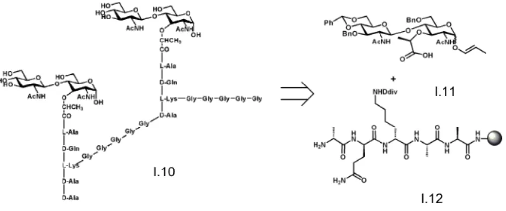
In a retrosynthetic overview, it can be divided in three fragments, a disaccharide NAG-NAM and two peptide chains, one that is directly bind to the NAM unit and the order that cross-link with other peptide chain as can be seen in Figure 1.6.
In our group, we have already synthetized a NAG-NAM disaccharide has already been synthesized with a pentapeptide attached D-Ala-L-Lys-D-Gln-L-Ala-L-Ala. The first
Figure 1.6 Retrosynthetic view of a dimeric PGN I.10
I.11
attempts were performed by preparing the NAG-NAM unit adapting the Fukase’s approach where the key step consisted on a stereoselective β-(1,4) glycosylationThe peptide was synthetized through Fmoc SPS (solid phase synthesis) with a global yield of 70%.20
As we can see in Figure 1.7, were used a 3:1 ratio (disaccharide:peptide) were used in the first step with a common coupling protocol, then the Div group of lysine was removed
an then coupled with a five glycines group, with the same coupling protocol. The final step was the cleavage of the resin with 1% TFA/DCM, giving the glycopeptide with a yield of 3%. Additionally we have developed the solid-phase synthesis of a glycopeptide, and monitoring by high resolution magic angle spinning NMR. 21
Recently the group have developed an efficient regioselective one-pot synthesis of Figure 1.7 Schematic NAG-NAM coupling to the peptide D-Ala-L-Lys-D-Gln-L-Ala, then coupled
with five glycines and finally cleavageof the resin
I.12 I.13
I.14 I.15
i) a) PhCHO, ZnCl2, 3 Å MS, RT, overnight; b) Ac2O, Pyridine, RT, overnight; ii) Morpholine, EtOAc, RT, overnight; iii) CCl3CN, CS2CO3, DCM, RT, 2h.
i) BnOH, AcCl, 80oC, 3h; ii) PhCHO, ZnCl2, overnight; iii) Ethyl L-(S)-2- trifluoromethanesulphonyloxy- propionate, NaH, DCM, RT, 3h; iv) Pd(PPh3)4, AcOH, TrocCl, RT; v) BH3Me2N, BF3OEt2, RT, 3h.
A
B
Figure 1.8: Synthesis of glucosamine Donor (A) and Acceptor (B)
I.16 I.17 I.18 I.19
I.20 I.21 I.22 I.23
glucosamine building blocks22 (Figure 1.8 and Figure 1.9). A properly N-protected glucosamine fully silylated at O-3, O-4 and O-6, was the key intermediate to achieve glucosamine building blocks, with a different substitution pattern, via a one-pot sequential procedure.
A one-pot strategy seems to be the most attractive way to achieve NAG-NAM disaccharides in opposition to the several synthetic strategies developed for the synthesis of carbohydrates and especially for the construction of complex oligosaccharides,23 Figure 1.9.
Such an approach allows the protection of the hydroxyl groups with suitable groups while avoiding multi-step sequences, difficult and challenging purification methods and it provides control for a regioselective glycosylation.19, 20, 22
Recently, the group has also reported the synthesis of a versatile intermediate towards NAM-NAG disaccharide, using a strategy that involves a multi-step approach with many purification steps.24 Despite the novelty of the approach and the key intermediate formed, the overall yield of the NAM-NAG moiety was only 2% in 11 steps. Due to the lack of resources, lack of pure and homogensous PGN, and an extremely hard-core synthesis providing PGN fragments with extremely low yields, our goal consists on finding the right strategy towards PGN synthesis. The use of a renewable starting material, such as a natural polymer would open a completely new and sustainable route to achieve PGN fragments of controlled degree of polymerization and high purity.
Chitin is a polymer that shares the same basic carbohydrate backbone as that of PGN. The advantage of the abundance and structure of chitin, a biopolymer having immense potential for chemical modification and for structural possibilities.
Figure 1.9 Synthesis of disaccharide NAG-NAM
i) TMSOTf, DCM, 3A MS, -15 oC; ii) Zn-Cu, THF: AcOH: Ac2O (1:1:1), then Ac2O/pyridine; iii) LiOH, THF:1,4-dioxane:H2O; iv) Pd(OH)2, H2, AcOH, RT.
I.19 I.25 I.26 I.27
1.4 Chitin and Chitosan
Chitin is the second most abundant polysaccharide after cellulose, can be obtain by extraction from crustaceans shells and can also be found in the cell wall of some fungi and algae. Chitin is constituted by of β-1-4-N-acetylglucosamine (GlcNAc) and D-glucosamine (GlcN) units, Figure 1.10.24-26 Chitosan is the copolymer, linear heteropolysaccharide, product of chitin’s deacetylation reaction27, with a DA (degree of acetylation) <50%, while chitin has DA=100%. Being a natural biopolymer, chitin has proprieties as non-toxic, biocompatible,
biodegradable and has been explored in many distinct areassuch, anti-bacterial agent28, fat-binding agent29, cell protection from carcinogenesis30, hypocholesterolemic agent31 , antioxidant 32, matrix for drug release33 artificial skin34 and wound dressing material35. Chitin has three different conformations α-, β- and ϒ-chitin that dependent on the animal source.36
Due to its poor solubility many strategies have been developed in order to obtain derivatives by chemical modification, with the objective of maximize their properties in the application field but this problem still the major setbackin the use of chitin and chitosan.37, 38 Depending on the source and consequently the conformation of the chitin, different solubilities are associated. α-Chitin has more intramolecular forces, hydrogen-bonds associated, than β -chitin that is more used due to this fact.38 Chitin showed to be soluble in water but its DA must be in a range between 45-55%.39 Several solvents have proved to dissolve chitin and chitosan, depending on its natural source has a different solubility, as shown in table 1.40
1
2
Figure 1.11- Antiparallel chain arrangment in α-chitin.(1); parallel arrangement in β-chitin. adapted from colocar aqui autores reference 33(e) (2) ou colocar A e B
O OH
O HO
NHR O
O HO
NHR
OH
n
Figure 1.10 Molecular skeleton of chitin (R=acetyl) and chitosan (R=H>50%) 1
Tabela 1-1: List of solvents and ionic liquids used to dissolve chitin and chitosan
Chitin Chitosan
N,N-dimethylacetamide, 5-10%(wt) LiCl,
Diluted acids
(phosphoric, sulfuric, citric, sebacic, acetic)
N,N-dimethylacetamide, N-methyl-2-pyrrolidone,
5-8%(wt) LiCl Dimethylsulfoxide
Methanol saturated with calcium chloride
dihydrate p-Toluene sulfonic acid
1-allyl-3-methylimidazolium bromide 10-Camphrosulfonic acid
1-butyl-3-methylimidazolium acetate 1-butyl-3-methylimidazolium chloride
1-butyl-3-methylimidazolium chloride 1-butyl-3-methylimidazolium acetate
hexafluoroacetone or hexofluoro-2-propanol 1-allyl-3-methylimidazolium acetate
In order to follow the degree of deacetylation or the depolymerization progress, and accomplish the characterization of the chitoligomers many technics have been used to provide this information, such as MALDI-TOF, 1H-NMR and chromatographic techniques as GPC.41-43 The degree of deacetylation can be calculated directly from the observation of the peak’s intensity on the 1H-NMR spectra as shown in equation 1.
𝐷!"#$ % = 1− ! !!!"! !
!!!!!!!
× 100 (1)
While the applications of chitin and chitosan in a wide range of areas, have been reviewed by several authors, the regiosselective and chemoselective modification of these polymers has been scarcely reported. 26, 44
Thelow molecular weight chitosan has greater potential for application in many different areas, due to the higher solubility.. In order to obtain low molecular weight chitosan, many strategies of hydrolysis have been explored. This depolymerization reaction can occur by chemical hydrolysis, by the use of a strong protic acid such as nitric acid45, diluted sulfuric acid46 and hydrochloric acid43, 47, 48, which is the most common way of hydrolysis of chitosan, or via enzymatic hydrolysis.49 Aditionally the physical mechanism of depolymerization via the ultrasonic depolymerization42, 50, which is directly affected by the geometry of the glass-wear used, has also been reported42. Apart from that there are also alternative methods, such as γ -irradiation depolymerization29 and depolymerization assisted by microwave51.
1.4.1 Chemical modification of chitin and chitosan
During the last few years, some developments on the chemoselective and regioselective modification of chitosan have been reported.
1.4.1.1 N-modifications
The protection of the amine group in chitosan is fundamental in order to achieve different
physical properties with consequently easier handling and to achieve different applications. Thus
several methods were developed, such the phtaloylation, acylation or carboxyacylation and
quaternization. These methods and the chemical methodology used will be discussed in the following
sections.
1.4.1.1.1 N-Phthaloylation
One method to prepare the N-phthaloyl chitosan consists on using aqueous acetic acid reaction media. Changes in the concentrations of aqueous AcOH to 10.0% maximum did not affect the regularity of the products, Figure 1.12. It should be emphasized that the irregular structure of the chitosan intermediate and acidic conditions caused by partial hydrolysis of phthalic anhydride allowed a homogeneous reaction in pure water. Since the N
-phthaloylation of chitosan is one of the most important reactions for the design of advanced materials, a reaction that requires no organic solvents will great contribute to the advancement of green chemistry.52
Figure 1.12 Scheme of amine moiety protection with phthalic anhydride
O OH
O
HO NH
2
O O
HO NH2
OH n
O OH
O HO
N O
O
HO NH
OH O
O
COOH O
Phthalic Anhydride
0-10% AcOH/H2O -H
2O
Reflux O
OH
O HO
N O
O
x y n
I.30
Other method consists on reacting fully deacetylated chitosan with phtalic anhydride in DMF with 5% of a co-solvent, such as ethanol, water, ethylene glycol and methyl cellosolve. Treatment of chitosan with phthalic anhydride, generally results in partial O -phthaloylation in addition to the N-phtaloylation. However in the present of 5% water, as co-solvent the partial O-6 phtaloylation are removed byhydrolysis, resulting in a 1.0 degree of N -substitution in chitosan, Figure 1.13.53
1.4.1.1.2 N-carboxylacylation
N-carboxymethylchitosan is the most typical amphiprotic chitosan with amine and carboxylic acid groups in the molecule. It is a unique macromolecular structure that provides
Figure 1.13 Schematic water-influence in the solvent composition on the chitosan and phthalic anhydride, reaction product
I.30
I.31
I.32
O OH
O HO
N O
n O
O
O Br
O HO
N O
n O
O O
OH
O HO
NH2 O
n
O N3
O HO
N O
n O
O
O N
O HO
N O
n O
O N
N R
O N
O HO
NH2 O
n N
N R
R= CO2H, PhMe
OH
n
a b
a
c d
Figure 1.14 Synhesis of a regioselective amphiprotic chitosan; (a)N-phtaloylation; (b) position 6 bromination; (c) position 6 azidation; (d) Husigen cycloadition; (e) phtaloyl group removal
I.30 I.32 I.33
specific properties and applications. However, preparing regioselective amphiprotic chitosan can be difficult by N- and O-carboxymethyl esterification using monochloroacetic acid. This derivative can be prepared via copper-catalyzed Huisgen cycloaddition, as it is shown further on Figure 1.14.54
N-carboxymethyl chitosan is obtained by using glyoxylic acid: the product is a glucan carrying pendant glycine groups. The solubility of chitosan in aqueous solutions of lithium and magnesium halides varies in the following order: LiCl < LiBr < LiI; MgCl2 < MgBr2 < MgI2. A
method for selective production of mono-N-(2-carboxyethyl)chitosan (NCE-chitosan) was developed via synthesis in gel (concentration of chitosan 4–20%) of the magnesium halides solution (1.1–3.5 M) using acrylic acid. The use of MgI2 or MgBr2 in the reaction, provides
relative greater amount of the monosubstituted amino groups (73–87%) in comparison with their absence. It is known that the bioactivity of carboxymethyl chitosan vary according to the ratio mono-:disusbtitution- of the amino groups.55
Water-soluble N-(4-carboxybutyroyl), Figure 1.15, chitosan derivatives with different degrees of substitution (DS) were synthesized to study the antimicrobial activity of chitosan. Chitosan in a solution of 2% aqueous acetic acid–methanol was reacted with different amounts of glutaric anhydride to give N-(4-carboxybutyroyl) chitosans at different DS. The chemical structures and DS were characterized NMR spectroscopy, which showed that acylation took place at the amine group. This synthesis gives a new chitosan derivative, soluble in water-, diluted acid or diluted base. The antimicrobial activity was investigated against the most economic plant pathogenic bacteria of Agrobacterium tumefaciens and Erwinia carotovora and fungi of Botrytis cinerea, Pythium debaryanum and Rhizoctonia solani. The antimicrobial activity of N-(4-carboxybutyroyl) chitosans revealed to be more efficient than the native chitosan with the increase of the DS. A compound of DS 0.53 was the most active one with minimum inhibitory concentration (MIC) of 725 and 800 mg/L against E. carotovora and A. tumefaciens, respectively and also in mycelial growth inhibiation against B. cinerea (EC50 = 899 mg/L), P. debaryanum (EC50 = 467 mg/L) and R. solani (EC50 = 1413
mg/L).56
A different approach to carboxyalkylation of chitosan consist on gel state approach by using aza-Michael addition and substitution reactions. Various reagents were applied including acrylic and crotonic acids, and halocarboxylic acids. The reaction of chitosan with Figure 1.15 Synthetic scheme of N-(4-carboxybutyroyl) chitosan derivatives using different mol
halocarboxylic acids showed no target product formation, either in solution or in the gel state. In the case of acrylic, crotonic, halocarboxylic acids, the reaction performed in the gel state (concentration of chitosan 20–40%) showed higher degree of substitution at lower reaction time and temperature than in diluted solutions (concentration of chitosan 0.5–2%). This method provided higher yield of the product per reaction volume, lower reaction time and temperature, lower consumption of solvents and reagents, and no need for stirring of the reaction mixture. All of these factors make the gel technique advantageous for large-scale production and turn the synthesis of these compounds more environmental friendly.57
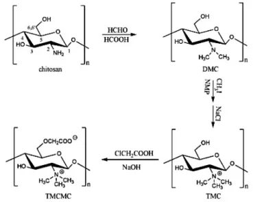
O-Methyl free N,N,N-trimethyl chitosan (TMC) can be synthesized by treating chitosan with formic acid and formaldehyde, followed by methylation with CH3I. Then TMC is
carboxymethylated by monochloroacetic acid to obtain N,N,N-trimethyl-O-carboxymethyl chitosan (TMCMC), Figure 1.16. Their antibacterial activity was investigated against Staphylococcus aureus and Escherichia coli. It seems to be a decreasing of the antibacterial activity of TMC as the degree of substitution increased at pH 5.5, however the structure activity relationship was the opposite at pH 7.2. TMCMC seems to have less activity than TMC, and also its activity decreased as the degree of carboxymethylation increased. The experimental results showed that the antibacterial activity of N,N,N-trimethyl amino group was lower than other non-quaternized amino groups, and carboxymethylation did not enhance the antibacterial activity directly.58
1.4.1.1.3 Quaternization
N,N,N-Trimethyl O-(2-hydroxy-3-trimethylammonium propyl) chitosans (TMHTMAPC) with different degrees of O-substitution can be prepared by reacting O-methyl-free N,N,N -trimethyl chitosan (TMC) with 3-chloro-2-hydroxy-propyl -trimethyl ammonium chloride (CHPTMAC). The products were investigated for antibacterial activity against Staphylococcus aureus and Escherichia coli at pH 5.5 and pH 7.2 conditions. TMHTMAPC exhibited enhanced antibacterial activity compared with TMC, and the activity of TMHTMAPC
increased as well as the DS increased. The use of divalent cations showed to be related to the lost of antibacterial activity of O-carboxymethyl chitosan and N,N,N-trimethyl-O -carboxymethyl chitosan. However it did not occur with such intensity in the repression on the antibacterial activity of TMC and TMHTMAPC. This indicated that the free amino group on chitosan backbone is the main functional group interacting with divalent cations.59
1.4.1.2 O-modification
Various protecting groups have been used for the protection of chitin’s and chitosan’s hydroxyl groups, in an attempt to modify chitosan derivatives properties, such as solubility, and to enable N-selective modifications. The most commonly used hydroxyl protecting groups are: acetyl, tosyl, trityl and silyl groups, such as trimethylsilyl (TMS) or tert-butyldimethylsilyl (TBDMS), Figure 1.17.61, 62
Usually O-modification at the O-6 position is commonly performed by the regioselective protection of the amine group with phthaloyl group.
The primary hydroxyl group in position 6 is very versatile, since is more reactive than the secondary hydroxyl in the position 3, enabling a wide variety of transformations. It can be regioselectively protected or transformed into different functionalities, such as a halide, an
a b
c < O OH O HO NHR1 O R1= Ac,Phth O OAc O AcO NHR1 O O OSiR3 O
R3SiO NHR
1 O O OTs O TsO NHR1 O
n n n
n
Figure 1.17 O-3 and O-6 N-acetyl/phtaloyl chitosan simultaneous protection; (a) Silylation; (b) Acetylation; (c) Tosylation
I.32 or I.37
I.38 I.39
I.40 O OH O HO NHAc O O OTs O HO NHAc O n n O O O HO NHAc O n O O HO NHAc O n O O HO NHAc O n Co2Et
EtO2C
CO2Et
P O OEt EtO a b c b d c
Figure 1.18 Selective O-6 Tosylation then FGI; (b)O-6-ethyl benzoate chitin; (c) 6-deoxy-diethyl malonate chitin; (d) 6-(deoxydiethyl) phosphitechitin
I.37 I.41
I.42
I.43
azide or an amine group.
Usually the O-6 position is selectively protected with bulky groups such as the triphenylmethyl group or the silyl groups, leaving the position O-3 capable of future modification. In these procedures the N-phthaloyl protected derivative is frequently used, increasesing the solubility in organic solvents and thereby enabling the introduction of the proper group at the primaryalcohol. At the end, the phthaloyl group can be easily cleaved by treatment with hydrazine.
The advantage of O-6-protected polymers, is the protection of the more reactive, primary hydroxyl, group leads to an increase of polymer solubility in organic solvents.
Trityl group is also placed in the polymer to increase the solubility in organic solvents. It is usually introduced reacting the N-protected chitin or chitosan with trityl chloride in pyridine.
Tosyl group is also very used as protecting group of O-6 position, since it has a good leaving character making tosylated polymer a very useful intermediate for further reactions at position O-6. Several nucleophiles such as sodium iodide, sodium borohydride, and potassium thioacetate63 were used to substitute the tosyl group to yield iodo-, deoxy-, and acetylthio- chitin derivatives, respectively, Figure 1.18.
The preparation of tosylated derivative could be achieved by reaction of chitin with
tosyl chloride (p-toluenesulfonyl chloride) in a DMAc/LiCl solvent system.63-65
Once functionalized, the resultant tosylated and fully N-acetylated chitin can react with the sodium salts of ethyl p-hydroxybenzoate, diethyl malonate, and diethyl phosphite, in DMAc, to afford the corresponding chitin derivatives of O-6-ethyl benzoate-chitin, 6-deoxy-diethyl malonate-chitin, and 6-(deoxy6-deoxy-diethyl) phosphitechitin, respectively, Figure 1.19.64, 66
O OH
O HO
N O
n
O
O
O OTBDMS
O HO
N O
n
O
O O
O
O HO
N O
n
O
O
O OTBDPS
O HO
N O
n
O
O
a b
c
Figure 1.19 O-6-N-phtaloyl chitosan selective protection; (a) Trytilation; (b) Silylation (TBDMS); (c) Silylation (TBDPS)
I.32
I.45 I.46
O-6 modification of N-protected chitosan is a strategy commonly employed to modify
chitosan and create an amphiprotic polymer. These modifications offer several advantages, since the resulting polymer possesses tunable solubility and a nanosized structure which enable several applications such as drug delivery systems. Usually, this amphiprotic character can be achieved by placing a suitable group in position O-6 such as a carboxylic acid, which can makes the polymer soluble under basic or acidic conditions. In neutral aqueous solution, a click chemistry approach has been developed, producing polymer nanoparticles, leading to the synthesis of a regioselective amphiprotic chitosan derivative.
The hydroxyl group can be converted to the brominated derivative that in turn is transformed in the azide derivative. The azide moiety at the C-6 position is then successfully converted to a 1,4-triazole linker with an appropriate R group, by a Huisgen cycloaddition between 6-azido-6-deoxy-N-pthaloyl-chitosan and an adequate propiolate in the presence of Cu(I) catalyst.54, 67
e O
OH
O HO
N O
n O
O
O Br
O HO
N O
n O
O O
OH
O HO
NH2 O
n
O N3
O HO
N O
n O
O
O N
O HO
N O
n O
O N
N R
O N
O HO
NH2 O
n N
N R
R= CO2H, PhMe
OH
n
a b
a
c d
Figure 1.20 Synhesis of a regioselective amphiprotic chitosan; (a) N-phtaloylation; (b) position 6 bromination; (c) position 6 azidation; (d) Husigen cycloadition; (e) phtaloyl group removal
I.30 I.32 I.33
The same group also verified that the chitin type (α or β) has a high influence on some chemical modifications. After an exhaustive study of reaction conditions, the authors verified that functionalization of hydroxyl groups of squid β-chitin with trityl or benzyl groups
proceeded much more easily than those of shrimp α-chitin. While in α−chitin, the O-6-trityl derivative is prepared by reaction of trimethylsilylated chitin, the β-chitin was found to exhibit higher reactivity and direct tritylation of β-chitin was achieved in a quantitative way, Figure 1.21.
3,6-O-Dibenzylation of β-chitin was also accomplished in simple one-step reaction, to afford the corresponding protected derivatives that exhibited good affinity for organic solvents.68
For the regioselective protection of chitin, Figure 1.21, the benzyl group was also explored as an efficient protecting group for O-3 postion, in combination with other protective groups including triphenylmethyl (trityl) for O-6 and acetyl or phthaloyl for N-2. Chitin was first silylated to form 3,6-O-trimethylsilyl derivative that was further reacted with trityl chloride to afford 6-O-trityl-chitin. This derivative was benzylated to give 3-O-benzyl-6-O-trityl-chitin, in which the trityl, benzyl, and acetyl groups could be selectively removed to afford three different derivatives with a free reactive hydroxyl of amine group at O-6, O-3, or N-2, respectively.60
Azide moiety in position 6 can also be transformed in an amine group via formation of a triphenylphosphinimine intermediate that is hydrolyzed using aqueous hydrazine, which also led to the removal of the N-phthaloyl group. This sequence gave 6-amino-6-deoxy-chitosan, which, unlike 6-amino-6-deoxy-chitosan, is soluble in water at neutral pH, Figure 1.22.69
O OH O HO N O n O O O OH O HO NH2 O n O OTr O HO N O n O O O OTr O BnO N O n O O O OH O BnO N O n O O O OTr O BnO NH2 O n O OTr O HO N O n O O
Figure 1.21 Selective protection/deprotection of chitosan; (a)N-phtaloylation; (b) O-6 tritylation; (c) O-3 benzylation; (d) O-6 trityl group removal; (e) N-phtaloyl group removal; (f) O-3 benzyl
group removal.
a b c
d
e
f
I.30 I.32 I.45
I.48
I.49
I.50
Conversion of the O-6 primary hydroxyls to carboxylate groups could be achieved using TEMPO/NaBr/NaClO oxidation systems making the insoluble a-chitin become water-soluble. 70
Chitosan is highly versatile biopolymer and can be easily converted in a wide range of functional groups, due to be possibility of chemoselective modification of its functional groups. Biological studies with PGN are limited by the difficulty in obtaining the pure, homogeneous and high molecular weight fragments of PGN. Adding these two facts we though “Is it possible to we convert chitosan into PGN’s sugar backbone?” and it is in this context that the present work was developed. We aimed to contribute to a sustainable and efficient route that enables the conversion of available chitin/chitosan into valuable PGN fragments.
O N3
O HO
N O
n O
O
O NH2
O HO
N O
n O
O
Figure 1.22 Conversion of the azide moiety into amine group
2
Results and Discussion
Since biological studies to investigate how the host receptors can recognize high molecular weight fragments of PGN at the surface of bacteria are limited by the amount and purity of natural PGN, we embraced the challenge of preparing pure and homogenous PGN
fragments in order to overcome the major setback in this research field.
To achieve this goal, we envisaged that a biopolymer, that shares the same basic
carbohydrate skeleton as PGN, could be used as starting material towards PGN. The aim
consisted on combining chemical and enzymatic methods to modify the polymer in order to
convert it into NAG-NAM units.
Chitin is a β-1,4-linked NAG biopolymer thus, to convert chitin into PGN, a lactyl unit must be introduced in alternating NAG units to which peptides may be connected. The main advantage of this proposal is the fact that chitin/chitosan already have the challenging β-1,4
glycosidic bond installed between NAG/glucosamine units and many orthogonal
protection/deprotection strategies to achieve enantioselective glycosylation as well as the
preparation of donors and acceptors will be avoided.
Chitin is very abundant in the biomass of shellfish discarded every year, is commercially available with different degrees of polymerization. However chitin’s low solubility represents a limitation to the required chemical modification. On the other hand chitosan, the
O OH HO AcHN O O OAcHN OH O HO O O OH HO AcHN O O OAcHN OH O HO O O OH HO AcHN O O OAcHN OH O n HO O O O HO AcHN O O HOAcHN OH O O O HO AcHN O O HOAcHN OH O O OR HO AcHN O O HOAcHN OR O O OR HO AcHN O O HOAcHN OR O X O OH HO H2N
O O HO H2N
OH O
O OH
HO H2N
O O HO H2N
OH
O O
OH
HO H2N
O O HO H2N
OH O
n
n n
Figure 2.1 PGN’s sugar backbone retrosynthetic plan II.1
II.2 II.3
alkaline derivative of chitin, has a better solubility profile and is amenable to chemical
modifications.
In an initial stage of the project we aimed at preparing small chitooligomers and
perform its structural elucidation. The idea was to establish a methodology for obtaining
chitooligomers of controlled molecular weight for further chemical modification towards PGN.
In a second stage of the project the main aims were:
- To perform chitin/chitosan regioselective chemical modification;
- To establish a methodology to introduce the lactyl unit into alternating units of NAG in the modified chitin/chitosan;
- To digest NAM-NAG polymers by lysozyme-like enzymes to release homogenous NAM-NAG polymers;
- To characterize the fragments obtained using different spectroscopic and
chromatographic techniques;
According to the retrosynthetic analysis depicted in figure XX, two different strategies
were explored to introduce the lactyl moiety in alternating protected glucosamine units. After
removal of the protecting groups, the NAG-NAM content was evaluated by using enzymatic
assays trough muramidases and lysozyme digestion. The amount of NAG-NAM units present
in the final polymers, and thus the success of the approaches, was analyzed by HPLC-UV
and HPLC-MS analyzes. Additionally PGRP affinity to the prepared samples was also
evaluated by pull-down assays and by SDS-PAGE electrophoresis.
This work and the results obtained will be presented and discussed in the present
chapter that is organized as follows:
• Preliminary studies;
• Polymeric chitosan chemical modification;
• Enzymatic studies of the final products
2.1 Preliminary studies
To date, X-ray crystallography of PGRPs has been performed with a NAM unit attached to a penta or a tripeptide stem, performed to achieve the structure of human PGRPs.71 Thus interaction studies of PGRPs with higher molecular fragments involving both NAM and NAG units have not been reported so far.