1317
MORPHOLOGICAL CHANGES IN LYMPH NODES AND SPLEEN UPON EAE INDUCTION IN C57BL/6 MIC
IRINA MASLOVARIĆ1, NATAŠA VUKOV1, ALEKSANDRA STOJKOVIĆ1, DEJANA KOSANOVIĆ1 and KATICA JOVANOVA-NEŠIĆ1,2
1 Biomedical Center of the Institute “Torlak”, 11152 Belgrade, Serbia
2 Institute of Virology, Vaccines and Sera “Torlak”, Immunology Research Center “Branislav Janković”,
11152 Belgrade, Serbia
Abstract - Myelin oligodendrocyte glycoprotein (MOG) is а protein widely used in the induction of experimental autoim-mune encephalomyelitis (EAE) for studying human multiple sclerosis (MS). In C57BL/6 female mice aged eight weeks, we administered subcutaneously MOG35-55 peptide in CFA (complete Freund’s adjuvant) along with pertussis vaccine injected intraperitoneally. We observed the sign of laccid tail as early as thirteen days post-immunization in ive of twelve animals. Hematoxylin and eosin staining of parain-embedded sections of lymph nodes and spleen revealed the presence of germinal centers in the immunized animals. In the control group of animals, lymphoid follicles without germinal cen-ters were observed. Immunohistochemical staining of spleen sections revealed an expression of MHC II molecules in the EAE-induced group. We would like to point out that even though the clinical signs are mild, the morphological changes are apparent in the lymph nodes and spleen of MOG35-55-immunized mice.
Key words: EAE, MOG, lymph nodes, spleen
INTRODUCTION
he immune system is functionally compartmental-ized into primary lymphoid organs responsible for the generation and diferentiation of naive T and B cells, and secondary lymphoid organs where immune responses are initiated. Only ater activation do T and B cells emigrate from secondary lymphoid organs to seek the antigen in the periphery. Secondary lym-phoid organs include the spleen, lymph nodes and organized lymphoid tissues associated with mucosal membranes such as the tonsils, appendix, and Peyer’s patches. hese highly organized secondary lymphoid organs provide the structures where the antigen is ef-iciently retained and presented and where ordered cellular interactions between antigen presenting cells
(APCs), T cells and B cells take place to initiate and promote eicient immune responses (Karrer et al., 1997, Abbas et al., 1997).
was found in mice with abated overall expression of MHC II molecules by plasmacytoid dendritic cells previously immunized with myelin oligodendrocyte glycoprotein (Irla et al., 2010).
We examined the morphological changes in lymph nodes and spleen tissue and the expression of MHCII molecules in sections of the spleen of C57BL/6 mice with EAE induced by MOG35-55.
MATERIALS AND METHODS
Animals
Female C57BL/6 8-week-old mice were purchased from the Military Medical Academy (Serbia). he mice were kept under standard laboratory conditions (room temperature 21±1°C, humidity 30%, 12/12 h light/dark cycle) with food and tap water ad libitum. he protocols for the animal experiment were con-ducted in accordance with the guidelines (86/609/ EEC) of the European Community Council Direc-tives and the Serbian Laboratory Animal Science As-sociation – SLASA. he study was performed on 19 mice divided in two groups: EAE-induced (mice im-munized with MOG in CFA; n=12) and an IC group (non-treated animals; n=7).
EAE induction
C57BL/6 mice were injected subcutaneously in the right side of the lank with 100 µg of myelin oli-godendrocyte glycoprotein peptide (MOG35-55, Sigma Aldrich) in complete Freund’s adjuvant (CFA) con-taining 1 mg/ml of heat-killed and dried
Mycobacte-riumtuberculosis H37Ra (Sigma Aldrich) according
to Suter et al. (2000). 1x10⁹ of heat-killed Bordetella
pertussis bacilli in 150 µl of PBS (pertussis vaccine, Institute of Virology, Vaccine and Sera “Torlak”) was injected intraperitoneally on the day of immuniza-tion and 48 h later.
Development of the clinical signs of EAE in the mice was monitored daily, starting at day 7 post im-munization according to the following criteria: (0), no clinical signs; (1) laccid tail; (2) hind leg paresis;
(3) full hind leg paralysis; (4) quadriplegia; (5) death. he mice were sacriiced on day 24 ater immuniza-tion. Over this period of time the mice showed the laccid tail sign that was less severe than typically ob-served, and were scored as 0.5 less than the indicated grade.
Tissue collection
All mice were placed under deep anesthesia, and then cardially perfused with PBS pH 7.4 followed by 4% PFA (paraformaldehyde) in 0.1 M phosphate bufer. he organs were preserved in 4% formalde-hyde for later histological and immunohistochemical examination. Formalin-ixed parain-embedded tis-sue was cut to 5-μm thick sections of the spleen and lymph nodes by a microtome (Microtome HM 450; Leica). he sections of each experimental group were then stained with hematoxylin and eosin to visualize cell morphology in the tissue sections.
Immunohistochemistry
Tissue analysis
he analysis of tissue sections was performed by capturing images of sections using a BH2 research microscope (Olympus Optical Co. LTD. Tokyo, Ja-pan) equipped with a Color View III digital camera (Olympus). Analysis Docu sotware (Olympus) was used to acquire images. All images were taken under
10x and 40x magniication.
RESULTS AND DISCUSSION
Clinical signs of EAE in C57BL/6 mice
All immunized animals were monitored for clinical signs of EAE development, starting at day 7
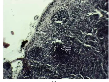
post-Fig. 1.Hematoxylin and eosin staining of the lymph node. Lymph node cortex of EAE-induced group and control group (photomi-croscopic images) (Fig. 1A, Fig. 1B). he germinal center of EAE-induced group and lymphoid follicles of control group is marked by arrows. Magniication x10.
Fig. 2 Hematoxylin and eosin staining of spleen sections. Spleen white pulp of EAE-induced group and control group (photomicro-scopic images) (Fig. 2A, Fig. 2B). he enlarged follicle of EAE-induced group with germinal center is marked by vertical white arrow. he follicle without germinal center of the control group is marked by a horizontal white arrow. Magniication x10.
1A 1B
MOG immunization. A clinical sign such as a laccid tail was observed in 5 of 12 animals on day 13 ater immunization. Aggravation of clinical signs was not observed on day 24 post immunization when the an-imals were killed. We further performed histological analysis of the lymph nodes and spleen, and immu-nohistochemical staining of the MHC II antigen in the sections of the spleen.
Presence of germinal centers in the lymph nodes and spleen in C57BL/6 mice
In the lymph nodes and spleen of both immunized and control groups, lymphoid follicles – B cell areas along with T cells located predominantly between the follicles, were observed. Histological examination of the lymph nodes and spleen from the EAE-induced mice showed an increased number of pyroninophilic cells in the germinal centers of the lymphoid follicles (Fig. 1A, Fig. 2A). T and B cell response to subcuta-neously introduced MOG35-55 peptide may trigger a cascade of events leading to germinal center forma-tion. Germinal centers that develop in response to antigen stimulation are the sites of B cell clonal ex-pansion. hey are functionally polarized into a dark zone in which the B cells divide and a light zone in which the B cells are activated, and selected based on their ainity for the antigen (Victora and
Nussenz-weig, 2012). In the non-treated group, the presence of lymphoid follicles without germinal centers in the lymph nodes and spleen was observed (Fig. 1B, Fig. 2B).Histological analysis of the spleen showed vis-ible changes in the lymphoid follicle of the spleen white pulp in the MOG35-55-immunized mice (Fig. 2A). he white pulp of the spleen consists of T cell areas and B cell follicles resembling the organization of the lymph nodes. his structure is considered one of the most important inductive sites of the immune system, where traicking and interactions among dendritic cells, T cells and B cells take place upon antigen challenge (Mebius and Kraal, 2005). We ob-served a positive correlation between the presence of germinal centers in the lymph nodes and spleen and circulating anti-MOG antibodies in the sera of the EAE-induced mice (unpublished results).
MHCII molecules with signiicantly higher expression in spleen sections of EAE-induced animals
A signiicantly higher expression of MHC II mol-ecules was detected in the sections of the spleen from the animals immunized with MOG35-55 peptide (Fig. 3A). he sections from the control group (IC) showed a lower expression of MHC class II mol-ecules (Fig. 3B). MHC II molmol-ecules play a crucial role in the regulation and induction of immune
re-Fig. 3 Immunohistochemical staining of MHC II molecules in spleen sections. EAE-induced group and control group (photomicro-scopic images) (Fig. 3A, Fig. 3B). MHC II expression in both groups is marked by arrows. Magniication x40.
sponse. Constitutive MHC II expression is the hall-mark of three distinct types of cells, dendritic cells, B cells and the cells of monocyte/macrophage line-age (Waldburger et al., 2001). Eicient priming of naive T lymphocytes requires both the presentation of an antigen in the context of MHC molecules and the delivery of accessory signals provided by antigen presenting cells (APCs), such as dendritic cells (DC) (Muraille et al., 2002).
Dendritic cells (DCs) play well-established roles in the induction of immunity and tolerance. Both functions require antigen (Ag)-speciic interactions between T cells and DCs in the secondary lymphoid tissues. he outcome of these interactions depends on the modulation and integration of three signals: T cell receptor engagement by peptide-MHC com-plexes, the recruitment of costimulatory and adhe-sion molecules, and the delivery of soluble mediators (Lebedeva et al., 2005). Signals associated with in-lammation, infections or tissue damage induce DC maturation, a process involving complex phenotypi-cal changes, including the upregulation of MHCII, costimulatory and adhesion molecules, the secretion of inlammatory mediators and altered migratory properties. Activation of naive T cells by mature DCs results in clonal expansion and diferentiation into efector and memory T cells (Irla et al., 2010).
Plasmacytoid DCs (pDCs) constitute a unique dendritic cell subtype. hey circulate in the blood and access secondary lymphoid organs from the blood, driven mainly by inlammatory stimuli. It is suggest-ed that they take part in both innate and adaptive im-munity (Colonna et al., 2004). However, pDCs also express MHCII molecules and undergo a maturation process similar to that of conventional DCs (Villa-dangos and Young, 2008). Furthermore, pDCs can internalize, process and present antigens to CD4+ T cells and cross-present antigens to CD8+T cells (Ho-efel et al., 2007; Sapoznikov et al., 2007; Di Pucchio et al., 2008; Young et al., 2008). hese indings have suggested that pDCs can function as APCs.
EAE induced by immunization with myelin oligodendrocyte glycoprotein (MOG) was found to
be severely exacerbated in mice exhibiting a selec-tive abrogation of MHCII expression by pDCs. EAE induction triggered the recruitment of pDCs to the lymph nodes, where they engaged in MHCII-de-pendent and MOG-speciic interactions with CD4+ T cells. It was suggested that MHCII expression by pDCs confers protection against EAE by inhibiting the priming of encephalitogenic T cells in second-ary lymphoid tissues but has no evident impact on the subsequent efector phase in the CNS (Irla et al., 2010).
MHC class II antigen positive cells, apparent in the sections of the spleen, might render a link between the mild clinical signs expressed in EAE-induced mice and the expression of MHCII molecules.
Acknowledgments - his work was supported by the Institute of Virology, Vaccines and Sera “Torlak” and the Ministry of Education, Science and Technology of the Republic of Serbia (Project No: M-175050).
REFERENCES
Abbas, A.K., Lichtman A.H. and J.S. Pober (1997). Cellular and molecular Immunology. 3rd edition. W. B. Sounders
Com-pany, Philadelphia.
Batoulis, H., Recks, MS., Addicks, K. and S. Kuerten (2011). Ex-perimental autoimmune encephalomyelitis achievements and prospective advances. APMIS; 119, 819-30.
Colonna, M., Trinchieri, G. and Y.J. Liu (2004). Plasmacytoid dendritic cells in immunity. Nat. Immunol. 5, 1219-1226.
Costa, O., Divouxc, D., Ischenkod, A., Trona, F. and M. Fontainae
(2003). Optimization of an animal model of experimental autoimmune encephalomyelitis achieved with a multiple MOG35–55 peptide in C57BL6/J strain of mice. J. Auto-immun.20, 51-61.
Di Puchino, T., Chatterjee, B., Smed-Sorensen, A., Clayton, S., Palazzo, A., Montes, M., Xue, Y., Mellman, I., Banchereau, J. and J.E. Connolly (2008). Direct proteasome-indepen-dent cross-presentation of viral antigen by plasmocytoid dendritic cells on major histocompatibility complex classI.
Nat. Immunol.9, 551-557.
Hoefel, G., Ripoche, A.C., Matheoud, D., Nascimbeni, M., Escriou, N., Lebon, P., Heshmati, F., Guillet, J.G., Gannagé, M. and S.
Caillat-Zucman (2007). Antigen crosspresentation by
Irla, M., Küpfer, N., Suter, T., Lissilaa, R., Benkhoucha, M., Skup-sky, J., Lalive, P. H., Fontana, A., Reith, W. and S. Hugues
(2010). MHC class II–restricted antigen presentation by plasmacytoid dendritic cells inhibits T cell–mediated au-toimmunity. J. Exp. Med.207, 1891-1905.
Jones, M. V., Nguyen, T.T., DeBoy, C.A., Griin, J. W., Whartenby, K.A., Kerr, D. A. and P.A. Calabresi (2008). Behavioral and pathological autcomes in MOG35-55 experimental
autoim-mune encephalomyelitis. J. Neuroimmunol.199, 83-93.
Karrer, U., Althage, A., Odermatt,B., Charles Roberts, W.M., Korsmeyer, Ch. R., Miyawaki, S.J., Hengartner,H. and R. M. Zinkernagel (1997). On the Key Role of Secondary Lymphoid Organs in Antiviral Immune Responses Stud-ied in Alymphoplastic (aly/aly) and Spleenless (Hox11−/−)
Mutant Mice. J. Exp. Med.185, 2157-2170.
Lebedeva, T., Dustin, M.L. and Y. Sykulev (2005). ICAM-1 co-stimulates target cells to facilitate antigen presentation.
Curr. Opin. Immunol. 17, 251-258.
MacKenzie-Graham, A., Tinsley, M.R., Shah, K.P., Aguilar, C., Strickland, L.V., Boline, J., Martin, M., Morales, L., Shat-tuck, D.W., Jacobs, R.E., Voskuhl, R.R. and A.W. Toga
(2006). Cerebellar cortical atrophy in experimental auto-immune encephalomyelitis. Neuroimage 32, 1016-1023.
Mebius R. E. and G. Kraal (2005). Structure and function of the spleen. Nat. Rev. Immunol.5, 606-616.
Muraille, E., De Trez, C., Pajak, B., Brait, M., Urbain, J. and O., Leo (2002).T Cell-dependent maturation of dendritic cells in response to bacterial superantigens. J. Immunol.168, 4352-4360.
Sapoznikov, A., Fischer, J.A., Zat, T., Krauthgamer, R., Dzionek, A. and S. Jung (2007). Organ-dependent in vivo priming of
naive CD4+, but not CD8+, T cells by plasmacytoid den-dritic cells. J. Exp. Med. 204, 1923–1933.
Suter, T., Malipijero, U., Otten, L., Ludewig, B., Muelethaler-Motter, A., Mach, B., Reith, W. and A., Fontana (2000) Dendritic cells and diferential usage of the MHC class II transactivator promoters in the central nervous system in experimental autoimmune encephalitis. Eur. J. Immunol
32, 794-902.
Stojkov, D., Lavrnja, I., Subasic, S., Bjelobaba, I., Pekovic, S, Gad-janski, I., Mostarica-Stojkovic, M., Stosic-Grujicic, S., Rakic, L. and M. Stojiljkovic (2006). herapeutic efects of nucle-oside analogues on experimental autoimmune encephalo-myelitis in Dark Agouti rats. Arch. Biol. Sci.58, 13-20.
Victora, G. D. and M. C. Nussenzweig (2012). Germinal centers.
Annu. Rev. Immunol.30, 429-57.
Villadangos, J.A. and L.Young. (2008). Antigen-presentation properties of plasmacytoid dendritic cells. Immunity. 29, 352-361.
Waldburger, J-M., Suter, T., Fontana, A., Acha-Orbea, H. and W. Reith (2001). Selective abrogation of MHCII expression on extrahematopoietic cells in mice lacking promoter IV of the class II transactivator gene. J. Experiment. Med.194, 393-406.
Young, L.J., Wilson, N.S., Schnorrer, P., Proietto, A., Broeke, ten T., Matsuki, Y., Mount, A.M., Belz, G.T., O`Keefe, M.
and M. Ohmura-Hoshino (2008). Diferential MHC class