Phosphotransferase System (PTS) of the Intracellular
Pathogen
Brucella melitensis
Marie Dozot1, Sandrine Poncet2, Ce´cile Nicolas1, Richard Copin1, Houda Bouraoui2, Alain Maze´2, Josef Deutscher2, Xavier De Bolle1, Jean-Jacques Letesson1*
1Research Unit in Molecular Biology (URBM), University of Namur, Namur, Belgium,2Laboratoire de Microbiologie et Ge´ne´tique Mole´culaire (INRA-CNRS-AgroParisTech), Thiverval-Grignon, France
Abstract
Background:In many bacteria, the phosphotransferase system (PTS) is a key player in the regulation of the assimilation of alternative carbon sources notably through catabolic repression. The intracellular pathogensBrucellaspp. possess four PTS proteins (EINtr, NPr, EIIANtrand an EIIA of the mannose family) but no PTS permease suggesting that this PTS might serve only regulatory functions.
Methodology/Principal Findings:In vitrobiochemical analyses andin vivodetection of two forms of EIIANtr(phosphorylated or not) established that the four PTS proteins ofBrucella melitensisform a functional phosphorelay. Moreover,in vitrothe protein kinase HprK/P phosphorylates NPr on a conserved serine residue, providing an additional level of regulation to theB. melitensisPTS. This kinase activity was inhibited by inorganic phosphate and stimulated by fructose-1,6 bisphosphate. The genes encoding HprK/P, an EIIAMan-like protein and NPr are clustered in a locus conserved amonga-proteobacteria and also contain the genes for the crucial two-component system BvrR-BvrS. RT-PCR revealed a transcriptional link between these genes suggesting an interaction between PTS and BvrR-BvrS. Mutations leading to the inactivation of EINtror NPr significantly lowered the synthesis of VirB proteins, which form a type IV secretion system. These two mutants also exhibit a small colony phenotype on solid media. Finally, interaction partners of PTS proteins were identified using a yeast two hybrid screen against the wholeB. melitensisORFeome. Both NPr and HprK/P were shown to interact with an inorganic pyrophosphatase and the EIIAMan-like protein with the E1 component (SucA) of 2-oxoglutarate dehydrogenase.
Conclusions/Significance:TheB. melitensiscan transfer the phosphoryl group from PEP to the EIIAs and a link between the PTS and the virulence of this organism could be established. Based on the protein interaction data a preliminary model is proposed in which this regulatory PTS coordinates also C and N metabolism.
Citation:Dozot M, Poncet S, Nicolas C, Copin R, Bouraoui H, et al. (2010) Functional Characterization of the Incomplete Phosphotransferase System (PTS) of the Intracellular PathogenBrucella melitensis. PLoS ONE 5(9): e12679. doi:10.1371/journal.pone.0012679
Editor:Edgardo Moreno, Universidad Nacional, Costa Rica
ReceivedApril 12, 2010;AcceptedAugust 15, 2010;PublishedSeptember 10, 2010
Copyright:ß2010 Dozot et al. This is an open-access article distributed under the terms of the Creative Commons Attribution License, which permits unrestricted use, distribution, and reproduction in any medium, provided the original author and source are credited.
Funding:This work was supported by Fonds de la Recherche Fondamentale Collective (FRFC, Belgium, convention 2.4521.04), by Action de recherche concertee (ARC, No. 04/09325 and 08/13015) and by an agreement with Commisariat general aux Relations internationales de la Communaute franc¸aise de Belgique -Fonds National pour la Recherche Scientifique - Centre National de la Recherche Scientifique (CGRI-FNRS-CNRS, reference PVB/ADK/FR/ad/2681). M. Dozot held a PhD fellowship from the Fonds pour la formation a` la Recherche dans l’Industrie et dans l’Agriculture (FRIA). The funders had no role in study design, data collection and analysis, decision to publish or preparation of the manuscript.
Competing Interests:The authors have declared that no competing interests exist. * E-mail: [email protected]
Introduction
In order to successfully colonize an ecological niche, bacteria have to integrate different signals indicating environmental changes, and subsequently trigger an adequate adaptative response by modulating their cellular activities. The appropriate response to changes in nutrient availability, for example, relies on diversified mechanisms, including global regulation systems such as the phosphoenolpyruvate (PEP): carbohydrate phosphotrans-ferase system (PTS). The PTS catalyzes the uptake and con-comitant phosphorylation of carbohydrates and is composed of several proteins forming a phosphorelay transferring the phos-phoryl group from PEP to the incoming sugar: (i) the general PTS proteins enzyme I (EI) and HPr are cytoplasmic components
usually common to all PTS carbohydrates; (ii) the enzyme II complex is specific for one or several sugars and is generally composed of at least three domains (or distinct proteins) including the cytoplasmic EIIA and EIIB, and the membrane-crossing EIIC (sometimes also EIID) that constitutes the permease of the system [1,2]. PTS proteins are usually phosphorylated on a conserved histidine, with the exception of most EIIB components that are phosphorylated on a cysteine. Besides its function in the transport and phosphorylation of carbon sources, the PTS plays a key role in the regulation of many aspects of bacterial physiology, including carbon catabolite repression (CCR) (for reviews see [1,2,3]).
called the nitrogen PTS (PTSNtr) [4,5,6,7]. The phosphoryl transfer chain of this system is composed of three proteins, EINtr (encoded byptsP), NPr (encoded byptsO) and EIIANtr(encoded by
ptsN) that are the respective paralogs of EI, HPr, and EIIA of the fructose PTS family; however, they are not associated with PTS permeases [4,5,6,7] but carry out multiple regulatory functions [8]. For example, the PTSNtris involved in the regulation of genes related to nitrogen metabolism [9,10,11,12]. Moreover, compared to EI, the EINtrpossesses an N-terminal extension homologous to the GAF N-terminal sensory domain of NifA from Azotobacter vinelandii, an activator that enhances transcription by s54
-associated RNA polymerase [13]. Finally, PTSNtr might favor the utilization of organic nitrogen compounds when bacteria are exposed to multiple carbon sources [4,11,12,14] and is involved in maintaining K+
homeostasis inE. coli[15].
Brucella spp. are Gram negative intracellular pathogens belonging to the a-proteobacteria group which includes other bacteria interacting with eukaryotic hosts, such as Agrobacterium tumefaciens or Sinorhizobium meliloti [16]. They are responsible for brucellosis, a worldwide zoonosis that affects a broad range of mammals [17], and can also infect humans where it may cause Malta fever, a serious debilitating chronic disease [18]. Large-scale screens aiming at the isolation of attenuated transpositional mutants of Brucella spp. led to the identification of many genes involved in carbon and nitrogen metabolism [19,20]. Moreover, genes encoding homologues of the three components of theE. coli
PTSNtrwere also isolated during these screens [19,20,21]. These data suggest that carbon and nitrogen metabolism might affect the virulence ofBrucella.
The availability of the genome sequence of several Brucella
species [22,23,24] allowed the identification of an additional PTS-related gene putatively encoding an EIIA belonging to the mannose PTS family. Moreover, a gene encoding a truncated homologue of HPr kinase/phosphorylase (HprK/P) was found in
Brucella genomes [25,26,27]. In most firmicutes (Gram positive bacteria with low GC content), HprK/P catalyses the phosphor-ylation and dephosphorphosphor-ylation of a conserved serine residue in HPr (usually Ser46) [28,29,30]. In these bacteria, HPr phosphor-ylated on this conserved serine (P-Ser-HPr) is a central regulator of carbon metabolism mediating among others inducer exclusion and acting as a co-repressor of the catabolite control protein A (CcpA) during CCR [1,31].
Similarly to Brucella spp, other a-proteobacteria including S. meliloti [32] and A. tumefaciens (S. Poncet, A. Khemiri and J. Deutscher, unpublished) possess the predicted PTSNtrproteins, as well as an EIIAMan-like protein and HprK/P, and lack PTS permeases. It was therefore suggested [25,26,27] thatBrucellaPTS proteins might form a phosphoryl transfer chain exclusively dedicated to regulatory functions. Interestingly, in these three bacteria, the genesptsO,ptsMandhprK(encoding respectively NPr, an EIIAMan-like protein and HprK/P) are localized close to genes encoding (i) a two-component system involved in virulence or symbiosis (BvrR-BvrS inBrucellaspp., ChvI-ChvG inA. tumefaciens
and ChvI-ExoS- inS. meliloti) [33,34,35], (ii) S-adenosyl homocys-teine hydrolase (SahH), an enzyme involved in the metabolism of methionine [36,37] and (iii) PEP carboxykinase, a key enzyme of gluconeogenesis [38,39,40]. As previously proposed by Hu and Saier [27], the conservation of this genomic locus in several a -proteobacteria suggests a functional link between PTS, HprK/P and the neighboring genes in regulating carbon/nitrogen metabolism in these organisms.
In this report, we demonstrate byin vitroandin vivoexperiments that the PTS proteins found in B. melitensis function in a phosphorelay. Moreover, we observed that NPr is phosphorylated
not only by EINtron His-30, but also by HprK/P on a conserved serine (Ser-61). This latter phosphorylation slows the in vivo
phosphotransfer to His66 in EIIANtr. We also demonstrated a transcriptional link between the PTS genes ptsO, ptsMand hprK
and the two component system genes bvrR/Sestablishing a link between virulence and metabolism that was reinforced by the observation that both,ptsP andptsO mutants, almost completely lost the synthesis of a type IV secretion system (T4SS). Finally by carrying out a yeast two hybrid screen against the whole B. melitensisORFeome we identified several interaction partners of PTS proteins allowing us to propose a preliminary model of regulation for carbon and nitrogen metabolism inB. melitensis.
Results
TheBrucella melitensis16M genome encodes four PTS proteins and HPr kinase/phosphorylase
The genome ofBrucella melitensis16M [22] contains three genes (ptsP/BMEI0190, ptsO/BMEI2031 and ptsN/BMEI1786) encod-ing homologues of the proteins composencod-ing the PTSNtr(EINtr, NPr and EIIANtr, respectively) and ptsM/BMEI2032 encoding a homologue of an EIIA of the mannose PTS family (EIIAMan -like). A homologue of HprK/P (encoded byhprK/BMEI2034) is also found inBrucella. All these genes are highly conserved in the genome of other sequencedBrucellaspecies. Sequence analyses and multiple aligments of these five proteins were carried out and allowed the prediction of the phosphorylatable histidine or serine residues (see Figures S1 to S5). Similar to its homologues inA. tumefaciensandS. meliloti,B. melitensisEINtrcontains a GAF domain resembling the sensory domain of the NifA protein ofA. vinelandii
[6]. The GAF domains are ubiquitous motifs present in many sensory proteins of eukaryotes and prokaryotes and are proposed to allosterically regulate catalytic activities of these proteins through the binding of small molecules [13]. When compared to HprK/Ps from firmicutes, HprK/Ps froma-proteobacteria lack about 130 N-terminal amino acids. The role of this domain is still unknown and artificially truncatedL. casei HprK/P (missing the first 127 amino acids) retained kinase and phosphorylase activities and all known regulatory properties [41]. Moreover, a carboxy-terminal conserved region is present in HprK/P from firmicutes and most b-, c- and d-proteobacteria, but absent from a -proteobacteria. This region was shown to be important for the phosphorylase activity of HprK/P, suggesting that in a -proteo-bacteria HprK/P might not be able to efficiently dephosphorylate P-Ser-NPr or dephosphorylate it by a different enzyme [30,42].
Based on the sequence analyses, we propose that EINtr autopho-sphorylates on His357 in the presence of PEP and subsequently transfers its phosphoryl group to His30 of NPr, which then phosphorylates EIIANtrand the EIIAMan-like protein on His66 and His9, respectively. Moreover, we suggest that HprK/P might phosphorylate NPr on the conserved Ser61. These hypotheses were tested by carrying outin vitrophosphorylation assays with purified proteins.
Phosphoryl group transfer from P,EI to the EIIAs via P,His-NPr
To carry outin vitrophosphorylation assays theptsP,ptsN,ptsM
with [32P]PEP (Fig. 1B, lane 3). In agreement with our prediction (Fig. S4), the additional presence of EI allowed the phosphory-lation of wild-type NPr (Fig. 1B, lane 4) and NPrS61A(data not shown), but not of NPrH30A (Fig. 1B, lane 2). We subsequently tested whetherB. melitensisEIIANtrand the EIIAMan-like protein were phosphorylated by P,His-NPr. Incubation of EIIANtr or the EIIAMan-like protein with [32P]PEP (data not shown) or [32P]PEP and EINtr (Fig. 1B, lanes 5 and 6) did not allow their phosphorylation. By contrast, EIIANtr and the EIIAMan-like protein were phosphorylated by [32P]PEP in the presence of both general PTS proteins EINtr and NPr, establishing that after its own phosphorylation on His30 by EINtr, P,His-NPr is able to transfer its phosphoryl group to EIIANtr and the EIIAMan-like protein (Fig. 1B, lanes 7 and 8). Purification of the EIIAMan-like protein provided always two distinct forms migrating to slightly different positions on SDS polyacrylamide gels (Fig. 1A), which apparently became both phosphorylated by P,His-NPr. The reason for the appearance of two EIIAMan-like forms is not known, but a similar observation has been reported for EIIABMan fromStreptococcus salivarius, which was also isolated in two distinct forms migrating to slightly different positions on SDS polyacryl-amide gels [43].
Serine 61 of NPr is the target of ATP-dependent phosphorylation catalyzed by HprK/P
The truncated HprK/P possibly adds an additional dimension of regulation to the B. melitensisPTS by phosphorylating NPr on a conserved serine residue (Ser61; see Fig S4). We therefore tested the ability of HprK/P to phosphorylate NPr on Ser61. As for thepts
genes, we cloned thehprK-coding sequence in an expression vector and purified the His6-tagged fusion protein. As shown in Fig. 2A, HprK/P phosphorylated wild type NPr and NPrH30Ain an ATP-dependent way, whereas NPrS61Awas not phosphorylated.
HPr kinase and P-Ser-HPr phosphorylase activities of HprK/P fromB. melitensis16 M
HprK/Ps of firmicutes possess antagonistic kinase (HPr phos-phorylation) and phosphorylase (P-Ser-HPr dephosphos-phorylation) activities, which are regulated by intracellular concentrations of inorganic phosphate (Pi) and glycolytic intermediates, such as fructose-1,6-bisphosphate (FBP) [28,29,44]. Indeed, the ATP-dependent kinase activity of HprK/P fromB. subtilisis stimulated by FBP, but inhibited by Pi, which is also one of the substrates in the phosphorylase reaction. Moreover, in addition to ATP, HprK/P can also use pyrophosphate (PPi), the product of the HprK/P-catalyzed phosphorylase reaction, as phosphate donor [45]. The effect of increasing concentrations of FBP on ATP- and PPi-dependent kinase activities ofB. melitensisHprK/P was tested. With both phosphoryl group donors, HprK/P was active as a kinase in the absence of FBP. Moreover, FBP has no stimulatory effect on the kinase activity in the absence of Pi (data not shown). Nevertheless, in the presence of 0.5 mM Pi, increasing concentrations of FBP (up to 10 mM) enhanced the ATP-dependent kinase activity, whereas under the same conditions almost no stimulatory effect was observed on the PPi-dependent activity (Fig. 2B). Similar results have been reported forL. caseiHprK/P [45]. Kinase activity assays were also carried out in the presence of increasing concentrations of Pi. As observed for all studied Gram-positive HprK/Ps, the addition of Pi resulted in an inhibition of both the ATP- and PPi-dependent kinase activities ofB. melitensisHprK/P (Fig. 2C).
We tested whetherB. melitensisHprK/P also exhibits Pi-requiring phosphorylase activity. Even at high Pi concentrations (25 mM), P,Ser-NPr was barely dephosphorylated byB. melitensisHprK/P (Fig. 3). In HprK/P of firmicutes, Pi binds to the same site as PPi and theb-phosphate of ATP, which is thought to be responsible for the inhibition of the ATP- and PPi-dependent kinase functions. However, althoughB. melitensisHprK/P seems to bind Pi, because
Figure 1. Purification and PEP-dependent phosphorylation ofB. melitensisPTS proteins.His-tagged proteins were purified as described in Materials and Methods and analyzed on 0.1% SDS-15% polyacrylamide gels before carrying out phosphorylation experiments. (A) Electrophoretic separation of MW standards, NPr, EIIANtrand the EIIAMan-like protein on a SDS gel stained with Coomassie Blue. (B) To carry out phosphorylation
experiments, samples containing 10mM [32P]PEP and the indicated proteins were incubated for 20 min at 37uC before they were separated on a 0.1%
SDS-15% polyacrylamide gel, which was dried and exposed to a storage phosphor screen (see Materials and Methods). Arrows indicate the migration positions of EINtr, EIIANtr, EIIAMan-like and NPr/NPrH30A. Preparations of the EIIAMan-like protein always gave two bands migrating to nearly identical
its ATP- and PPi-dependent kinase activities are inhibited by Pi (Fig. 2C),B. melitensisHprK/P failed to promote efficient P, Ser-NPr dephosphorylation. This seems to be the case for HprK/P from other proteobacteria, such asA. tumefaciens(I. Mijakovic, A. Khemiri and J. Deutscher, unpublished) andNeisseria meningitidis(S. Poncet, M.-K. Taha, M. Larribe and J. Deutscher, unpublished).
P,EIIANtris formed in anhprK mutant, but not inpts mutants or wild-typeB. melitensis
To ascertain that the PTS phosphorylation cascade is also functionalin vivowe used Western blots in order to demonstrate
the presence of P,EIIANtrin B. melitensiscrude extracts. This was possible because we demonstrated with purified proteins that EIIANtr and P,EIIANtr can be separated on non-denaturing polyacrylamide gels, with P,EIIANtrmigrating significantly faster than EIIANtr (data not shown). Extracts were prepared from the wild-type strain and theDptsP,DptsOandDhprKmutants grown in rich medium to exponential phase (OD600= 0.8). and aliquots containing 60mg of protein were loaded on a non-denaturing polyacrylamide gel. EIIANtr and P,EIIANtr were separated by electrophoresis and detected by Western blotting with anti-EIIANtr polyclonal antibodies. Under the conditions employed, only the slower migrating EIIANtrband could be detected in extracts of the wild-type strain and the DptsP and DptsO mutants (Fig. 4).
Figure 2. NPr kinase assays withB. melitensisHprK/P.(A) The NPr kinase assay was carried out with 200 ng of HprK/P and 2mg of either
wild-type NPr (WT), NPrH30A(H30A) or NPrS61A(S61A) in the presence of 25mM [c-32P]ATP and in the absence of FBP and potassium phosphate (KPi). (B)
Kinase assay with 200 ng of HprK/P and 3mg of wild-type NPr, in the presence of 0.5 mM KPi, 25mM [c-32P]ATP or [32P]PPi and increasing
concentrations of FBP (0, 1, 2.5, 5, 10 mM, lanes 1 to 5). (C) Kinase assay with 200 ng of HprK/P and 3mg of wild-type NPr in the presence of 25mM
[c-32P]ATP or [32P]PPi and increasing concentrations of potassium phosphate (0, 0.2, 1, 5, 25 mM, lanes 1 to 5).
doi:10.1371/journal.pone.0012679.g002
Figure 3. P-Ser-NPr dephosphorylation assay withB. melitensis HprK/P.Phosphorylase assays were carried out with 3mg of P-Ser-NPr,
450 ng of HprK/P and increasing concentrations of KPi (2, 5, 10 and 20 mM, lanes 4 to7). Lane 1, 3mg of NPr. Lane 2, phosphorylase assay
with 100 ng HprK/P and no Pi. Lane 3, 3mg of P-Ser-NPr.
doi:10.1371/journal.pone.0012679.g003
Figure 4. Detection of EIIANtrand P,EIIANtrby Western blot in wild-type strain andDptsandDhprKmutants.Extracts from the wild-type,DptsP, DptsOandDhprKstrains grown in 2YT (exponential phase; OD600 about 0.8) were loaded on non-denaturing
polyacryl-amide gels and subsequent electrophoresis allowed the separation of phospho and dephospho EIIANtr (independently established with purified proteins; data not shown) and the corresponding bands of the two EIIANtrforms were subsequently detected by Western blot with the anti-EIIANtrpolyclonal antibody. Identical results were obtained in a
second independent experiment. doi:10.1371/journal.pone.0012679.g004
However, in theDhprKmutant an additional faster migrating band corresponding to P,EIIANtrwas present. The absence of P-Ser-NPr, which is probably a poor substrate for the PEP-dependent phosphorylation, apparently allows significant phosphoryl transfer to EIIANtr.
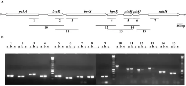
The PTS ofB. melitensisis transcriptionally linked to the BvrR/S two component system
The gene order around B. melitensis hprK, ptsM and ptsO is conserved in other a-proteobacteria and is as follows: (i) a
transcriptional response regulator and (ii) a sensor kinase of a two-component system known to be involved in host-symbiont (chvI
-exoSinS. meliloti[34]) or host-pathogen interaction (chvI-chvGinA. tumefaciens[33];bvrR-bvrSinB. abortus[35]), (iii)hprK, (iv)ptsM, (v)
ptsO, and finally (vi)sahH, which encodes an enzyme involved in the biosynthesis of methionine [36] (Fig. 5A). An additional gene called pckA that encodes PEP carboxykinase, a key enzyme of gluconeogenesis [39,40] is oriented in opposite direction to this cluster (Fig. 5A). In order to see whether this conserved organization reflects a functional link between these genes, we tried to determine whether they were transcriptionally linked. For that purpose, we performed PCR assays using cDNA of B. melitensis16M as template (Fig. 5B). Positive and negative control experiments were performed by using as template either genomic DNA or DNase-treated RNA in the absence of reverse transcriptase, respectively. When using cDNA as template we camplified intragenic regions of each gene of the cluster (Fig. 5A and B, bars and lanes 2 to 7), confirming that these genes are expressed in cells tat have grown in rich medium to late exponential phase. PCR products were also obtained for the intragenic regions of ptsP and ptsN (Fig. 5B, lanes 8 and 9, respectively), and for the neibhouring pckAgene (Fig. 5A and B, bar and lanes 1). The use of appropriate primers and cDNA as template also allowed the amplification of intergenic regions (lanes 11 to 15 in Fig. 5B), demonstrating that the following pairs of genes are co-transcribed:bvrR-bvrS,bvrS-hprK,hprK-ptsM,ptsM-ptsO
andptsO-sahH(Fig. 5B). As expected we could not amplify by RT-PCR the intergenic region between pckA and bvrR, two genes
oriented in opposite directions (Fig. 5A and B, bar and lanes 10). In conclusion, we demonstrated that theB. melitensis ptsgenes and
hprKare expressed during vegetative growth and thathprK, ptsM
andptsOcan be co-transcribed withbvrR/SandsahH.
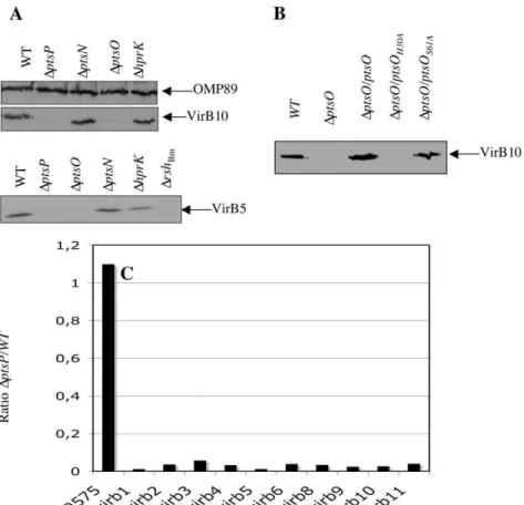
DptsPandDptsO mutants barely produce VirB5 and VirB10
Knowing that transpositional PTS mutants ofB. melitensis are attenuated [19,20,21] and having demonstrated a transcriptional link with several pts genes and the genes for the BvrR/S two component system, which regulates major virulence determinants [35], we wanted to investigate a possible link between PTS and virulence by constructing deletion mutants of the corresponding genes. Since hprK, ptsM and ptsO are probably organized in an operon withbvrR, bvrSandsahH, we chose to construct the mutants by allelic replacement using the non-polar cassette aphA4 as previously described [46]. Mutants were obtained for the ptsP,
ptsO,ptsNandhprKgenes. Despite numerous attempts, we were not able to deleteptsM.
VirB is a major virulence factor of Brucellaspp. composed of twelve subunits encoded in thevirBoperon [47,48,49,50], that is induced in response to nutrient availability [46,51,52] and controlled by (p)ppGpp, a bacterial alarmone that mediates global physiological control in response to starvation [46]. Since PTS proteins are also involved in global regulation in response to nutrient supply (for review see [1]), we examined the role ofpts
genes and hprK in the control of virB expression. Western blot analyses using anti-VirB5 and anti-VirB10 antisera [53] were performed to determine the relative amounts of VirB5 and VirB10 proteins inDptsP, DptsO, DptsNandDhprKmutants compared to the wild-type strain B. melitensis16M. Crude extracts of bacteria grown in 2YT to late exponential – early stationary phase (OD600 of 0.8–1.2) were prepared and analyzed by Western blot. The
DptsNandDhprKmutants produced VirB5 and VirB10 in amounts similar to those of the wild-type strain (Fig. 6A). However, no or very little VirB5 and VirB10 were detected in extracts prepared fromDptsPandDptsOmutants, suggesting that EINtrand NPr are required for production or stability of several B. melitensis VirB
Figure 5. Transcriptional link betweenpts genes and the genes encoding the two-component system BvrR/BvrS. (A) Schematic representation of the genomic region encodinghprK,ptsMandptsOinB. melitensis16M. The regions amplified (1–15) by RT-PCR are indicated and the primers are listed in Table S2. (B) Agarose gel of the RT-PCR amplified products. For each primer pair, three lanes are shown: a, positive control usingB. melitensis16M genomic DNA as template; b, RT-PCR; and c, a negative control using RNA as template (without RT). Identical results were obtained in several independent experiments.
subunits (Fig. 6A). Complementation of the DptsO mutant with wild-type ptsOconstitutively expressed from a low copy plasmid fully restored VirB10 (Fig. 6B) and VirB5 production (data not shown). For unknown reasons, plasmid-encoded ptsP did not restore VirB5 or VirB10 production in theDptsPmutant, although the plasmid was functional because it complemented the ‘‘small colony’’ phenotype of the DptsP mutant (see Fig. 7B). Knowing that EINtris strictly required for P
,His-NPr formation we tried to complement the DptsO mutant with the mutant allelesptsOH30A
andptsOS61A. Constitutive expression ofptsOandptsOS61AinDptsO
restored VirB10 production, whereas theDptsO/ptsOH30A strain
failed to produce VirB10 (Fig. 6B). This confirms that P,His-NPr is needed for VirB 10 synthesis and consequently that EINtris also required.
In order to confirm the impact of theptsPmutations on T4SS expression we carried out a transcriptional analysis of the whole
virBoperon with the wild-type strain and theptsPmutant. Indeed, the expression level of the individualvirB genes was more than thirty times lower in theptsPmutant than in the wild-type strain (Fig. 6C)
Colony size heterogeneity ofpts andhprK deletion mutants plated on rich medium
When plated on 2YT rich medium, theDptsP,DptsO,DptsNand, to a lesser extent, theDhprKmutant displayed a heterogeneity in
colony size compared to the wild-type strain B. melitensis 16M (Fig. 7A). Small colonies were detected only 8 to 10 days after inoculation, whereas the larger colonies were visible after 3 to 4 days as usually observed for the wild-type strain. To ensure that the colony size heterogeneity ofptsandhprKmutants resulted from the deletion of the corresponding genes, we first carried out a complete typing of these strains confirming that they all derived fromB. melitensis16M wild-type, exhibited a smooth phenotype, and were not contaminated with other strains (data not shown). Next, we complemented the two mutants with the most marked phenotype (DptsPandDptsO) by constitutively expressing wild-type copies of theptsPandptsOgenes in the corresponding mutants. As shown in Fig. 7B, the complemented strainsDptsP/ptsPandDptsO/ ptsOdisplayed bigger colonies than the DptsPandDptsOmutants transformed with the empty vector-pMR10cat. Their colonies resembled those of the wild-type strain carrying the empty vector-pMR10cat.
Finally, we measured growth of the four mutants when cultivated in liquid 2YT medium (Fig. 7C). No differences were observed between growth of the mutants and the wild-type strain, suggesting that the growth heterogeneity observed on solid medium might not result from the composition of the medium, but rather from parameters that distinguish liquid and solid cultures, such as oxygen supply, nutrient or water availability.
Figure 6. Synthesis of VirB proteins inDhprKandDptsmutants.(A) Detection of VirB10 (top) and VirB5 (bottom) by Western blot analysis in the wild-type (Bm16M),DptsP,DptsO,DptsNandDhprKstrains following growth in 2YT to late-exponential phase. An established negative control (DrshBm) was included in the anti-VirB5 Western blot analysis [46] (B) Western blot analysis of VirB10 withBm16M (+pMR10cat),DptsO(+pMR10cat)
and the complemented strainDptsO/ptsO,DptsO/ptsOH30A,andDptsO/ptsOS61A. Identical results were obtained in a second independent experiment.
(C) Transcription analysis ofvirB gene expression in wild-type andptsPmutant. The values presented by the bars correspond to the ratio of normalized and averaged microarray data (n = 263) obtained for 10virBORFs in theptsPmutant and the wild-type strain grown under the same conditions. BMEI0575 is a control ORF whose expression is not modulated whatever the strain considered.
doi:10.1371/journal.pone.0012679.g006
Yeast two hybrid assays reveal oligomerization of EINtr,
the EIIAMan-like protein and HprK/P, and interaction between NPr and HprK/P
Since in B. melitensis 16M two pts genes, hprK and the two-component system genes bvrR/bvrS are co-transcribed and functionally linked we wanted to test if there existed any physical interactions between PTS components, HprK/P and the BvrR/S proteins. A yeast two hybrid (Y2H) interaction matrix of 64 interactions was performed with the four PTS proteins, HprK/P, BvrR and BvrS fused to the Gal4 DNA binding domain (BD) and tested against the same proteins fused to the Gal4 activating domain (AD). Each BD and AD fusion was also tested against Gal4-AD and Gal4-BD alone. The previously evidenced interac-tion between BvrR and BvrS [54] was used as a positive control. The results presented in Fig. 8 and S6 show that two PTS proteins (EINtr, EIIAMan-like) and HprK/P interacted with themselve, suggesting that these proteins form oligomers similar to some well-studied EI, EIIAMan and HprK/P homologues [55,56,57]. Additionally, a bidirectional interaction was evidenced between NPr and HprK/P (Fig. 8 and S6) confirming the results of thein
vitro phosphorylation test. No interaction was observed between other PTS proteins that were shown to phosphorylate each other
in vitro. Finally, an interaction between BvrR and BvrS could be demonstrated (Fig. S6), but no interaction was detected between any of the PTS proteins or HprK/P and the two-component partners.
A yeast two-hybrid screen against theB. melitensis ORFeome reveals interaction partners of the EIIAMan-like protein and NPr
Having demonstrated that the incomplete PTS ofB. melitensisis functional and knowing that PTS-dependent regulations are mediated either by allosteric interaction or by direct phosphory-lation of target proteins [1], we performed a Y2H screen to detect interaction partners of the EIIAMan-like protein and NPr of B. melitensis16M to get some preliminary clues about the functional role of this PTS. Briefly, these PTS proteins were fused to the Gal4 DNA binding domain and used as baits to identify interaction partners in an « ORFeomic » library. Two clones provided a positive signal with at least two of the three reporter genes and the
Figure 7.Dptsmutants mutants display colony size heterogeneity on solid medium.(A) Colony size heterogeneity displayed by the mutantsDptsP,DptsO,DptsNandDhprKin comparison to the wild-type strain. Late log phase cultures were diluted and plated on 2YT medium and grown at 37uC for 8 to 10 days. (B) Complementation of the colony size heterogeneity phenotype for theDptsPandDptsOmutants. Wild-type,DptsP
andDptsOstrains (carrying the empty vector pMR10-cat; see Table S1) and the complemented mutantsDptsP/ptsPandDptsO/ptsO(carrying vectors pRH001-ptsPand -ptsO, respectively; see Table S1) were plated on 2YT supplemented with 20mg/ml of chloramphenicol as described in (A) and
grown at 37uC for 8 to 10 days. (C) Growth ofDptsandDhprKmutants in 2YT liquid cultures in comparison to the wild-type strain. Overnight 2YT cultures of the five strains were back diluted to an OD600(optical density at 600 nm) of 0.05, and growth was monitored by measuring the OD600at
corresponding proteins were identified as Ppa and SucA by sequencing the inserts in the pVV213 vector. NPr interacts with the inorganic pyrophosphatase PPa (BMEI0076) and the EIIAMan -like protein with the E1 component (SucA) (BMEI0140) of 2-oxoglutarate dehydrogenase.
In order to validate these interactions, the ORFeome entry clones forptsO(NPr),ppa,hprK,sucAandptsM(EIIAMan-like) were checked by sequencing and the coding sequences were subcloned in the Y2H vectors pVV212 and pVV213. Three interaction matrices were designed and the interactions between NPr and Ppa and the EIIAMan-like protein and SucA were confirmed (Fig. 8). In addition, a new interaction between PPa and HprK was established. SucA is the E1 component of the 2-oxoglutarate dehydrogenase complex, which contains also the mide succinyltransferase SucB (E2 component) and dihydrolipoa-mide dehydrogenase (E3 component) and plays a crucial role in the TCA cycle by converting 2-oxoglutarate to succinyl-CoA and CO2. Knowing that the PTSNtr presumably links regulation of carbon and nitrogen metabolism [1,2] and that 2-oxoglutarate is
at the cross-road between the TCA cycle and nitrogen assimilation, we tried to confirm the interaction between the EIIAMan-like protein and SucA by another independent method.
SucA, the E1 component of the enzymatic 2-oxoglutarate dehydrogenase complex physically interacts with the EIIAMan-like protein
DivIVA is attracted to and remains at cell poles not only in its native organism,B. subtilis, but also inE. coliand other bacteria [58]. In addition, DivIVA fused to a ‘‘bait’’ protein X can target an interacting GFP tagged ‘‘prey’’ protein Y to the pole [59]. To confirm the interaction between the EIIAMan-like protein and SucA we fused DivIVA to SucA and the EIIAMan-like protein to GFP.
After arabinose induction, strain DH10B[pSKoriTcat
-pBad-divIVA-gfp] synthesizing DivIVA-GFP exhibits fluorescens mainly at the cell poles (positive control; data not shown), whereas DH10B[pMR10kan-ptsM-gfp] producing EIIAMan-GFP (with or without arabinose induction) was uniformly fluorescent (Fig. 9B; negative control). DH10B bearing the two plasmids (pSKoriTcat
-Figure 8. Detection of interaction partners forBrucellaPTS proteins by Y2H assays.Interaction between NPr, HprK/P and PPa (top row). Interaction between EIIAMan-like and EIIANtr(middle row). Interaction between EIIAMan-like and SucA (bottom row). AD fusion = protein of interest
fused with activating domain of Gal4; BD fusion = protein of interest fused with DNA binding domain of Gal4. The reporter used is indicated by the letter in the upper right corner of each picture:b-galactosidase activity (G); growth test without uracil (U); growth test without histidine in the presence of 40 mM of 3AT (H). ‘‘-’’ indicates an empty vector. Identical results were obtained in three independent experiments.
doi:10.1371/journal.pone.0012679.g008
pBad-divIVA-sucA and pMR10kan-ptsM-gfp) showed a bipolar fluorescence pattern only when arabinose was present (Fig. 9C). This illustrates that SucA was targeted to the pole by DivIVA and able to recruit the EIIAMan-GFP fusion at the same location, thus confirming the interaction detected in the Y2H experiments.
Discussion
Since PTS permeases are lacking in a-proteobacteria, their soluble PTS proteins are not involved in carbohydrate transport and phosphorylation, but probably participate only in a regulatory phosphorelay (Fig. 10) [25,26,27]. In this paper, we present the first extensive biochemical and genetic characterization of the PTS components in an organism that lacks PTS permeases. A few studies on similar systems have previously been carried out, but were either limited to biochemical studies of PTS protein phosphorylation [60] or to genetic studies of mutants [61]. First we established that in B. melitensisthe phosphoryl group transfer from PEP to the EIIAs is fully functional. Second, several pieces of evidence allow us to propose a link between the PTS and the virulence ofB. melitensis. Finally, we report a connection between the PTS and systems likely to maintain the N/C balance. These three points are discussed in detail hereunder.
1- The PTS phosphorelay ofB. melitensisis fully functional and senses the metabolic state
B. melitensisEINtrautophosphorylates with PEP and transfers the phosphoryl group to the conserved His30 of NPr before it is passed on to either EIIANtr or the EIIAMan-like protein (Fig. 10). EINtr probably senses the PEP availability (PEP/pyruvate ratio), that is translated into relative levels of phosphorylated vs. non-phosphor-ylated forms of NPr and EIIAs [62]. In addition to His30, NPr is also phosphorylated on a serine residue. Similar to firmicutes,B. melitensispossesses an HprK/P using ATP or PPi as phosphoryl
donor. Only phosphorylation of NPr with ATP is stimulated by FBP (Fig. 2B). Identical observations were made forL. caseiHprK/ P [45]. However, Brucella spp lack a 6-phosphofructokinase and consequently hexose catabolism does not occur via the Embden-Meyerhof-Parnas pathway, but is redirected through the pentose phosphate and perhaps the Entner-Doudorof pathway. Accord-ingly, the only pathway that is expected to produce FBP is gluconeogenesis [63]. We therefore propose that, in contrast to firmicutes, the FBP signal sensed byB. melitensisHprK/P reflects gluconeogenetic instead of glycolytic activity. High gluconeoge-netic flux will probably activate HprK/P (via FBP) (Fig. 2B), which in turn will slow the PEdependent phosphoryl transfer from P-Ser-NPr to the EIIAs (Fig. 4) [1,64,65].
Interestingly, an inorganic pyrophosphatase named PPa inter-acts with NPr and HprK/P in Y2H tests (Fig. 8). PPi serves as substrate for the kinase reaction and is formed during P-Ser-HPr dephosphorylation [45]. Hydrolysis of PPi by PPa not only lowers the PPi concentration, but also produces inorganic phosphate (Pi), which inhibits both, ATP- and PPi-dependent kinase activities of
B. melitensis HprK/P (Fig. 2C). Elevated PPa activity might therefore reduce phosphorylation on Ser61 of NPr. Similarly, Mijakovic [45] proposed that theB. subtilispyrophosphatase YvoE (theyvoEgene is located in thehprKoperon) indirectly decreases the kinase activity of HprK/P, and stimulates P-Ser-HPr dephosphorylation by HprK/P. The physical interaction of PPa with NPr and HprK/P might allow efficient regulation of HprK/P activity inB. melitensis. Alternatively, a link might exist between the PTS and the ppGpp production/degradation system (also called stringent response) as was demonstrated inE. coli[66]. NPr might affect the PPa-catalyzed conversion of PPi to Pi and thus modulate the PPi-producing ppGpp degrading activity of Rsh (RelA/SpoT homologue).
Purified B. melitensis HprK/P barely dephosphorylated P-Ser-NPr underin vitro conditions (Fig. 3). Similar observations were
Figure 9. The EIIAMan-like protein interacts with SucA.DIC and corresponding fluorescent images were taken fromE.coli(pSKoriTcat
–pBad-divIVA-sucAand pMR10kan-ptsM-gfp) grown in different conditions: (A) without IPTG and arabinose (no synthesis of EIIAMan-GFP and SucA-DivIVA). (B) with IPTG and no arabinose (synthesis of EIIAMan-GFP) and (C) with both IPTG and arabinose (EIIAMan-GFP and SucA-DivIVA are synthesized). Only
made for HprK/P from Treponema denticola [60], M. pneumoniae
[67], N. meningitidis (S. Poncet, M.-K. Taha, M. Laribe and J. Deutscher, unpublished) and A. tumefaciens (I. Mijakovic, A. Khemiri and J. Deutscher, unpublished). In the case of a -proteobacteria, the poor phosphorylase activity of HprK/P might be due to the absence of a C-terminal conserved region required for P-Ser-HPr dephosphorylation (Fig. S5) [30,41,42]. In M. pneumoniae and possibly other bacteria, dephosphorylation of P-Ser-HPr seems to be catalyzed by a protein phosphatase of the PP2C family [67].
2- A link between PTS and virulence ofBrucella
It seems that PTS-mediated carbon source utilization can affect host-bacteria interactions [61,68]. B. melitensis pts mutants were previously shown to be attenuated [19,20,21] but the underlying mechanism remained unknown. During this work two lines of evidence for a link between PTS and virulence emerged. First, we demonstrate a transcriptional link between the PTS genes hprK, ptsM and ptsO and the bvrR/S genes (Fig. 5) encoding a two component system crucial for virulence of B. melitensis. In all pathogenic or symbiotica-proteobacteria theptsgenes are located downstream from the two-component system genes essential for
infection or symbiosis [33,34,35]. In addition, a recent transcrip-tome analysis withB. abortusshowed thathprK(BAB1_2094) was downregulated in the bvrR:Tn5 mutant [69]. It is therefore tempting to assume that the PTS might also be involved in virulence regulation, possibly via a cross-talk between PTS proteins and the two-component system. This concept is also supported by the finding that deletion ofptsPorptsO, but notptsN
orhprK, lowers the production of a major virulence factor, the type IV secretion system VirB, by reducingvirBgene expression (Fig. 6). The expression ofBrucellaspp.virBhas previously been shown to be controlled by nutrient availability via an unknown mechanism [46,48,51]. The Bartonella henselae BvrR/S homologues BatR/S, whose genes are also followed byhprK,ptsMand npr, have been reported to control virB expression and BatR binds to thevirB
promoter region [70]. It is therefore tempting to assume that theB. melitensisPTS communicates the metabolic state of the cell to the
virB promoter by phosphorylating or interacting with the two component system BvrR/S. However, we cannot exclude the possibility that PTS components interact with one of the two other transcriptional regulators known to bind to thevirBpromoter: the quorum sensing regulator VjbR [52,71] and HutC, a transcrip-tional repressor of the histidine utilization (hut) genes [72].
Figure 10. Model proposed for the role of theBrucellaPTS in connecting C and N metabolisms.In agreement with the results of thein vitroandin vivophosphorylation assays (Fig. 1–4), we postulate that a phosphoryl group is sequentially transferred from PEP to EINtr, NPr, and finally to the EIIAMan-like protein. By binding both an unknown ligand (possibly 2-oxoglutarate) through its GAF domain and autophosphorylating in
response to the PEP/pyruvate ratio, EINtrmight sense the metabolic status of the cell and communicate it to the EIIAMan-like protein that would regulate the 2-oxoglutarate dehydrogenase activity accordingly. In addition, HprK/P is expected to slow phosphorylation of the EIIAMan-like protein
by hindering the phosphotransfer through the PTS in response to changes in the FBP concentration or other metabolites. Solid arrows indicate metabolic reactions, large open arrows represent phosphoryl transfer between PTS proteins, and dashed arrows show putative regulatory processes. The enzyme HprK/P was also included in this scheme since it phosphorylates NPr on a conserved serine. The names of the genes encodingB. melitensisPTS and HprK/P proteins are put between brackets. The pentose phosphate pathway, which connects glucose-6-phosphate (G6P) to glycolysis inB. melitensis, and the connection between TCA cycle and nitrogen metabolism starting from 2-oxoglutarate are indicated in grey. FBP, fructose-1,6-bisphosphate; PEP, phosphoenolpyruvate; CoA, coenzyme A; TCA cycle, tricarboxylic acid cycle; NH4+
, ammonium. P,H-X and P,S-X
indicate PTS enzymes phosphorylated on histidine or serine, respectively. doi:10.1371/journal.pone.0012679.g010
While no differences were observed between growth of the four mutants and the wild-type strain when cultivated in liquid 2YT medium (Fig. 7C), theDptsO,DptsPand to a lesser extent theDptsN
and DhprK mutants displayed a heterogeneity in colony size compared to the wild-type strain when plated on solid 2YT rich medium (Fig.7A and C). This is reminiscent of a similar defect described forptsmutants ofS. melilotigrown on solid media [61]. These small colonies resemble a phenotype called small colony variant (SCV). The presence of SCVs in pathogenic bacteria, includingB. abortus, has often been associated with the persistence in the host [73,74,75,76,77]. It will be interesting to test whether
ptsandhprKmutants persist longer than the wild-type during mice infection, as was described for a SCV of the B. abortus vaccinal strain S19 [73].
3- Carbon catabolite repression and the coordination of carbon and nitrogen metabolism
In many bacteria the PTS is linked to carbon catabolite repression (CCR). Interestingly, in thea-proteobacteriumS. meliloti
HprK/P regulates succinate-mediated CCR [32]. In Brucella, erythritol is the most favoured carbon source and is able to inhibit glucose incorporation [78], but to our knownledge the underlying mechanism is not known and diauxic growth has not been reported. CcpA as well as Crp [79] and adenylate cyclase seem to be absent fromBrucella spp. If general CCR exists in Brucella, it should therefore differ from the E. coli and B. subtilis CCR mechanisms.
The PTSNtrproteins (EINtr, NPr and EIIANtr) have previously been suggested to provide a regulatory link between carbon and nitrogen metabolism [3,4,5,6,11,80,81]. Additionally, recent ‘‘in silico’’ analyses suggest that some of the diverse regulatory PTS functions acquired during evolution serve to assure an appropriate balance in C and N supply [82]. Key signals of C and N supply in
E. coliappear to be the levels of glutamine and 2-oxoglutarate, the latter being at the crossroad between carbon and nitrogen metabolism [83]. Several results reported in our paper converge on these metabolites and prompted us to propose a model linking the PTS to the maintenance of the carbon and nitrogen balance in
B. melitensis16M:
First, the EIIAMan-like protein interacts with the SucA subunit of 2-oxoglutarate dehydrogenase (Fig. 8 and 9).
Second, the enzyme EINtr possesses an N-terminal GAF domain (Fig. S1). This domain is known to regulate the activity of NifA fromA. vinelandiiby binding 2-oxoglutarate [84,85]. Finally, three PTS genes are transcriptionaly linked to the genes encoding the two component system BvrR/S (Fig. 5).
The latter finding supports a link between regulation of C and N metabolism and PTSNtrcomponents because a proteomic study with a B. abortus bvrR mutant [86] revealed that two 2-oxoglutarate-dependent proteins are regulated by BvrR-BvrS: the first is the PII sensor protein that controls nitrogen metabolism and that was shown to bind 2-oxoglutarate [83,87]; the second is the oxoglutarate dehydrogenase complex that converts 2-oxoglutarate into succinyl-CoA in the TCA cycle. This is the very same enzyme whose subunit SucA interacts with the EIIAMan-like protein (Fig. 8 and 9).
We therefore propose a model in which EINtr senses the metabolic status of the cell via the PEP/pyruvate ratio [62]. The existence of a GAF domain in EINtrprovides a link between GAF-sensed signals (a-ketoglutarate [84,85] or other ligands [88]) and PTS phosphoryl transfer. The signals (EI phosophorylation state and HprK/P activity) are transmitted to the EIIAMan-like protein,
which in turn regulates the activity of 2-oxoglutarate dehydroge-nase (Fig. 10). In this model, the dephospho EIIAMan-like protein is predicted to interact with and to inhibit 2-oxoglutarate dehydrogenase. Indeed, exclusively dephospho EIIAMan is prob-ably present in yeast during two-hybrid tests, where the EIIAMan/ SucA interaction was first detetcted. Finally, one can envisage that HprK/P might control EIIAMan-dependent regulation of 2-oxoglutarate dehydrogenase. Similar as observed for HPr from firmicutes [1,64,65], HprK/P-catalyzed phosphorylation of Ser61 of NPr probably slows phosphorylation of His30 and thus increases the amount of dephospho EIIAs (Fig. 4).
It will also be interesting to test whether the N-terminal domain of EINtris able to bind 2-oxoglutarate and whether this ligand can modulate the phosphotransfer activity of the PTS protein. It might also be worth studying the enzymatic activity of 2-oxoglutarate dehydrogenase in different mutant backgrounds. Finally, our model should be tested with other a-proteobacteria possessing homologues of the PTS regulatory proteins and the crucial two component system encoded by genes arranged in a strictly conserved genomic context.
Materials and Methods
Ethics statement
Animal handling and experimental protocol was in accordance with European (DOCE 86/609/EEC), and National (AR25/04/ 2004) directives, and was supervised and authorized by the Ethical Committee of the University of Namur (FUNDP) (Commission d’e´thique en experimentation animale approval Nu FUNDP08/ 106).
Bacterial strains and growth conditions
AllBrucellastrains used in this study were derived fromBrucella melitensis16M NalR (Table S1) and were routinely cultivated in 2YT complex medium (10% yeast extract, 1% tryptone and 0.5% NaCl).E. colistrains (Table S1) were cultivated in Luria Bertani (LB) medium. Antibiotics were used at the following concentra-tions when appropriate: nalidixic acid, 25mg/ml; kanamycin, 50mg/ml; chloramphenicol, 20mg/ml; ampicillin, 100mg/ml;
gentamycin, 50mg/ml.
To observe the colony size heterogeneity of pts and hprK
mutants, overnight cultures were adjusted to an OD600of 0.05 in 2YT complex medium and grown at 37uC with constant shaking until late log phase (OD600of 1.0). Dilutions of these cultures were plated on 2YT agar supplemented with appropriate antibiotics and incubated for 8 to 10 days at 37uC.
To evaluate growth of the pts and hprK mutants in liquid cultures, overnight cultures were diluted to an OD600 of 0.05 in 2YT complex medium and grown at 37uC with constant shaking. The experiment was carried out twice.
Construction of overexpression plasmids
For the construction of overexpression plasmids, theB. melitensis
ORFeome entry vectors [89] bearing ptsN, ptsM and hprK
(pDONR201-ptsN, ptsM and hprK, respectively – Table S1) were checked by DNA sequencing before they were used to amplify by PCR theptsN, ptsMandhprKgenes with oligonucleotide pairs SP67-SP68, SP69-SP70, and SP74-SP75 respectively (Table S2). TheptsN-,ptsM- andhprK-PCR products were digested with BamHI and KpnI and cloned into pQE30 (Table S1) digested with the same enzymes, resulting in plasmids pQE30-ptsN, -ptsM -and –hprK and encoding (His)6-EIIA
Ntr
, (His)6-EIIA Man
The published genome sequence predicts aptsOgene starting with a GUG codon and lacking a ribosome binding site (RBS) [22]. We therefore assumed that NPr might be 4 amino acids longer and that the gene starts with an ATG preceded by a RBS. Accordingly, a new pDONR201 entry vector (pDONR201-ptsO) bearing a longer version of theptsOgene was constructed. Briefly, theB. melitensis16MptsOCDS (BMEI2031) was amplified by PCR with genomic DNA with and the GatewayTMprimers GWnprF and GWnprR (Table S2) and cloned in the entry vector pDONR201 (Invitrogen Life-technologies) as previously described [89]. Directed mutagenesis of ptsO was performed with the QuickChangeTMSite Directed Mutagenesis kit (Stratagene) using plasmid pDONR201-ptsO as a template. Primers used to obtain the ptsOH30A and ptsOS61A alleles are listed in Table S2. The correct sequence of all PCR products was confirmed by DNA sequencing. Plasmids pQE30-ptsO, -ptsOH30A and -ptsOS61A encoding (His)6-NPr and its two mutant forms, were obtained by amplification of the corresponding allele using oligonucleotides SP65 and SP66, and plasmids pDONR201-ptsO, -ptsOH30A and
-ptsOS61A respectively, as templates. The PCR products were digested withBamHI and KpnIand cloned into pQE30 digested with the same enzymes.
Overexpression and purification of PTS proteins
The E. coli NM522 (Stratagene) transformants (Table S1) harboring the various pQE30-derivatives were grown in 500 ml of LB medium supplemented with 100mg/ml ampicillin to an OD600 of 0.7. The synthesis of (His)6-fusion proteins was induced with 0.1 mM isopropyl-b-D-thiogalactopyranoside and growth was continued for 3 hours at 37uC. Protein extracts were prepared and loaded on a 1 ml Ni-NTA column (Qiagen); purification was carried out under native conditions by following the recommen-dations of the manufacturer. (His)6-tagged EI
Ntr
, NPr, NPrH30A, NPrS61A, EIIANtr, EIIAMan-like and HprK/P were recovered as soluble proteins.
Protein phosphorylation and dephosphorylation assays [32P]PEP was synthesized by following the PEP-pyruvate isotope exchange method in the presence of pyruvate kinase and [c-32P]ATP [90]. Transfer of the phosphoryl group from [32P]PEP via EINtr, NPr or NPrH30A to EIIANtror EIIAMan-like was tested at 37uC in 30ml reaction mixtures containing 50 mM Tris-HCl pH 7.4, 5 mM MgCl2, 10mM [
32
P]PEP, 1.5mg of EINtr, 3mg of
NPr or NPrH30A, 4.5mg of EIIANtr or EIIAMan-like. Samples were incubated for 20 min at 37uC and reactions were stopped by addition of SDS sample buffer. Proteins were separated by electrophoresis on 0.1% SDS-15% polyacrylamide gels, which were subsequently dried and exposed overnight to a storage phosphor screen (STORM).
ATP-dependent NPr phosphorylation assays were performed in 50ml reaction mixtures containing 50 mM Tris-HCl pH 7.4,
5 mM MgCl2, 25mM [c-32P]ATP or [32P]PPi and varying amounts of either FBP or potassium phosphate. The assay mixtures were incubated for 20 min at 37uC and the reaction was stopped by addition of SDS sample buffer. Proteins were separated on 0.1% SDS-15% polyacrylamide gels. After electro-phoresis, gels were boiled for 10 min in 0.5 N HCl, dried and exposed overnight to a storage phosphor screen.
For P-Ser-NPr dephosphorylation assays, P-Ser-NPr was obtained by incubating B. melitensis (His)6-NPr with (His)6 -HprK/P for 30 min at 37uC in 50 mM Tris-HCl pH 7.4, 5 mM MgCl2, 5 mM ATP and 25 mM FBP. HprK/P was subsequently inactivated by keeping the reaction mixture for 10 min at 65uC. P-Ser-NPr was then loaded on a PD-10 column
(GE Healthcare), eluted with 20 mM NH4HCO3 to eliminate ATP, and lyophilized. Dephosphorylation assays were carried out in reaction mixtures containing 50 mM Tris-HCl pH 7.4, 5 mM MgCl2, 3mg of P-Ser-NPr, 0.45mg of HprK/P and various
concentrations of potassium phosphate. The assay mixtures were incubated for 30 min at 37uC before HprK/P was heat-inactivated for 10 min at 65uC. The different forms of NPr were separated by electrophoresis on non-denaturing 12.5% polyacryl-amide gels, followed by staining with Coomassie Blue.
RNA isolation and RT-PCR assays
Extraction of B. melitensis 16M total RNA was performed on cultures (40 ml) grown to late exponential growth phase in 2YT. Bacterial cells were harvested by centrifugation for 10 min at 3500 rpm, and resuspended in 100ml 10% SDS, 20ml proteinase
K (20 mg/ml) and RNaseOUTTM(Invitrogen Life-Technologies), and incubated for 1 hour at 37uC. Total RNA was then extracted using TRIzolH reagent. Contaminating genomic DNA was digested with DNase I DNA-free (Ambion) before the enzyme was inactivated by DNase Inactivation Reagent (Ambion). Reverse transcriptions (RT) were performed as follows: random primers (200 ng/ml) (Invitrogen Life-Technologies) and dNTP mix (10 mM each dNTP) (Invitrogen Life-Technologies) were added to 3–4mg of DNase-treated total RNA and the mixture was
incubated at 65uC for 10 min. 5X First-Strand buffer, DTT (0.1 M) and RNaseOUTTM (Invitrogen Life-Technologies) were added to the solution, which was incubated at 25uC for 2 min. Finally, SuperScriptTM reverse transcriptase (Invitrogen Life-Technologies) was added and incubated for 10 min at 25uC and 50 min at 42uC. The enzyme was inactivated by heating to 70uC for 15 min. To remove RNA hybridized to the cDNA, E. coli RNase H (Invitrogen Life-Technologies) was added to the RT reaction. A control reaction containing the same components but no reverse transcriptase was included to check for DNA contamination. The cDNA products (2ml) were then used in a PCR performed in a final volume of 30ml and containing 1.25 U of GoTaq DNA polymerase (Promega), dNTP mix (5 mM each), and 10 pmol of each primer. A PCR control in which B. melitensis 16M genomic DNA was used as template was included. The amplification consisted of one cycle of 5 min at 95uC, followed by 35 cycles of 30 sec at 95uC, 30 sec at annealing temperature (depending on the primers used), 90 sec at 72uC, and a final step of 10 min at 72uC. Primers used in this experiment are listed in Table S2.
Concerning the microarray data for thevirBexpression, RNA was reverse transcribed, labeled and hybridized by NimbleGenTM Systems, Inc using the catalogue design for B. melitensis 16M chromosomes I (NC_003317) and II (NC_003318) with 20 probes per gene (10 perfect matches and 10 mismatches). Each probe (24 mer) was replicated three times on a chip at a random position (design includes random GC probes). Duplicate samples of each strain were processed. Analysis of the data were performed ‘‘mutatis mutandis’’ as described previously [71].
GatewayH cloning of genes of interest in Y2H vectors For Y2H interaction tests, each protein of interest (YFP) was fused with both AD and BD domains of the transactivator Gal4. Entry vectors pDONR201 of the ORFeome [89] corrresponding to detected genes of interest (YFG) (Table S1) were subcloned in Y2H destination vectors pVV212 and pVV213 (Table S1) [91]. LR recombination procedure was performed as recommended by the manufacturer (Invitrogen Life-Technologies) to fuse YFP with both Gal4-BD (in pVV212) and Gal4-AD (in pVV213) generating plasmids pVV212-YFG and pVV213-YFG [54].
Yeast two hybrid assay
Haploı¨d yeast Mav103 and Mav203 [92] were transformed with pVV212-YFG and pVV213-YFG respectively, and selected on SD-W (tryptophan omission medium) and SD-L (leucin omission medium) respectively. Mating of two plasmid-carrying yeasts was then carried out, and SD-LW (leucin and tryptophan omission medium) was then used to select diploids containing both pVV212 and pVV213. Two growth tests can be used to detect physical interactions between proteins, i.e. (i) SD-HLW+3-AT (medium without histidine and with 20 to 50 mM triaminotriazole (3-AT) and (ii) SD-ULW (medium without uracil). The additional lacZ
reporter gene was used to detect interactions by performing b -galactosidase coloration assays. For all Y2H assays used in this study, except for the interaction test between PTS proteins and BvrR and BvrS,b-galactosidase coloration tests were performed as follow. Diploid yeasts were plated on a nitrocellulose filter laid on a yeast peptone dextrose (YPD) plate and grown overnight at 30uC. The filter was then placed in liquid nitrogen to lyse the cells, transferred on a new plate containing two Whatman papers saturated withb-galactosidase assay solution (for each plate 5 ml of Z-buffer, 120ml of 4% X-gal and 13ml of 100% b -mercaptoethanol), and finally incubated at 37uC. In the case of interaction tests between PTS proteins and BvrR or BvrS, b -galactosidase coloration tests were performed using an overlay plate assay as described in [93].
Y2H screen against the ORFeome ofB. melitensis16M Briefly, entry vectors pDONR201 of the ORFeome [89] were pooled by 48 (half of a 96-wells plate) to obtain 69 pools borne in a single 96-well plate. Each pool was subcloned in the Y2H vector pVV213 in order to fuseB. melitensisproteins to the Gal4 activating domain [91] using LR. Pools of pVV213 were used to transform the haploı¨d yeast Mav203. To select interacting partners of our proteins of interest, mating was performed using the pools of Mav203 containing pVV213 plasmids and Mav103 strains containing pVV212 bearing our genes of interest. Diploı¨ds were selected using SD-LW medium. As a first screen for selecting interactions, an overnight culture of the diploı¨ds was grown in SD-HLW medium at 30uC under shaking, and plated on SD-HLW with 20 mM 3-AT. Five diploid controls were used for this Y2H assay containing: (i) empty pVV212 and pVV213 (negative control), (ii) a weak interaction (BD-Rb and AD-E2F), (iii) a strong interaction (BD-Fos and AD-jun), (iv) complete Gal4 with empty pVV213 and (v) a strong interaction (BD-DP and AD-E2F) [94]. For each pool that showed growth, a maximum of four clones was cultivated in SD-HLW and plated on SD-LW (for a back-up), SD-HLW with 20 mM of 3-AT, on SD-ULW and on nitrocellulose filters placed on a YPD plate for b-galactosidase coloration tests. Clones that were positive for at least two Y2H tests were selected and PCR was carried out with primers iGAl4AD and Gal4term to amplify the inserts cloned in the pVV213-derived plasmids. Finally, the PCR products were sequenced using primer iGAl4AD to identify the putative interacting partner. Interactions between our proteins of interest and newly detected partners were confirmed as described in the Y2H assay.
DivIVA interaction test
The plasmids used for the experiments were obtained as follows. The pKD46 vector was used to amplify the pBad promoter sequence with oligonucleotide pairs (Fpbad and Rpbad) (Table S2). The pBad-PCR product was cloned into pSKoriTcatdigested withEcoRV. The pZD6 vector was used to amplify thedivIVA-gfp
fusion with oligonucleotide pairs (FdivIVA and Rgfp) (Table S2).
The divIVA-gfp PCR product was cloned into the pGEMTeasy vector. The pGEMT-divIVA-gfpvector was digested withNheI and
KpnI and the fused genes were cloned into pSKoriTcat –pBad digested with the same enzymes. The plasmid pSKoriTcat
–pBad-divIVA-gfpwas used as positive control.
The entry vector bearing theptsMgene (pDONR201- ptsM– Table S1) was taken from theB. melitensisORFeome. This vector was used to amplify by PCR theptsMgene with oligonucleotide pairs (FptsM and RptsM) (Table S2). TheptsM-PCR product was cloned into vector pSKoriTcatdigested withEcoRV, giving plasmid pSKoriTcat–ptsMencoding the EIIAMan-like protein. The pZD6 vector was used to amplify thegfpgene with oligonucleotide pairs (Fgfp and Rgfp) (Table S2). Thegfp-PCR product was cloned into the pGEMTeasy vector. The pGEMT-gfpvector was digested with
BglII andKpnI to getgfpwhich was cloned into pSKoriTcat–ptsM
digested with the same enzymes. The pSKoriTcat–ptsM-gfpvector was digested with HindIII and KpnI and cloned into pMR10kan
digested with the same enzymes.
The pZD6 vector was used to amplify the divIVA gene with oligonucleotide pairs FdivIVA and RdivIVA (Table S2). The
divIVA-PCR product was cloned into pSKoriTamp digested with
EcoRV. The pSKoriTamp-divIVAvector was digested withNheI and
HindIII anddivIVAwas cloned into pSKoriTcat-pBad digested with the same enzymes.
B. melitensisgenomic DNA was used to amplify by PCR thesucA
gene with oligonucleotide pairs (FsucA and RsucA) (Table S2). The sucA-PCR product was cloned into the pGEM11Zf vector. The pGEM11Zf-sucAvector was digested withHindIII andXhoI and cloned into pSKoriTcat–pBad-divIVAdigested with the same enzymes. The correct sequence of all PCR products was confirmed by DNA sequencing.
The two plasmids, pSKoriTcat –pBad-divIVA-sucA and pMR10kan-ptsM-gfp, were used to co-transform E. coli DH10B competent cells. The resulting strain was cultivated in 10 ml SOB medium (tryptone 2%, yeast extract 0.5%, NaCl 0.058%, KCl 0.019% and MgCl20.19%) with chloramphenicol (20mg/ml) until the OD600 reached 0.1. Arabinose (10 mM) induction was performed during three hours before the microscopic observation.
Rabbit immunization
In order to produce monospecific polyclonal antisera against EIIANtr, rabbits were immunized with the purified protein (50mg
per dose), initially in the presence of complete Freund’s adjuvant and on days 30 and 60 with incomplete Freund’s adjuvant. Rabbits were bled 1 week after the last injection.
Detection ofin vivophosphorylated EIIANtr
Construction ofDptsmutants and complementation strains
B. melitensis16Mpts knock out mutants were obtained by gene replacement as previously described [46]. For eachptsgene, upstream and downstream regions (about 500 bp) flanking the gene were PCR amplified fromB. melitensis16M genomic DNA by using appropriate primers (Table S2). A second PCR was used to associate the two PCR products by cohesive ends. The final PCR product that carries aBglII site between the upstream and the downstream regions was inserted into theNotI site of pSKoriTcat(Table S1). TheaphA4cassette [46] was excised from pUC4aphA4 (Table S1) with BamHI and subsequently cloned into the BglII site to generate plasmid pSKoriTcat-Dpts(or -DhprK) (Table S1). These constructs were used to transformE. colistrain S17-1 and subsequently introduced intoB. melitensis16M by mating. Clones exhibiting a double recombination phenotype (Cms Kanr) were selected and their genotypes were verified by PCR and by Southern blot analysis using appropriate probes. The complementation plasmids pRH001-ptsP and -ptsO
(Table S1) were constructed by using the GatewayTM technique (Invitrogen Life-Technologies). LR recombination cloning was carried out as recommended by the manufacturer (Invitrogen Life-Technologies) in order to insert selected genes in pRH001 using pDONR201-ptsPand -ptsO, -ptsOH30Aand -ptsOS61Aas entry vectors (Table S1). The resulting vectors pRH001-ptsPand-ptsO,-ptsOH30A and -ptsOS61A(Table S1) were transferred by mating into theDptsPor DptsO mutants to generate the complemented strains DptsP/ptsP,
DptsO/ptsO, DptsO//ptsOH30A and DptsO/ptsOS61A. In parallel, pMR10cat(Table S1) was transfered intoB. melitensis16M wild-type,
DptsPandDptsOstrains by mating.
Detection of VirB5 and VirB10 proteins by Western blot analyses
For VirB detection in total lysates ofB. melitensis16M and various mutants, strains were grown overnight at 37uC in 2YT complex medium and then diluted and grown at 37uC until late log phase (OD6000.8–1.2). Aliquots of the cultures were kept for 1 hour at 80uC in order to inactivate cell functions and then adjusted to the same OD600. Following SDS-polyacrylamide gel electrophoresis and Western blot analysis, immunodetection of VirB5 and VirB10 in total lysates was performed with rabbit polyclonal anti-VirB5 and -VirB10 antisera [53] at respective dilutions of 1/5000 and 1/2000. Immunodetection with a monoclonal antibody anti-Omp 89 [95] was used as loading control.
Supporting Information
Figure S1 Multiple sequence alignment of N-terminal portion of enzyme INtr. The predicted PEP-dependent phosphorylated histidine of enzymes INtr regarding multiple alignment with paralogous enzymes I is marked by an asterisk and shaded, and the conserved region surrounding it is boxed. The predicted N-terminal GAF domain homologous to the NifA-sensory domain of
Azotobacter vinelandiiis underlined and limited by two vertical bars. Red residues are identical for the five proteins, whereas green and blue residues are strongly or weakly similar, respectively. (EInSme), Sinorhizobium meliloti, (EInAtu) Agrobacterium tumefaciens, (EInBme)Brucella melitensisand (EInEco)Escherichia coli.
Found at: doi:10.1371/journal.pone.0012679.s001 (0.86 MB TIF)
Figure S2 Multiple sequence alignment of enzyme IIANtr. Conserved histidine predicted to be phosphorylated by NPr in
E. coli, S. meliloti, A. tumefaciensand B. melitensisis marked by an asterisk and shaded. The well-conserved region surrounding the putative phosphorylation site is boxed. Red residues are identical
for the five proteins, whereas green and blue residues are strongly or weakly similar, respectively. (EInSme), Sinorhizobium meliloti, (EInAtu) Agrobacterium tumefaciens, (EInBme) Brucella melitensis and (EInEco)Escherichia coli.
Found at: doi:10.1371/journal.pone.0012679.s002 (0.43 MB TIF)
Figure S3 Mutiple sequence alignment of enzyme IIAMan. Conserved histidine phosphorylated by HPr inE. colithat is predicted to be phosphorylated by NPr inS. meliloti, A. tumefaciensandB. melitensis
is marked by an asterisk and shaded. Red residues are identical for the five proteins, whereas green and blue residues are strongly or weakly similar, respectively. Sinorhizobium meliloti (IIAmSme), Agrobacterium tumefaciens(IIAmAtu),Brucella melitensis(IIAmBme) and domain IIA of enzyme IIABMan fromEscherichia coli(IIAmEco).
Found at: doi:10.1371/journal.pone.0012679.s003 (0.38 MB TIF)
Figure S4 Multiple sequence alignment of NPr proteins. The conserved histidine residue phosphorylated by enzyme I on HPr fromB. subtilisandE. coli, that is predicted to be phosphorylated by enzyme INtron NPr proteins fromE. coli, S. meliloti, A. tumefaciens
andB. melitensisis marked by an asterisk and shaded. Similarly, the conserved serine residue phosphorylated by HprK/P on HPr protein fromB. subtilis, that is predicted to be phosphorylated by HprK/P on NPr proteins from S. meliloti, A. tumefaciens and B. melitensis is marked by an asterisk and shaded. The consensus sequences surrounding these two predicted phosphorylation sites are boxed. Red residues are identical for the five proteins, whereas green and blue residues are strongly or weakly similar, respectively.Sinorhizobium meliloti(NPrSme),Agrobacterium tumefaciens
(NPrAtu),Brucella melitensis(NPrBme),Escherichia coli(NPrEco) and HPr proteins fromE. coli(HPrEco) andBacillus subtilis(HPrBsu). Found at: doi:10.1371/journal.pone.0012679.s004 (0.42 MB TIF)
Figure S5 Multiple sequence alignment of HprK/P proteins. The conserved Walker A motif which binds ATP, PPi and Pi in HprK/P proteins is boxed (155-GDSGVGGKS-162 in L. casei HprK/P)). The HprK/P signature sequence, whose consensus is (I,L,M)E(I,V)RG(I,L,M,V)G(I,V)(I,L,M) (residues 203 to 211 inL. casei HprK/P), is also boxed. An additional conserved region present in HprK from Gram positive bacteria and playing an important role in phosphorylase activity of the protein is underlined. This region is not conserved in HprK/P from a -proteobacteria. Shaded residues are amino acids that were shown to be required either for kinase or phosphorylase activities. Red residues are identical for the five proteins, whereas green and blue residues are strongly or weakly similar, respectively.Sinorhizobium meliloti (HprKSme), Agrobacterium tumefaciens (HprKAtu), Brucella melitensis(HprKBme) and C-terminal portion of HprK/P proteins fromLactobacillus casei(HprKLca) andBacillus subtilis(HprKBsu). Found at: doi:10.1371/journal.pone.0012679.s005 (0.58 MB TIF)
Figure S6 Interaction matrix for PTS proteins, HprK/P and the two-component system BvrS/BvrR. AD-P = protein of interest fused with the activating domain (AD) of Gal4; BD-P = protein of interest fused with the DNA binding domain (BD) of Gal4. Interactions demonstrated with one or two reporter genes (lacZor
HIS3) are shown in grey and black respectively.
Found at: doi:10.1371/journal.pone.0012679.s006 (0.04 MB DOC)
Table S1 Strains and plasmids used in this study.
Found at: doi:10.1371/journal.pone.0012679.s007 (0.10 MB DOC)
Table S2 List of the primers used in this study.
Found at: doi:10.1371/journal.pone.0012679.s008 (0.11 MB DOC)
![Figure 2. NPr kinase assays with B. melitensis HprK/P. (A) The NPr kinase assay was carried out with 200 ng of HprK/P and 2 mg of either wild- wild-type NPr (WT), NPr H30A (H30A) or NPr S61A (S61A) in the presence of 25 mM [c- 32 P]ATP and in the absence o](https://thumb-eu.123doks.com/thumbv2/123dok_br/18276565.345121/4.918.91.571.87.510/figure-kinase-assays-melitensis-kinase-carried-presence-absence.webp)