RAPID COMMUNICATION
Malignant peripheral nerve sheath tumors:
clinico-pathological aspects, expression of p53 and survival
Karin S. G. Cunha,I,IIAnabela C. Caruso,IVPaulo A. S. de Faria,IVLicı´nio E. da Silva,IAndre´a R. C. Pires,I Mauro Geller,III,V,VIVaˆnia S. Lopes,IRodrigo S. de Moura-NetoVIIIUniversidade Federal Fluminense (UFF), School of Medicine, Post-graduate Program in Pathology, Niteroi/RJ, Brazil.IIUniversidade Federal Fluminense (UFF), Nova Friburgo University Pole, School of Dentistry, Nova Friburgo/RJ, Brazil.IIICarlos Chagas Institute, Post-graduation Program of Medicine, Rio de Janeiro/RJ, Brazil.IVInstituto Nacional do Caˆncer (INCA), Pathology Division, Rio de Janeiro/RJ, Brazil.VTereso´polis Medical School (UNIFESO), Department of Microbiology and Immunology, Tereso´polis/RJ, Brazil.VIUniversidade Federal do Rio de Janeiro (UFRJ), Martaga˜o Gesteira Institute of Puericulture and Pediatrics, Department of Clinical Genetics, Rio de Janeiro/RJ, Brazil.VIIUniversidade Federal do Rio de Janeiro (UFRJ), Department of Botany, Biology Institute, Rio de Janeiro/RJ, Brazil.
Email: [email protected] Tel.: 55 21 32531990/26299128
INTRODUCTION
Malignant peripheral nerve sheath tumors (MPNSTs) are rare and highly aggressive neoplasms, representing only 5% of soft tissue sarcomas (1,2). Approximately half of MPNST cases occur in association with neurofibromatosis type 1 (NF1) (3). MPNSTs may appearde novoor develop from the malignant transformation of a benign neural neoplasm, generally a plexiform neurofibroma (1). Solitary (unasso-ciated with NF1) and localized (or discrete; multiple in NF1) neurofibromas do not have malignant transformation potential (1,3).NF1loss of heterozygosity (LOH) has been demonstrated in NF1-associated and sporadic MPNSTs. Although NF1 LOH is believed to be sufficient for neu-rofibroma development, MPNST pathogenesis has been suggested to be a multistage process that includes other molecular alterations (4,5).TP53mutations have been found in a subgroup of MPNSTs, indicating that a p53-mediated pathway is involved in their development (5,6).
Some clinicopathological features (e.g., the presence of NF1, high histological grade, necrosis, and rhabdomyoblastic differentiation) have been indicated to be important factors for lower survival in MPNST cases in some studies but not in others (2,7–10). The clinical significance of p53 expression in MPNSTs is also a controversial issue. We aimed to study p53 expression in MPNSTs and investigate its impact, as well as the impacts of the clinicopathological features of MPNSTs, on the survival rates. We also compared p53 expression in MPNSTs with their clinicopathological features and with p53 expression in neurofibromas.
MATERIALS AND METHODS
The Ethical Committee of the National Institute of Cancer (INCA), RJ, Brazil, approved this study.
Case Selection
MPNSTs diagnosed from 1996-2005 were obtained from the pathology files of INCA. The following inclusion criteria were used: available medical records and preserved paraffin blocks from the resected primary tumor with a sufficient quantity of material (in patients submitted to radiotherapy and/or chemotherapy prior to primary tumor resection, the biopsy material was used if it had the same histological grade as the resected material). Tumors with one of the following features were included: arose within a peripheral nerve; arose during the transition from a benign neural tumor; developed in a NF1 patient and exhibited the same histological features of most MPNSTs originating from a nerve; and developed in a non-NF1 patient, exhibited the same histological features as most MPNSTs and expressed S-100 (Dako Corp., Carpinteria, CA, USA, 1:4,000) and/or CD57 (clone TB01; Dako, 1:50). All samples were immunoreactive for anti-vimentin (clone V9; Dako, 1:800) and negative for anti-cytokeratin (clone AE1/ AE3; Dako, 1:400), anti-melanosome, (clone HMB-45, 1:200), anti-actin (smooth muscle; clone 1A4; Dako, 1:250), anti-actin/ muscle (clone HHF35; Dako, 1:1,000), and anti-desmin (clone D33; Dako, 1:100) antibodies, except the malignant triton tumors, which exhibited anti-actin/muscle and desmin immunopositive areas. The immunohistochemistry (IHC) was performed after reviewing the H&E sections.
Plexiform neurofibromas diagnosed from 1996-2005 were obtained from the pathology files of INCA. The following inclusion criteria were used: available medical records, preserved paraffin blocks with sufficient quantity of material and presence of heterogeneous and diffuse expression of S-100 protein. The other selected neurofibro-mas had been used in two previous studies (11,12).
The diagnoses of all the MPNSTs and neurofibromas were confirmed by two pathologists.
Histological Analysis of Malignant Peripheral Nerve Sheath Tumors
Tables 1 and 2 show the clinical and pathological features, respectively, of the MPNSTs analyzed in this research. The tumors were classified as low- or high-grade according to the Armed Forces Institutes of Pathology criteria (1). Copyrightß2012CLINICS– This is an Open Access article distributed under
the terms of the Creative Commons Attribution Non-Commercial License (http:// creativecommons.org/licenses/by-nc/3.0/) which permits unrestricted non-commercial use, distribution, and reproduction in any medium, provided the original work is properly cited.
No potential conflict of interest was reported.
CLINICS 2012;67(8):963-968 DOI:10.6061/clinics/2012(08)18
Table 1 -Clinical data of the patients with malignant peripheral nerve sheath tumors.
Patient
Age at
diagnosis Gender Race NF1 Site
Free surgical
margins Treatment
Local
recurrence Metastasis
Site of
metastasis Death
Overall survival (months)
Disease-free survival (months)
1 27 F — Yes Lumbar No Resection Yes No No 21.60 18.23
2 44 F W No Left elbow No Resection+Rxt No No No 13.60 13.60
3 29 F — Yes Right flank No Resection+Rxt No No Yes 12.47 12.47
4 23 F B Yes Pelvis No Resection No No Yes 4.87 4.87
5 28 F W Yes Right flank Yes Resection No No No 76.13 76.13
6 32 F W No Left lower limb Yes Resection No Yes Lung No 12.10 .00*
7 34 F W No Right lower limb Yes Resection No No No 44.77 44.77
8 40 F W No Breast No Resection+Rxt No No No 96.90 96.90
9 20 F — Yes Lumbar — Rxt No No Yes 1.83 1.83
10 53 F B Yes Left shoulder Yes Resection No No Yes 10.87 10.87
11 21 F W Yes Left lower limb Yes Resection No No No 67.50 67.50
12 23 M W Yes Left lower limb — Ct No Yes Lung Yes 2.73 .00*
13 42 F W No Pelvis No Resection+Rxt No Yes Liver and
pancreas
Yes 11.30 9.60
14 68 M W No Right upper limb — Resection+Rxt Yes No No 9.50 7.00
15 19 M W No Right upper limb — Rxt No Yes Lung No 16.10 .00*
16 23 F W Yes Sacrum and spine — Rxt No Yes Lung Yes 7.73 7.67
17 34 F B Yes Abdomen No Resection Yes No No 10.10 5.43
18 60 F B Yes Right lower limb Yes Resection No Yes Lung No 40.80 22.83
19 40 F B No Thorax Yes Resection+Rxt No No No 61.10 61.10
20 78 F W No Face sinus No Resection+Rxt Yes No Yes 77.90 50.20
21 24 M W Yes Right upper limb — Biopsy+Ct No Yes Lung Yes 12.80 6.60
22 45 M B Yes Abdomen -— Resection Yes Yes Lung Yes 14.70 13.07
23 63 M B Yes Supraclavicular Yes Resection No No Yes 60.30 60.30
24 85 M W No Head (temporal) Yes Resection No No Yes 20.20 20.20
25 72 F W No Left lower limb Yes Resection+Rxt No Yes Lung Yes 42.00 39.93
26 80 F W No Right foot Yes Resection Yes No Yes 13.40 8.10
27 30 F B No Thorax Yes Resection+Rxt No Yes Bone No 35.70 4.67
28 41 M B Yes Right lower limb — Rxt No Yes Cervical region Yes 7.20 6.97
peripheral
nerve
sheath
tumors
KSG
et
al.
CLINICS
2012;67
(8):963-968
The epithelioid MPNSTs included in this study were com-posed predominantly of epithelioid cells and exhibited spindle cells identical to those of conventional MPNSTs. The malig-nant triton tumors had areas of cells with rhabdomyoblast morphology, which expressed desmin and/or actin/muscle.
Construction of Tissue Microarray Paraffin Blocks
Two tissue microarray (TMA) paraffin blocks containing samples from all tumors were constructed. Five morpholo-gically representative regions of each tumor were marked with a colored pen on the glass slides of H&E sections. Areas of necrosis and severe inflammatory infiltration were avoided. From each corresponding original paraffin block, five tissue cores (1.1 mm in diameter) were sampled from the marked areas in the donor block and mounted onto a recipient paraffin block, using the alternative method for the manual construction of TMAs (13).
Immunohistochemistry
Sections of 3mm in thickness were cut and collected on
silane-coated slides. After dewaxing, the p53 protein expression was assessed by IHC (anti-p53 antibody; clone DO-7; Dako Corporation, 1:100) using a protocol described elsewhere (14). A metastatic carcinoma was used as the positive control, and the omission of the primary antibody was used to establish the negative control.
The quantification of the p53 staining was performed with computerized digital image analysis (Image-Pro Plus software v4.5; Media Cybernetics), as previously described (14). The quantification was expressed as the positivity
index (PI), which was defined as the p53-positive area divided by the tissue area. The tumors were classified as having low or high PIs (cut-off value = 0.0020). The intensity of the p53 expression (weak, moderate, or intense) was also evaluated.
Statistical Analysis
Clinicopathological and immunohistochemical variables were compared using the chi-squared, Fisher’s exact, Student’s t-, and Mann-Whitney U tests. The Kaplan-Meier method was used to evaluate the survival curves. The statistical significance of the clinicopathological variables was determined with the log-rank test. Multivariate analysis was performed using the Cox regression model. SPSS software v.11 was used for the statistical analyses. Differences were considered signifi-cant ifp,0.05.
RESULTS
Twenty-eight MPNSTs and thirty-eight neurofibromas were included in this study.
Figures 1A–1C show some examples of MPNST included in the study (malignant triton tumor and MPNST with chondrosarcoma differentiation).
The overall and disease-free five-year survival rates of the MPNST patients were 46 and 39%, respectively. The data regarding the p53 expression in the MPNSTs and neurofibromas are described in Tables 2 and 3. p53 expression was more common in the MPNSTs than in the
Table 2 -Pathological data of the malignant peripheral nerve sheath tumors.
Case number
Size
(cm) Grade Presence of heterologous differentiation Necrosis
Mitotic index (mitotic figures in
10 high-power fields)* p53 PI values
1 4.6 high no no 7.0 (s.d. = 2.0) 0.00070
2 17.0 high rhabdomyosarcomatous areas yes 9.3 (s.d. = 1.5) 0.00
3 15.0 high no yes 5.3 (s.d. = 1.2) 0.00020
4 21.0 high rhabdomyosarcomatous areas yes 17.0 (s.d. = 2.0) 0.01597
5 7.0 low chondrosarcomatous areas no 3.7 (s.d. = 1.5) 0.00010
6 21.0 high no yes 7.0 (s.d. = 3.5) 0.00
7 10.0 high no no 9.0 (s.d. = 2.0) 0.00
8 16.0 low no no 0.3 (s.d. = 0.6) 0.00090
9 — high no no 18.0 (s.d. = 4.0) 0.00160
10 28.0 high no yes 19.0 (s.d. = 1.0) 0.00050
11 17.0 high no yes 13.0 (s.d. = 2.6) 0.00280
12 — high no yes 10.7 (s.d. = 1.5) 0.00040
13 13.0 high chondrosarcomatous areas yes 10.3 (s.d. = 5.7) 0.00
14 — high no no 10.3 (s.d. = 4.0) 0.00
15 — low epithelioid no 3.0 (s.d. = 0.0) 0.00
16 — high no yes 7.0 (s.d. = 2.6) 0.00170
17 13.0 high epithelioid yes 7.3 (s.d. = 1.2) 0.00
18 12.0 high chondrosarcomatous area no 6.0 (s.d. = 2.0) 0.00
19 6.0 low no no 2.3 (s.d. = 1.5) 0.00200
20 — low no no 3.0 (s.d. = 1.0) 0.00030
21 — high no yes 7.0 (s.d. = 2.0) 0.00
22 23.0 high no yes 13.3 (s.d. = 3.2) 0.00060
23 5.0 high chondroma area no 7.0 (s.d. = 2.0) 0.00010
24 10.0 low no no 3.7 (s.d. = 1.5) 0.01610
25 13.0 low no no 3.7 (s.d. = 1.2) 0.00320
26 3.0 high no no 6.7 (s.d. = 0.6) 0.00050
27 11.0 high no no 13.0 (s.d. = 3.0) 0.00640
28 — high no yes 8.0 (s.d. = 1.0) 0.00
— information not available (the tumor resection was performed at another institution); PI, positivity values *, in each case, at least 30 fields were analyzed, and the mean value of the sum of the mitotic figures in 10 high-power fields is shown in the table.
CLINICS 2012;67(8):963-968 Malignant peripheral nerve sheath tumors
Cunha KSG et al.
neurofibromas (x2p,0.001). The only p53-positive
neurofi-broma (plexiform neurofineurofi-broma) presented weak nuclear immunoreactivity. In the MPNSTs, the nuclear p53 expres-sion varied from weak to intense.
Figures 1D–1F show the expression of p53 in the plexi-form neurofibroma and in two MPNSTs.
The p53 expression was not correlated with any of the following clinicopathological features of MPNSTs (Fisher’s exact test): NF1 (p =0.433), local recurrence (p =0.642), metastasis (p =0.125), high grade (p =0.364), high mitotic index (p =0.278), and necrosis (p =0.249).
In the p53-positive MPNSTs, there was no correlation of high PI with the clinicopathological variables (Mann-Whitney test): NF1 (p =0.179), local recurrence (p =0.643), metastasis (p =0.353), high grade (p =0.208), high mitotic index (p =0.156), and necrosis (p =0.387).
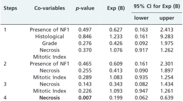
The epithelioid variant (p =0.036) was the only significant variable in the log-rank analysis of disease-free survival (Table 4). The Cox regression analysis showed that necrosis (p =0.024) was an independent prognostic factor for lower overall survival (p =0.007) (Table 5).
DISCUSSION
The five-year overall survival rate of patients with MPNSTs has been reported to range from 23% to approxi-mately 50% (2,15–17), similar to the rate of 46% found in this study. In our study, seven patients censored in the overall survival calculation had recurrence or metastasis, and three patients were lost to follow-up when the disease became terminal. Their deaths most likely went unobserved; there-fore, their overall survival time may have been overesti-mated. This issue is usually a bias inherent in retrospective studies that intend to measure overall survival.
Although some authors have demonstrated the correla-tion of various clinicopathological parameters with the biological behavior of the MPNSTs, there is no consensus with regard to the importance of these factors in the prognosis of MPNST (2,7–10). In our study, the presence of necrosis was an independent predictor of mortality.
In the present work, only one (2.6%) neurofibroma (plexiform neurofibroma) expressed p53. Other studies have also demonstrated that p53 expression is rare or absent in neurofibromas (18–21). In contrast, the majority (64.3%) of MPNSTs expressed p53. Our results are similar to those of previous studies that used the same anti-p53 antibody clone, and the results of these studies showed that p53 expression in the MPNSTs varies from 42 to 100% (6,9,16,18–20,22). To our knowledge, there are only two previous studies that used TMA technology to investigate p53 expression in MPNSTs (16,22).
In some studies (9,20,21), an association between p53 expression and histological grade was observed, which differs from our research and that of others (18,19). One explanation for these divergent results could be the different criteria adopted for histological gradation. Although the United States National Institutes of Cancer and Fe´de´ration Nationale des Centres de Lutte Contre le Cancer (FNCLCC) grading systems are the most commonly used systems for sarcomas, the histological grading systems for sarcomas have no prognostic value for some histological subtypes, including MPNSTs (23). Therefore, we preferred to use a
Figure 1 -Malignant triton tumor: case 4 (A and B).A.Area of differentiation in the rhabdomyosarcoma (arrows); H&E, 40x.B.
Cells immunopositive for desmin; immunohistochemistry (diami-nobenzidine), 40x.C.Malignant peripheral nerve sheath tumor with heterologous differentiation in a chondrosarcoma: case 13; H&E, 20x. D. p53-immunopositive plexiform neurofibroma (arrow); immunohistochemistry (diaminobenzidine), 40x. E.
Malignant peripheral nerve sheath tumor immunoreactive for p53: case 24; immunohistochemistry (diaminobenzidine), 40x.F.
Malignant peripheral nerve sheath tumor immunoreactive for p53: case 4; immunohistochemistry (diaminobenzidine), 40x. OBS: positive cells in brown.
Table 3 -Immunohistochemical data of neurofibromas and malignant peripheral nerve sheath tumors.
Group % of positive cases (n) Mean PI
Standard
deviation Median PI PI min/max IQ range
Neurofibromas 2.6% (n = 1) 0.0005 — — 0.0000/0.0005 —
Plexiform neurofibromas 6.3% (n = 1) 0.0005 — — 0.0000/0.0005 —
Neurofibromas (from NF1 and non-NF1 patients)
0.0% (n = 0) — — — — —
MPNSTs 64.3% (n = 18) 0.001931 0.0042199 0.000350 0.0000/0.01597 0.0017
simple system in which MPNSTs are classified into two grades: low and high (1).
We could not observe any influence of p53 expression on survival rates, similar to the results of a previous study (24). In contrast, other researchers showed that p53 expression was an important predictive factor for lower survival rate (16,19). In some studies (16,22), p53 expression was more common in neurofibromas associated with NF1 than in those not associated with NF1, but other studies (20) did not observe this association, similar to our results. There are a few possible explanations for these divergent results. First,
some authors considered all cases that had any immuno-positive cells as being immuno-positive, whereas others established cut-off points, varying from 3 to 10% of positive cells (9,20– 22,24). Another important factor is that all previous studies used conventional pathologist-based manual scoring to quantify the p53 staining, which increases the inter-observer and intra-observer variabilities. Our study was the first to use computerized image analysis to calculate the p53 expression. Moreover, technical considerations, such as the storage time of the tissue sections on glass slides, can influence p53 immunoreactivity.
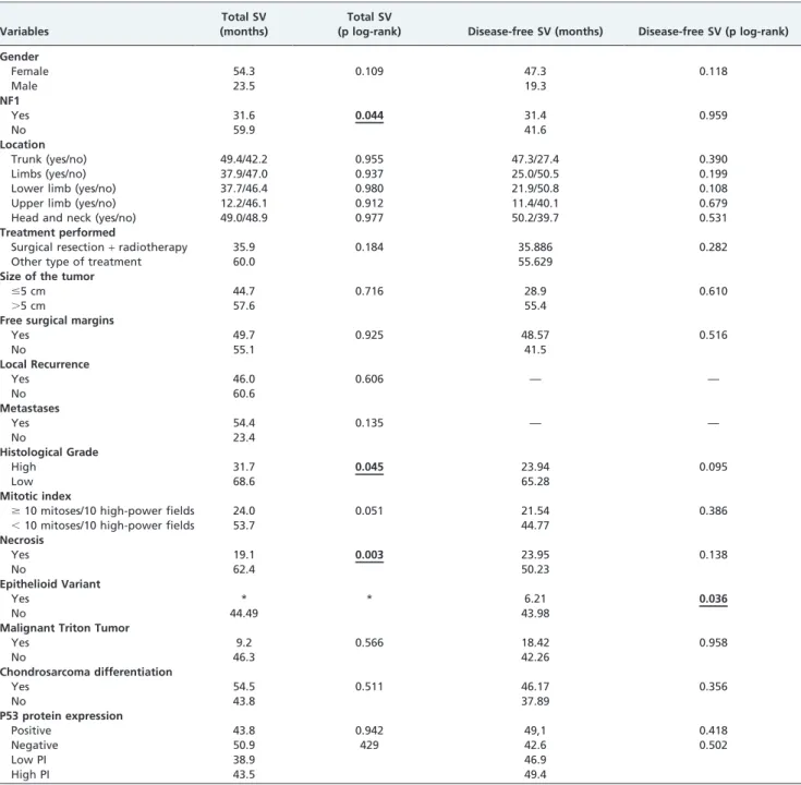
Table 4 -Results of the influence of the clinicopathological variables on the overall and disease-free survival in patients with malignant peripheral nerve sheath tumors.
Variables
Total SV (months)
Total SV
(p log-rank) Disease-free SV (months) Disease-free SV (p log-rank)
Gender Female Male 54.3 23.5 0.109 47.3 19.3 0.118 NF1 Yes No 31.6 59.9 0.044 31.4 41.6 0.959 Location Trunk (yes/no) Limbs (yes/no) Lower limb (yes/no) Upper limb (yes/no) Head and neck (yes/no)
49.4/42.2 37.9/47.0 37.7/46.4 12.2/46.1 49.0/48.9 0.955 0.937 0.980 0.912 0.977 47.3/27.4 25.0/50.5 21.9/50.8 11.4/40.1 50.2/39.7 0.390 0.199 0.108 0.679 0.531 Treatment performed
Surgical resection+radiotherapy
Other type of treatment
35.9 60.0
0.184 35.886
55.629
0.282 Size of the tumor
#5 cm
.5 cm
44.7 57.6
0.716 28.9
55.4
0.610 Free surgical margins
Yes No 49.7 55.1 0.925 48.57 41.5 0.516 Local Recurrence Yes No 46.0 60.6
0.606 — —
Metastases Yes No
54.4 23.4
0.135 — —
Histological Grade High Low 31.7 68.6 0.045 23.94 65.28 0.095 Mitotic index
$10 mitoses/10 high-power fields
,10 mitoses/10 high-power fields
24.0 53.7 0.051 21.54 44.77 0.386 Necrosis Yes No 19.1 62.4 0.003 23.95 50.23 0.138 Epithelioid Variant Yes No * 44.49 * 6.21 43.98 0.036
Malignant Triton Tumor Yes No 9.2 46.3 0.566 18.42 42.26 0.958 Chondrosarcoma differentiation Yes No 54.5 43.8 0.511 46.17 37.89 0.356 P53 protein expression
Positive Negative Low PI High PI 43.8 50.9 38.9 43.5 0.942 429 49,1 42.6 46.9 49.4 0.418 0.502
SV, survival; PI, positive index; *, the value could not be calculated because all cases were censored.
CLINICS 2012;67(8):963-968 Malignant peripheral nerve sheath tumors
Cunha KSG et al.
Necrosis was an important prognostic factor for lower overall survival, and the epithelioid variant was an important prognostic factor for shorter disease-free survival. p53 expression was not associated with any clinicopatholo-gical features and did not have an impact on the survival rates of the MPNST patients. p53 expression was rare in the neurofibromas and common in the MPNSTs, showing that the p53 pathway most likely plays an important role in the tumorigenesis of MPNSTs.
AUTHOR CONTRIBUTIONS
Cunha KS designed and conducted the research, analyzed the data, wrote the paper and had primary responsibility for the final content. Faria PA, Geller M and Moura-Neto RS designed the research and wrote the paper. Lopes VS designed and conducted the research. Caruso AC and Pires AR conducted the research. Silva LE analyzed the data. All authors read and approved the final version of the manuscript.
REFERENCES
1. Scheithauer BW, Woodruff JM, Erlandson RA. Primary malignant tumors of peripheral nerve. Tumors of the peripheral nervous system. Washington, DC: Amer Registry of Pathology; 1999. p.303-72. 2. Ducatman BS, Scheithauer BW, Piepgras DG, Reiman HM, Ilstrup DM.
Malignant peripheral nerve sheath tumors. A clinicopathologic study of 120 cases. Cancer. 1986;57(10):2006-21, http://dx.doi.org/10.1002/1097-0142(19860515)57:10,2006::AID-CNCR2820571022.3.0.CO;2-6. 3. Friedman JM, Gutmann DH, Maccollin M, Riccardi VM.
Neurofibro-matosis: phenotype, natural history, and pathogenesis. Johns Hopkins University Press; 1999.
4. Serra E, Puig S, Otero D, Gaona A, Kruyer H, Ars E, et al. Confirmation of a double-hit model for the NF1 gene in benign neurofibromas. Am J Hum Genet. 1997;61(3):512-9, http://dx.doi.org/10.1086/515504. 5. Karube K, Nabeshima K, Ishiguro M, Harada M, Iwasaki H. cDNA
microarray analysis of cancer associated gene expression profiles in malignant peripheral nerve sheath tumours. J Clin Pathol. 2006;59(2):160-5, http://dx.doi.org/10.1136/jcp.2004.023598.
6. Mawrin C, Kirches E, Boltze C, Dietzmann K, Roessner A, Schneider-Stock R. Immunohistochemical and molecular analysis of p53, RB, and PTEN in malignant peripheral nerve sheath tumors. Virchows Arch. 2002;440(6):610-5, http://dx.doi.org/10.1007/s00428-001-0550-4.
7. Kar M, Deo SVS, Shukla NK, Malik A, DattaGupta S, Mohanti BK, et al. Malignant peripheral nerve sheath tumors (MPNST)-clinicopathological study and treatment outcome of twenty-four cases. World J Surg Oncol. 2006;4:55, http://dx.doi.org/10.1186/1477-7819-4-55.
8. Angelov L, Davis A, O’Sullivan B, Bell R, Guha A. Neurogenic sarcomas: experience at the University of Toronto. Neurosurgery. 1998;43(1):56-64; discussion 64-65.
9. Zhou H, Coffin CM, Perkins SL, Tripp SR, Liew M, Viskochil DH. Malignant peripheral nerve sheath tumor: a comparison of grade, immunophenotype, and cell cycle/growth activation marker expression in sporadic and neurofibromatosis 1-related lesions. Am J Surg Pathol. 2003;27(10):1337-45, http://dx.doi.org/10.1097/00000478-200310000-00006.
10. Evans DGR, Baser ME, McGaughran J, Sharif S, Howard E, Moran A. Malignant peripheral nerve sheath tumours in neurofibromatosis 1. J Med Genet. 2002;39(5):311-4, http://dx.doi.org/10.1136/jmg.39.5.311. 11. Cunha KSG, Barboza EP, Fonseca EC da. Identification of growth
hormone receptor in plexiform neurofibromas of patients with neurofi-bromatosis type 1. Clinics (Sao Paulo). 2008;63(1):39-42, http:// dx.doi.org/10.1590/S1807-59322008000100008.
12. Cunha KSG, Barboza EP, da Fonseca EC. Identification of growth hormone receptor in localised neurofibromas of patients with neurofi-bromatosis type 1. J. Clin. Pathol. 2003;56(10):758-63, http://dx.doi.org/ 10.1136/jcp.56.10.758.
13. Pires ARC, Andreiuolo F da M, de Souza SR. TMA for all: a new method for the construction of tissue microarrays without recipient paraffin block using custom-built needles. Diagn Pathol. 2006;1:14, http:// dx.doi.org/10.1186/1746-1596-1-14.
14. Cunha KSG, Caruso AC, Gonc¸alves AS, Bernardo VG, Pires ARC, da Fonseca EC, et al. Validation of tissue microarray technology in malignant peripheral nerve sheath tumours. J Clin Pathol. 2009;62(7):629-33, http:// dx.doi.org/10.1136/jcp.2008.063081.
15. Wong WW, Hirose T, Scheithauer BW, Schild SE, Gunderson LL. Malignant peripheral nerve sheath tumor: analysis of treatment outcome. Int J Radiat Oncol Biol Phys. 1998;42(2):351-60, http://dx.doi.org/ 10.1016/S0360-3016(98)00223-5.
16. Brekke HR, Kolberg M, Skotheim RI, Hall KS, Bjerkehagen B, Risberg B et al. Identification of p53 as a strong predictor of survival for patients with malignant peripheral nerve sheath tumors. Neuro-oncology. 2009;11(5):514-28.
17. Hruban RH, Shiu MH, Senie RT, Woodruff JM. Malignant peripheral nerve sheath tumors of the buttock and lower extremity. A study of 43 cases. Cancer. 1990;66(6):1253-65.
18. Kindblom LG, Ahlde´ n M, Meis-Kindblom JM, Stenman G. Immunohistochemical and molecular analysis of p53, MDM2, prolifer-ating cell nuclear antigen and Ki67 in benign and malignant peripheral nerve sheath tumours. Virchows Arch. 1995;427(1):19-26.
19. Halling KC, Scheithauer BW, Halling AC, Nascimento AG, Ziesmer SC, Roche PC et al. p53 expression in neurofibroma and malignant peripheral nerve sheath tumor. An immunohistochemical study of sporadic and NF1-associated tumors. Am J Clin Pathol. 1996;106(3):282-8.
20. Liapis H, Marley EF, Lin Y, Dehner LP. p53 and Ki-67 proliferating cell nuclear antigen in benign and malignant peripheral nerve sheath tumors in children. Pediatr Dev Pathol. 1999;2(4):377-84, http://dx.doi.org/ 10.1007/s100249900138.
21. McCarron KF, Goldblum JR. Plexiform neurofibroma with and without associated malignant peripheral nerve sheath tumor: a clinicopathologic and immunohistochemical analysis of 54 cases. Mod Pathol. 1998;11(7): 612-7.
22. Sabah M, Cummins R, Leader M, Kay E. Immunoreactivity of p53, Mdm2, p21(WAF1/CIP1) Bcl-2, and Bax in soft tissue sarcomas: correlation with histologic grade. Appl Immunohistochem Mol Morphol. 2007;15(1):64-9.
23. Fletcher CD, Unni KK, Mertens F. World Health Organization classification of tumours. Pathology and genetics of tumours of soft tissue and bone. Lyon: IARC Press. 2002;47:91.
24. Watanabe T, Oda Y, Tamiya S, Kinukawa N, Masuda K, Tsuneyoshi M. Malignant peripheral nerve sheath tumours: high Ki67 labelling index is the significant prognostic indicator. Histopathology. 2001;39(2):187-97, http://dx.doi.org/10.1046/j.1365-2559.2001.01176.x.
Table 5 -Results of the Cox regression model for significant variables in the univariate analysis of overall survival.
Steps Co-variables p-value Exp (B) 95% CI for Exp (B)
lower upper
1 Presence of NF1 Histological
Grade Necrosis Mitotic Index
0.497 0.846 0.276 0.370
0.627 1.233 0.426 1.076
0.163 0.161 0.092 0.917
2.413 9.283 1.975 1.262 2 Presence of NF1
Necrosis Mitotic Index
0.465 0.255 0.289
0.609 0.413 1.083
0.161 0.090 0.935
2.301 1.897 1.254
3 Necrosis
Mitotic Index
0.143 0.226
0.343 1.093
0.082 0.947
1.434 1.261