Human papillomavirus infection and
p53 protein expression in vulvar
intraepithelial neoplasia and invasive
squamous cell carcinoma
Departamento de Patologia Anatômica, Faculdade de Ciências Médicas, Universidade Estadual de Campinas, Campinas, SP, Brasil
D.E.S. Engelman, L.A.L.A. Andrade and J. Vassallo
Abstract
The etiopathogenesis of vulvar intraepithelial neoplasia (VIN III) and invasive squamous cell carcinoma are largely unknown. Since there are few studies on Brazilian patients, our purpose was to determine the frequency of human papillomavirus (HPV) infection and the expres-sion of p53 in these leexpres-sions, and associate them with other factors such as age, morphological subtypes, multicentric and multifocal disease. Thirty-eight cases of VIN III, nine of superficially invasive carcinoma, and 55 of invasive vulvar carcinoma were retrospectively evaluated from 1983 to 1995 for the presence of HPV by immunohistochemistry and in situ hybridization, and for p53 protein expression by immuno-histochemistry on paraffin sections. All cases for whom material (slides and paraffin blocks) and clinical data were available were included. HPV and p53 were detected in 57.9 and 21.1% of the VIN III lesions, 33.3 and 66.7% of superficially invasive carcinomas, and 7.3 and 58.2% of invasive squamous cell carcinomas, respectively. HPV infection was associated with younger age in the VIN III and invasive carcinoma groups. In the latter, HPV infection was associated with the basaloid variant. p53 expression rate was higher in superficially invasive and invasive lesions and was not related to HPV infection. Our findings are similar to others and support the hypothesis that there are two separate entities of the disease, one associated with HPV and the other unrelated, with p53 inactivation possibly being implicated in some of the cases.
Correspondence
D.E.S. Engelman
Avenida Princesa D’Oeste, 1180 Apto 42
13095-010 Campinas, SP Brasil
Fax: +55-19-3232-7567 E-mail: [email protected] Publication supported by FAPESP.
Received September 9, 2002 Accepted May 7, 2003
Key words
•Vulvar intraepithelial neoplasia
•Vulvar carcinoma •Human papillomavirus •p53 protein
Introduction
Several lines of evidence have shown the involvement of human papillomavirus (HPV) in cervical carcinogenesis (1-4). Its onco-genic role has been attributed to a transform-ing capacity of the oncogenic types 16/18, encoding two transforming gene products,
A number of epidemiological studies have linked vulvar squamous cell carcinoma to some of the risk factors described for cervi-cal carcinoma, and it has been shown that a proportion of the former are associated with HPV infection (8,9). It seems that HPV is related to a minority of cases, especially in younger women, with the basaloid variant of squamous carcinoma and associated vulvar intraepithelial neoplasia (VIN) (1,2,9,10). HPV DNA is detected in about 10-20% of all vulvar squamous cell carcinomas (6,11,12), and in 50-80% of the basaloid subtype of invasive carcinoma (2,5,8,11). However, most vulvar carcinomas occur in older pa-tients, are predominantly of the usual kerati-nizing type and are associated with lichen sclerosus and squamous hyperplasia, sug-gesting that vulvar carcinomas may have different etiologies, with HPV and other fac-tors possibly playing a significant role and are still a matter of debate (1,2,9,10-14).
Current data point to the p53 gene as the leading factor in the surveillance of DNA integrity. Inactivation of its tumor suppress-ing activity seems to be an almost universal step in the development of human malignan-cies and may also be related to tumor pro-gression (2,4,6,7).
p53 inactivation in vulvar carcinomas can occur either by interaction with HPV E6 oncoprotein, or by p53 gene mutation in the absence of the virus. In addition, several cellular proteins and events can also interfere with or inhibit p53-mediated transactivation functions (2-7,13). p53 expression has been described in 50 to 88% of cases of vulvar squamous cell carcinoma (2,5,6,15-18).
The purpose of the current study was to determine the frequency of HPV infection and p53 expression in primary VIN and in-vasive squamous cell carcinoma in Brazilian patients and to associate them with other factors such as age and morphologic sub-types. Since there are few studies on vulvar neoplasia in Brazil, our intention was to compare our findings with those for the
worldwide population described in the lit-erature, and to contribute additional cases.
Material and Methods
In a retrospective study conducted from 1983 to 1995, 38 cases of VIN grade III, nine of superficially invasive carcinoma, and 55 of invasive squamous cell carcinoma of the vulva were collected and reviewed from the files of the Department of Pathology of the State University of Campinas.
Biopsies were taken from areas found to be altered upon physical examination or that were positive for the Collins test. Our selec-tion was based on the most representative fragment of the lesion, the presence of histo-logical signs consistent with HPV infection, and the presence of altered and normal epi-thelium adjacent to the neoplasia.
Multiple neoplastic lesions in the vulvar region separated by non-neoplastic epithe-lium were considered to be multifocal dis-ease. The presence of intraepithelial neopla-sia or invasive carcinoma at other sites of the lower female genital tract such as the vagina and uterine cervix was considered to be mul-ticentric disease. Superficially invasive car-cinoma was defined as stromal invasion of 1 mm or less (19).
Patient age, morphological type of the neoplasia, the presence of multifocal and multicentric lesions, and the epithelial alter-ations adjacent to the neoplasia were ana-lyzed and correlated with HPV detection and p53 expression.
New sections of paraffin-embedded speci-mens were placed on sylanized slides for immunohistochemistry and in situ hybrid-ization. The specimens were deparaffinized and rehydrated.
detects both the wild-type and mutant forms of the p53 protein, after inhibition of endog-enous peroxidase with 3% hydrogen peroxi-dase in methanol solution. Antigen retrieval was performed by heating with a household microwave oven (2 cycles of 5 min each at 750 W for HPV detection and 3 cycles of 7 min each at 750 W for p53) in 10 mM citrate buffer, pH 6.0. After rinsing with PBS solu-tion, the slides were exposed to a biotinyl-ated secondary antibody (polyclonal, 1:200; Multilink, Dako, Carpinteria, CA, USA). The reaction was visualized by the streptavidin-biotin-peroxidase method (Dakopatts). The slides were stained with diaminobenzidine substrate solution and counterstained with Mayer’s hematoxylin.
In situ hybridization was performed with biotinylated probes for HPV types 6/11, 16/ 18, and 31/35/51 (Dakopatts), after diges-tion with a 0.08% (w/v) pepsin soludiges-tion (Sigma, St. Louis, MO, USA) at 37ºC for 10 min. After overnight incubation of tissue with the probes at 37ºC and rinsing with stringent solution (1:50, Dakopatts), the re-action was visualized using the streptavidin-biotin-peroxidase detection system (CSA System, Dakopatts). The slides were stained with diaminobenzidine substrate solution and counterstained with Mayer’s hematoxylin.
Positive and negative controls were in-cluded in each protocol. Immunostaining for p53 protein was assessed by a semiquantita-tive method, considering the number of posi-tive cells and their intensity within the nega-tive epithelium, and graded as follows: - (negative), + (less than 10.0% of positive cells), ++ (10.0% or more, and less than 50.0% of positive cells), and +++ (50.0% or more of positive cells) (17,19,20). At least 200 cells were analyzed in each case. HPV was considered to be absent (-) or present (+) regardless of the number of positive nuclei for both methods.
Statistical analysis was performed using the Statistical Analysis System software pack-age (SAS Institute Inc., Cary, NC, USA) and
“EpiInfo, 6.02b” (Centers for Disease Con-trol and Prevention, Atlanta, GA, USA) and one of the following tests when appropriate: chi-square test to analyze the association between the variables in the three groups of carcinomas, Fisher exact test when the ex-pected value was less than five, and Student t-test when comparing ages. Univariate and multivariate logistic regression analysis was performed to verify which of the variables analyzed were associated with HPV detec-tion and p53 expression. A P value of 0.05 or less was considered to be statistically sig-nificant.
The present study was approved by the Research Ethics Committee of the Faculty of Medical Sciences, State University of Cam-pinas.
Results
Vulvar intraepithelial neoplasia
Invasive squamous cell carcinoma
This group included 55 patients with a mean age of 67.8 ± 12.5 years. Multicentric disease was present in 7.3% and multifocal lesions in 23.6% of the cases. Histological subtypes were: 78.2% usual keratinizing, 10.9% basaloid, 7.3% warty and 3.6% verru-cous. Associated epithelial changes corre-sponded mostly to dystrophic lesions (60.0%), but VIN III (25.5%) and HPV-associated le-sions (7.3%) were also present. The remaining cases (7.2%) contained normal adjacent epi-thelium. Inguinal nodal metastases were pres-ent in 56.8% of the cases and in 16.4%, the disease was recurrent. HPV capsid antigen was detected in three cases and HPV DNA in two. Overall, HPV was detected by one of the methods in 4 of 55 cases (7.3%), all of them HPV type 16/18. p53 was expressed in 58.2% of the cases, being graded as ++/+++ in 47.4%. HPV detection was associated with younger age, presence of other neoplastic lesions in the female lower genital tract, and presence of the basaloid subtype of invasive carcinoma. How-ever, by logistic regression analysis, only age (46 vs 69.5 years, P = 0.05) and the presence of multicentric disease (P = 0.04) remained sig-nificant. p53 expression was only related to
depth of invasion (P = 0.04). Patients with the basaloid subtype of invasive carcinoma were younger (mean age, 56.8 years), and this sub-type was associated with VIN III in 83.3% and with dystrophic lesions in the adjacent epithe-lium in 16.7%. HPV was present in 50.0% and p53 in 33.3% of the cases. For the usual keratinizing type, mean age was 68.9 years, and this subtype was associated with VIN III in 20.9% and with dystrophic lesions in 60.5%. HPV was detected in 2.3% and p53 in 60.5% of these cases. In five patients (11.6%) there was no abnormality in the adjacent epithelium and in three (7.0%), no adjacent epithelium was available for evaluation.
Lymph node metastasis was associated with involvement of the clitoris (P = 0.005), tendency to a tumor size larger than 2 cm (P = 0.05), and grade of the neoplasia (P = 0.05).
When more than one exam for the same patient was available in our files we com-pared the presence of relapse in the invasive vulvar carcinoma group. Relapse was asso-ciated with younger mean age (54.6 ± 13.8 vs 70.3 ± 10.5 years, P = 0.0002) and was not related to the presence of multicentric and multifocal disease, or to HPV detection and p53 expression.
Superficially invasive carcinoma
The mean age of the nine patients with superficially invasive carcinoma was 57.9 ± 16.6 years. Multicentric disease was present in five cases and multifocal lesions in five. In eight cases, the warty type of VIN III was present in adjacent epithelium and HPV-associated lesions were present in two. HPV was detected in three cases (33.3%) and p53 in six (66.7%).
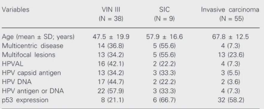
Table 1 summarizes the main clinicopatho-logical data of the three groups of lesions.
Discussion
In agreement with other reports, we show Table 1. Clinicopathological findings of patients with three types of lesions.
Variables VIN III SIC Invasive carcinoma
(N = 38) (N = 9) (N = 55)
Age (mean ± SD; years) 47.5 ± 19.9 57.9 ± 16.6 67.8 ± 12.5
Multicentric disease 14 (36.8) 5 (55.6) 4 (7.3)
Multifocal lesions 13 (34.2) 5 (55.6) 13 (23.6)
HPVAL 16 (42.1) 2 (22.2) 4 (7.3)
HPV capsid antigen 13 (34.2) 3 (33.3) 3 (5.5)
HPV DNA 17 (44.7) 2 (22.2) 2 (3.6)
HPV antigen or DNA 22 (57.9) 3 (33.3) 4 (7.3)
p53 expression 8 (21.1) 6 (66.7) 32 (58.2)
that HPV infection associated with VIN III lesions occurred predominantly in younger patients, and was not related to p53 expres-sion (6,15,16,21).
In spite of having detected only nine patients with superficially invasive vulvar carcinomas between 1983 and 1995, our intention was to identify any similarity with the VIN III or with the invasive carcinoma group. Their clinicopathological character-istics resembled those of the VIN III group, except for the p53 protein, with a rate similar to that of the invasive carcinoma group. The association with VIN III in the adjacent epi-thelium in eight of nine superficially inva-sive vulvar carcinomas indicates that VIN III lesions may progress to invasive disease (10,14,22).
HPV infection is related to VIN III le-sions and to a small subset of younger pa-tients with invasive vulvar carcinoma of the basaloid variant, which is frequently associ-ated with adjacent VIN III (1,2,9-11). Most patients with invasive carcinomas have the usual histological type, which is related nei-ther to HPV infection nor to VIN III lesions, and is associated with higher rates of dystro-phic lesions (1,2,9-14).
The finding of high rates of p53 expres-sion in invasive carcinoma supports the role of p53 inactivation, either by mutation or by binding with HPV E6 oncoprotein (2-7). Nevertheless, as also reported elsewhere (17,23), p53 expression was not related to HPV infection, being negative in 20 of the 55 cases and both (p53 and HPV) positive in only one case. This indicates that other fac-tors besides HPV infection and p53 inactiva-tion may be involved in the development of vulvar carcinoma (1,2,9,11-13).
Our low rate of HPV and the finding of the oncogenic type 16/18 in most of the positive cases were similar to that reported in the literature for vulvar carcinoma (9,12). However, the rate was lower than in other studies in which a more sensitive technique (PCR) was used (5,8,12). Our study was
performed on formalin-fixed and paraffin-embedded tissues. Although the more sensi-tive methods can be performed on this kind of specimens, DNA damage reduces their sensitivity. In our experience (data not shown), DNA extraction from paraffin-em-bedded tissues is successful in only a quarter of the cases, a fact that would reduce even more our series. In addition, topographic relations are lost using these methods. For this reason, in situ hybridization was adopted in the present study.
The condylomatous subtype of squamous cell carcinoma presented characteristics simi-lar to those of the usual type and was not associated with the presence of HPV infec-tion. This finding is in contrast to data re-ported for other series (9). However, this could be due to the small number of this carcinoma variant in our study, or to limita-tions of the method applied, i.e., in situ hybridization.
The low rate of p53 expression in VIN III lesions, and the higher rate in superficially invasive and invasive carcinoma suggest that p53 expression occurs as a late event in vulvar carcinogenesis, and is related to the capacity of stromal invasion by the neopla-sia, as described for other sites (23). In our study, among the patients with invasive car-cinoma, p53 detection was related only to the depth of invasion, and was not related to the presence of lymph node metastases or recurrent disease. These findings are similar to some studies (2) and contrast with others (6,8,16).
abnormal cells (13,14). Although more re-search is necessary, it seems that p53 might be useful as a progression marker for intra-epithelial neoplastic lesions (13,14).
In order to reduce surgical morbidity, to identify which patients will develop recur-rent disease and metastasis, several prognos-tic markers have been proposed, including p53 (12,25,26). There are also studies inves-tigating its role as a predictor of radiotherapy and chemotherapy resistance (7,25). How-ever, the results are contradictory. Some re-ports associate the presence of p53 mutation or protein expression with a worse outcome, high rates of metastasis and advanced stage (6,16), while others do not (2,13,26), al-though the latter studies reported that p53 was associated with the disease-free inter-val. According to some reports, recurrent disease is not associated with HPV or p53 protein detection (2,12,13,25,26). More re-search is needed to determine the role of p53 in the progression of the disease, as a prog-nostic marker and as a predictor of the tumor response to different treatments.
The characteristics of our patients are
similar to those described in the literature and support the hypothesis that there are two separate entities of the disease, with HPV infection being responsible for the develop-ment of VIN III lesions and for a few cases of vulvar carcinomas (1,2,9,11,12). On the other hand, p53 alterations may be implicated in the pathogenesis of most cases of invasive vulvar carcinomas (5,6,13,15). However, 36.4% of the cases were negative for both HPV and p53, indicating that about one-third of the vulvar carcinomas might arise through a still unknown mechanism inde-pendent of p53 inactivation or HPV infec-tion (12-14).
The number of cases in our study was higher than in several published reports. Until recently, vulvar studies were scarce and the etiology of vulvar carcinoma and its path-ways are still a matter of debate. In addition, few vulvar studies are available in Brazil and our intention was to contribute additional cases, to compare our findings with pub-lished reports, and to stimulate further inves-tigations on vulvar diseases.
References
1. Costa S, Syrjänen S, Vendra C, Chang F, Guida G, Hippeläinen M, Terzano P, Tervahauta A, Yliskoski M & Syrjänen K (1995). Human papillomavirus infections in vulvar precancerous lesions and can-cer. Journal of Reproductive Medicine, 40: 291-298.
2. Kagie MJ, Kenter GG, Tollenaar RAEM, Hermans J, Trimbos JB & Fleuren GJ (1997). P53 protein overexpression is independent of human papillomavirus infection in squamous cell carcinoma of the vulva. Cancer,80: 1228-1233.
3. Thomas M, Pim D & Banks L (1999). The role of the E6-p53 interac-tion in the molecular pathogenesis of HPV. Oncogene, 18: 7690-7700.
4. Stoler MH (2000). Human papillomaviruses and cervical neoplasia: a model for carcinogenesis. International Journal of Gynecological Pathology,19: 16-28.
5. Lee YY, Wilczynski SP, Chumakov A, Chih D & Koffler UP (1994). Carcinoma of the vulva: HPV and p53 mutations. Oncogene, 9: 1655-1659.
6. Pilotti S, D’Amato L, Della Torre G, Donghi R, Langoni A, Giarola M, Sampietro G, De Palo G, Pierotti MA & Rilke F (1995). Papillomavi-rus, p53 alteration, and primary carcinoma of the vulva. Diagnostic Molecular Pathology, 4: 239-248.
7. Velculescu VE & El-Deiry WS (1996). Biological and clinical
impor-tance of the p53 tumor suppressor gene. Clinical Chemistry, 42: 858-868.
8. Hørding U, Junge J, Daugaard S, Lundvall F, Poulsen H & Bock JE (1994). Vulvar squamous cell carcinoma and papillomaviruses: indi-cations for two different etiologies. Gynecologic Oncology, 52: 241-246.
9. Trimble CL, Hildensheim A, Brinton LA, Shah KV & Kurman RJ (1996). Heterogeneous etiology of squamous carcinoma of the vulva. Obstetrics and Gynecology, 87: 59-64.
10. ACOG Technical Bulletin (1994). Vulvar cancer. International Journal of Gynecology and Obstetrics,44: 79-86.
11. Andersen WA, Franquemont DW, Williams J, Taylor PT & Crum CP (1991). Vulvar squamous cell carcinoma and papillomaviruses: two separate entities? American Journal of Obstetrics and Gynecology, 165: 329-336.
12. Pinto AP, Lin MC, Mutter GL, Sun D, Villa LL & Crum CP (1999). Allelic loss in human papillomavirus-positive and -negative vulvar squamous cell carcinomas. American Journal of Pathology, 154: 1009-1015.
Mo-lecular Morphology, 9: 150-163.
14. Yang B & Hart WR (2000). Vulvar intraepithelial neoplasia of the simplex (differentiated) type. American Journal of Surgical Patholo-gy, 24: 429-441.
15. Hietanen S, Grénman S, Syrjänen K, Lappalainen K, Kauppinen J, Carey T & Syrjänen S (1995). Human papillomavirus in vulvar and vaginal carcinoma cell lines. British Journal ofCancer, 72: 134-139. 16. Kohlberger PD, Kirnbauer R, Bancher D, Gitsch G, Reinthaller A, Leodolter S, Tschachler E, Kainz C & Breitenecker G (1998). Ab-sence of p53 protein overexpression in precancerous lesions of the vulva. Cancer, 82: 323-327.
17. Kim JW, Cho YH, Lee CG, Kim JH, Kim HK, Kim EJ, Han KT & Namkoong SE (1997). Human papillomavirus infection and TP53 gene mutation in primary cervical carcinoma. Acta Oncologica, 36: 295-300.
18. Tervahauta AI, Syrjänen SM, Väyrynen M, Saastamoinen J & Syrjänen KJ (1993). Expression of p53 protein related to the pres-ence of human papillomavirus (HPV) DNA in genital carcinomas and precancer lesions. Anticancer Research, 13: 1107-1111.
19. Benedet JL, Bender H, Jones 3rd H, Ngan HY & Pecorelli S (2000). FIGO staging classifications and clinical practice guidelines in the management of gynecologic cancers. FIGO Committee on Gyneco-logic Oncology. International Journal of Gynecology and Obstetrics, 70: 209-262.
20. Vassallo J, Derchain SF, Pinto GA, Martinez EZ, Syrjänen KJ & Andrade LA (2000). High risk HPV and p53 protein expression in cervical intraepithelial neoplasia. International Journal of Gynecology
and Obstetrics, 71: 45-48.
21. Edwards CL, Tortolero-Luna G, Linares AC, Malpica A, Baker VV, Cook E, Johnson E & Mitchell MF (1996). Vulvar intraepithelial neoplasia and vulvar cancer. Obstetrics and Gynecology Clinics of North America, 23: 295-324.
22. Herod JJO, Shafi MI, Rollason TP, Jordan JA & Luesley DM (1996). Vulvar intraepithelial neoplasia with superficially invasive carcinoma of the vulva. British Journal of Obstetrics and Gynaecology, 103: 453-456.
23. Chetty R, Bramdev A, Aguirre-Arteta A, Pegoraro RJ & Sataar N (1997). Relation between retinoblastoma and p53 proteins in human papilloma viruses 16/18 positive and negative cancers of the uterine cervix. Journal of Clinical Pathology, 50: 413-416.
24. Junge J, Poulsen H, Horn T, Hørding U & Lundvall F (1997). Progno-sis of vulvar dysplasia and carcinoma in situ with special reference to histology and types of human papillomavirus (HPV). Acta Pathologica, Microbiologica et Immunologica Scandinavica, 105: 963-971.
25. Kagie MJ, Kenter GG, Tollenaar RAEM, Hermans J, Trimbos JB & Fleuren GJ (1997). P53 protein overexpression, a frequent observa-tion in squamous cell carcinoma of the vulva and in various synchro-nous vulvar epithelia, has no value as a prognostic parameter.
International Journal of Gynecological Pathology, 16: 124-130. 26. McConnell DT, Miller ID, Parkin DE & Murray GI (1997). P53 protein