Comparative cephalometric study of Class II
malocclusion treatment with Pendulum and Jones jig
appliances followed by fixed corrective orthodontics
Mayara Paim Patel1, José Fernando Castanha Henriques2, Renato Rodrigues de Almeida3, Arnaldo Pinzan4, Guilherme Janson2, Marcos Roberto de Freitas2
How to cite this article: Patel MP, Henriques JFC, Almeida RR, Pinzan A, Janson G, Freitas MR. Comparative cephalometric study of Class II malocclusion treatment with Pendulum and Jones jig appliances followed by ixed corrective orthodontics. Dental Press J Orthod. 2013 Nov-Dec;18(6):58-64.
» The authors report no commercial, proprietary or inancial interest in the prod-ucts or companies described in this article.
Contact address: Mayara Paim Patel
Rua Francisco Leitão, 115 – Bairro Pinheiros – São Paulo/SP — Brazil CEP: 05414-025 – E-mail: [email protected]
1 PhD in Orthodontics, College of Dentistry – Bauru/ University of São Paulo
(USP).
2 Full professor, Department of Pediatric Dentistry, Orthodontics and Collective
Health, College of Dentistry – Bauru/ University of São Paulo (USP).
3 Full professor, Department of Orthodontics, College of Dentistry – Bauru/
University of São Paulo (USP) and UNOPAR
4 Full professor, Department of Pediatric Dentistry, Orthodontics and Collective
Health, College of Dentistry – Bauru/ University of São Paulo (USP).
Submitted: July 01, 2011 - Revised and accepted: January 28, 2012
Objective:The purpose of this study was to cephalometrically compare the skeletal and dentoalveolar efects in the treatment of Class II malocclusion with Pendulum and Jones jig appliances, followed by ixed corrective orthodon-tics, and to compare such efects to a control group. Methods: The sample was divided into three groups. Group 1: 18 patients treated with Pendulum, Group 2: 25 patients treated with Jones jig, and Group 3: 19 young subjects with untreated Class II malocclusions and initial mean age of 12.88 years. The chi-square test was applied to assess severity and gender distribution. Groups 1 and 2 were compared to the control group by means of the one-way ANOVA and Tukey tests in order to diferentiate treatment changes from those occurred by craniofacial growth. Results: There were no signiicant changes among the three groups with regard to the components of the maxilla and the mandible, maxillomandibular relationship, cephalometric and tegumental pattern. Buccal tipping of mandibular incisors was sig-niicantly greater in the experimental groups and increased mesial angulation of the maxillary second molars was found in the Jones jig group. In the experimental groups, dental relationship, overbite and overjet were corrected. Conclusion:
It can be stated that the distalization achieved its purpose of correcting the Class II.
Keywords:Angle Class II malocclusion. Corrective orthodontics. Molar tooth.
Objetivo:a proposta desse estudo foi comparar, cefalometricamente, os efeitos esqueléticos e dentoalveolares no tra-tamento da má oclusão de Classe II com os distalizadores Pendulum e Jones jig, seguidos de aparelho ixo corretivo, e compará-los ao grupo controle. Métodos: a amostra foi dividida em três grupos. Grupo 1: 18 pacientes tratados com o Pendulum; grupo 2: 25 pacientes tratados com o Jones jig; e grupo 3: 19 jovens com má oclusão de Classe II não tratada. Empregou-se o teste qui-quadrado para avaliar a distribuição da severidade e do sexo. Os grupos 1 e 2 foram comparados ao controle pela ANOVA a um critério; também empregou-se o teste de Tukey com o intuito de diferenciar as alterações do tratamento daquelas ocorridas pelo crescimento craniofacial. Resultados: não foram observadas alterações signii-cativas entre os três grupos quanto aos componentes da maxila, mandíbula, relação maxilomandibular, padrão cefálico e tegumentar. Veriicou-se uma vestibularização signiicativamente maior dos incisivos inferiores nos grupos experimentais e maior angulação mesial dos segundos molares superiores no grupo 2 (Jones jig). Nos grupos experimentais, a relação dentária e os trespasses horizontal e vertical foram corrigidos. Conclusão: pode-se airmar que os distalizadores alcan-çaram seus objetivos de correção da Classe II.
Figure 1 - A) Lines and Planes: A = Line E; B = Frankfort Plane; C = Palatal Plane; D = Mandibular Plane (Go-Me); E = Mandibular Plane (Go-Gn); F = Pterygoid vertical line (PTVI); G = Occlusal Plane; B) Dental angular measurements: N = SN.1; R = SN.4; O = SN.5; P = SN.6; Q = SN.7. C) Dental linear measurements: A = PTVI-1; B = PTVI-4; C = PTVI-5; D = PTVI-6; E = PTVI-7; F, = PP-1; G, = PP-4; H = PP-5; I = PP-6; J = PP-7; K = PTVI- ; L = GoMe - , M = Overjet; N = Overbite. INTRODUCTION
Intraoral distalizers difer in terms of insertion site,4
mechanism of action and anchorage reinforcement.15
The Jones jig appliance is inserted buccally and acts through a nickel titanium spring anchored in the second
premolars.14 The Pendulum appliance is palatally
posi-tioned, anchored in the irst and second premolars and
its force is dissipated through TMA springs.12
The intraoral distalization performed with ixed in-traoral devices is only the irst phase of a treatment that will be inalized with ixed corrective mechanics. There are few studies in the literature that scientiically assess
the results of both phases of treatment;3,6,7,20 most studies
only assess the results of distalization.9,10,12,15,16,17,21 There-fore, it is essential to perform a study assessing and com-paring the results of corrective orthodontic treatment initiated by intraoral maxillary molar distalization with diferent intraoral distalization appliances.
MATERIAL AND METHODS
Initially, the research project was evaluated and
ap-proved by the College of Dentistry – Bauru/ University
of São Paulo (USP) Institutional Review Board . Three groups with Class II malocclusion were com-pared: Group 1: comprised 18 patients (initial mean age of 13.92 years), 6 males and 12 females. A normal molar relationship was obtained from maxillary molar distalization performed with the Pendulum appliance and maintained by the nightly use of cervical headgear (KHG) associated with corrective ixed appliances.
The mean treatment time was 4.55 years (Table 1). Group 2: comprised 25 patients (initial mean age of 12.09 years), 14 males and 11 females. Class II correc-tion was achieved with the Jones jig appliance and main-tained by the nightly use of medium-high headgear traction (helmet jeans), during corrective orthodontic treatment. The mean duration of orthodontic treatment was 4.09 years (Table 1).
Group 3: comprised 19 young subjects with un-treated Class II malocclusion (control group), 10 males and 9 females (initial mean age of 12.88 years) and fol-lowed up for a mean period of 3.71 years (Table 1). This sample was selected from a group of young subjects that had been annually radiographed and accompanied by
the Department of Orthodontics, School of
Dentist-ry – Bauru/ University of São Paulo (USP). All patients had been referred for orthodontic treatment, however, some of them opted for late intervention and others had no interest in the treatment.
The cephalometric variables analyzed were based on the orthodontic literature3,6,8,11,22 and aimed at pro-moting a comparative study, allowing discussion of the results obtained (Fig 1).
At irst, chi-square tests were used to assess sever-ity and gender distribution (Tables 2 and 3). The three groups were assessed and cephalometrically compared in order to observe the efects of orthodontic treatment and to diferentiate them in terms of the changes pro-moted by craniofacial growth and development (Fig 1). Thus, one-way ANOVA and Tukey tests were used.
B
A
Q P O R N J
E D CB
K
A
N
M
L I H PTVI
PTVI
PTVI-A
PTVI-B C
G
F
E D
F G
RESULTS
Initially, the groups were compared in order to quantify any potential diferences existing prior to orth-odontic treatment. Out of the 43 variables analyzed, only 10 presented statistically signiicant diferences, demonstrating that the sample had approximately 77% of initial cephalometric compatibility (Table 4).
Changes during treatment as well as changes oc-curring during the growth and development period were obtained by means of establishing the diference between treated patients’ initial and inal mean values. Table 4 shows the results of one-way ANOVA and Tukey tests performed among the initial cephalomet-ric measurements mean values of the three groups.
The components related to the maxilla, mandible, maxillomandibular relationship, vertical pattern and sot tissue did not present statistically signiicant dif-ferences (Table 5).
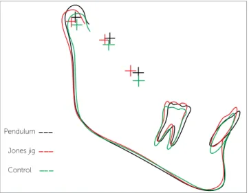
Greater mesial movement of maxillary second mo-lars was observed in the Jones jig group. Buccal tipping of mandibular incisors was greater in the Pendulum group than in the control group. Additionally, greater protrusion of these incisors was observed in the ex-perimental groups (Fig 2). The mandibular irst mo-lars showed similar mesial movement for all the three groups; however, greater extrusion was observed in the Jones jig group when compared to the Pendulum and control groups (Table 5).
There was a signiicant diference in molar relation-ship, with a signiicant change for the experimental groups, which resulted in correction of the Class II. Conversely, the initial malocclusion remained in the control group (Table 5).
DISCUSSION
There are few comparatives studies assessing the irst (maxillary molars distalization) and the second phase of treatment (corrective orthodontic treatment).3,6,7,8 Thus, the aim of this study was to compare the changes at the end of the corrective orthodontic treatment, which was initialized by the distalization of the maxillary molars by two diferent intraoral distalization appliances. Addi-tionally, it compared such changes to the control group. Assessment of the characteristics related to the groups proved that there was compatibility in terms of initial age and treatment/observation times. On the other hand, the inal age was statistically and
Figure 2 - Comparison between Pendulum and Jones jig appliances and the control group.
* Statistically signiicant for P < 0.05
Diferent letters stand for statistically signiicant diference.
Table 1 - Compatibility of the mean initial and inal ages as well as the obser-vation mean time of the young patients in the three groups (ANOVA).
VARIABLE (Y)
Group 1 (Pendulum)
N = 18
Group 2 (Jones jig)
N = 25
Group 3 (Control)
N = 19 P
Mean ± SD Mean ± SD Mean ± SD
Pretreatment age 13.92A 1.71 12.90A 1.43 12.88A 1.47 0.063
Posttreatment
age 18.48
A 1.33 16.99B 1.87 16.60B 2.31 0.008*
Observation time
(T3 -T1) 4.55
A 0.79 4.09A 0.99 3.71A 1.63 0.110
* Statistically signiicant diference for P < 0.05
Diferent letters stand for statistically signiicant diference.
Table 2 - Number of female and male subjects for each group and result of the chi-square test.
Group Sex Total
Male Female
1 – Pendulum 6 (33.3%) 12 (66.7%) 18
2 – Jones jig 14 (56%) 11 (44%) 25
3 – Control 10 (52.6%) 9 (47.4%) 19
Total 30 32 62
c2 = 2.35; gl = 2; P = 0.3087
* Statistically signiicant diference for P < 0.05
Diferent letters stand for statistically signiicant diference.
Table 3 - Comparison of Class II malocclusion severity among groups and chi-square test results.
Group
Molar relationship
¼ Class II
½ Class II
¾ Class II
Full-cusp
Class II Total
1 – Pendulum 1 (5.6%) 7 (38.8%) 5 (27.8%) 5 (27.8%) 18
2 – Jones jig 11 (44%) 7 (28%) 3 (12%) 4 (16%) 25
3 – Control 9 (47.4%) 5 (26.3%) 3 (15.8%) 2 (10.5%) 19
Total 21 19 11 11 62
c2 = 9.76; gl=6; P = 0.1350
Pendulum
Jones jig
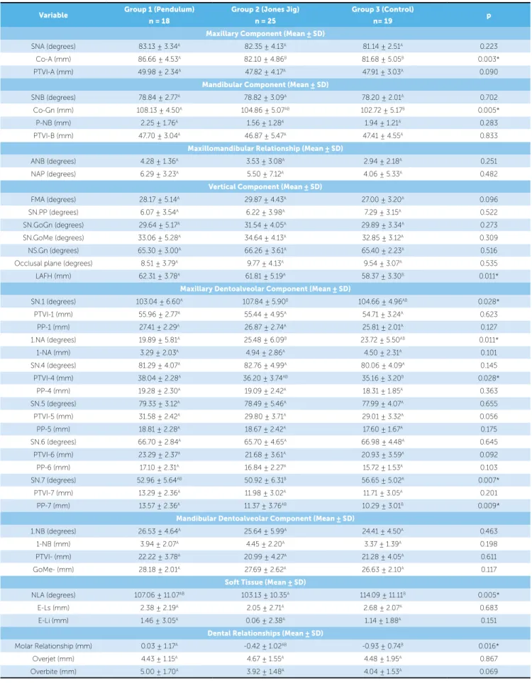
Table 4 - ANOVA and Tukey test results: means and standard deviation of initial cephalometric measurements mean values taken to assess compatibility among groups as well as values of the signiicance probability level (P) - (T1).
Variable Group 1 (Pendulum)
n = 18
Group 2 (Jones Jig) n = 25
Group 3 (Control)
n= 19 p
Maxillary Component (Mean ± SD)
SNA (degrees) 83.13 ± 3.34A 82.35 ± 4.13A 81.14 ± 2.51A 0.223
Co-A (mm) 86.66 ± 4.53A 82.10 ± 4.86B 81.68 ± 5.05B 0.003*
PTVI-A (mm) 49.98 ± 2.34A 47.82 ± 4.17A 47.91 ± 3.03A 0.090
Mandibular Component (Mean ± SD)
SNB (degrees) 78.84 ± 2.77A 78.82 ± 3.09A 78.20 ± 2.01A 0.702
Co-Gn (mm) 108.13 ± 4.50A 104.86 ± 5.07AB 102.72 ± 5.17B 0.005*
P-NB (mm) 2.25 ± 1.76A 1.56 ± 1.28A 1.94 ± 1.21A 0.283
PTVI-B (mm) 47.70 ± 3.04A 46.87 ± 5.47A 47.41 ± 4.55A 0.833
Maxillomandibular Relationship (Mean ± SD)
ANB (degrees) 4.28 ± 1.36A 3.53 ± 3.08A 2.94 ± 2.18A 0.251
NAP (degrees) 6.29 ± 3.23A 5.50 ± 7.12A 4.06 ± 5.33A 0.482
Vertical Component (Mean ± SD)
FMA (degrees) 28.17 ± 5.14A 29.87 ± 4.43A 27.00 ± 3.20A 0.096
SN.PP (degrees) 6.07 ± 3.54A 6.22 ± 3.98A 7.29 ± 3.15A 0.522
SN.GoGn (degrees) 29.64 ± 5.17A 31.54 ± 4.05A 29.89 ± 3.34A 0.273
SN.GoMe (degrees) 33.06 ± 5.28A 34.64 ± 4.13A 32.85 ± 3.12A 0.309
NS.Gn (degrees) 65.30 ± 3.00A 66.26 ± 3.61A 65.40 ± 2.23A 0.516
Occlusal plane (degrees) 8.51 ± 3.79A 9.77 ± 4.13A 9.54 ± 3.07A 0.535
LAFH (mm) 62.31 ± 3.78A 61.81 ± 5.19A 58.37 ± 3.30B 0.011*
Maxillary Dentoalveolar Component (Mean ± SD)
SN.1 (degrees) 103.04 ± 6.60A 107.84 ± 5.90B 104.66 ± 4.96AB 0.028*
PTVI-1 (mm) 55.96 ± 2.77A 55.44 ± 4.95A 54.71 ± 3.24A 0.623
PP-1 (mm) 27.41 ± 2.29A 26.87 ± 2.74A 25.81 ± 2.01A 0.127
1.NA (degrees) 19.89 ± 5.81A 25.48 ± 6.09B 23.72 ± 5.50AB 0.011*
1-NA (mm) 3.29 ± 2.03A 4.94 ± 2.86A 4.50 ± 2.31A 0.101
SN.4 (degrees) 81.29 ± 4.07A 82.76 ± 4.99A 80.06 ± 4.09A 0.145
PTVI-4 (mm) 38.04 ± 2.28A 36.20 ± 3.74AB 35.16 ± 3.20B 0.028*
PP-4 (mm) 19.28 ± 2.30A 19.09 ± 2.42A 18.31 ± 1.85A 0.363
SN.5 (degrees) 79.33 ± 3.12A 78.49 ± 5.46A 77.99 ± 4.07A 0.655
PTVI-5 (mm) 31.58 ± 2.42A 29.80 ± 3.71A 29.01 ± 3.32A 0.056
PP-5 (mm) 18.81 ± 2.28A 18.67 ± 2.42A 17.60 ± 1.67A 0.175
SN.6 (degrees) 66.70 ± 2.84A 65.70 ± 4.65A 66.98 ± 4.48A 0.645
PTVI-6 (mm) 23.29 ± 2.37A 21.68 ± 3.61A 20.93 ± 3.59A 0.092
PP-6 (mm) 17.10 ± 2.31A 16.84 ± 2.27A 15.72 ± 1.53A 0.103
SN.7 (degrees) 52.96 ± 5.64AB 50.92 ± 6.31B 56.65 ± 5.02A 0.007*
PTVI-7 (mm) 13.29 ± 2.36A 11.98 ± 3.02A 11.71 ± 3.05A 0.201
PP-7 (mm) 13.57 ± 2.36A 11.37 ± 3.76AB 10.29 ± 3.01B 0.009*
Mandibular Dentoalveolar Component (Mean ± SD)
1.NB (degrees) 26.53 ± 4.64A 25.64 ± 5.99A 24.41 ± 4.50A 0.463
1-NB (mm) 3.94 ± 2.07A 4.45 ± 2.20A 3.37 ± 1.39A 0.198
PTVI- (mm) 22.22 ± 3.78A 20.99 ± 4.27A 21.28 ± 4.05A 0.611
GoMe- (mm) 28.18 ± 2.01A 27.69 ± 2.62A 26.63 ± 2.10A 0.117
Soft Tissue (Mean ± SD)
NLA (degrees) 107.06 ± 11.07AB 103.13 ± 10.35A 114.09 ± 11.11B 0.005*
E-Ls (mm) 2.38 ± 2.19A 2.05 ± 2.71A 2.68 ± 2.07A 0.683
E-Li (mm) 1.46 ± 3.05A 0.06 ± 2.38A 1.14 ± 1.88A 0.151
Dental Relationships (Mean ± SD)
Molar Relationship (mm) 0.03 ± 1.17A -0.42 ± 1.02AB -0.93 ± 0.74B 0.016*
Overjet (mm) 4.43 ± 1.15A 4.67 ± 1.55A 4.48 ± 1.95A 0.867
Overbite (mm) 5.00 ± 1.70A 3.92 ± 1.48A 4.04 ± 1.53A 0.069
* Statistically signiicant diference for P < 0.05.
Table 5 - ANOVA and Tukey test results: means and standard deviation of cephalometric measurements means.(ANOVA – T3-T1)
* Statistically signiicant diference for P < 0.05
Diferent letters stand for statistically signiicant diference.
Variable Group 1 (Pendulum)
n = 18
Group 2 (Jones Jig) n = 25
Group 3 (Control)
n = 19 P
Maxillary Component (Mean ± SD)
SNA (degrees) -1.07 ± 1.75A 0.02 ± 1.85A -0.67 ± 3.43A 0.329
Co-A (mm) 1.22 ± 3.27A 1.41 ± 3.59A 3.20 ± 3.43A 0.151
PTVI-A (mm) 0.73 ± 2.40A 1.15 ± 2.29A 1.08 ± 3.62A 0.878
Mandibular Component (Mean ± SD)
SNB (degrees) -0.26 ± 1.73A 0.74 ± 2.28A -0.35 ± 2.24A 0.169
Co-Gn (mm) 4.77 ± 5.82A 5.98 ± 4.21A 4.92 ± 3.31A 0.626
P-NB (mm) 0.61 ± 0.98A 0.46 ± 0.84A 0.25 ± 0.81A 0.473
PTVI-B (mm) 1.36 ± 3.62A 2.03 ± 2.70A 1.69 ± 5.09A 0.851
Maxillomandibular Relationship (Mean ± SD)
ANB (degrees) -0.81 ± 2.02A -0.72 ± 2.19A -0.11 ± 3.03A 0.796
NAP (degrees) -2.30 ± 4.68A -2.00 ± 4.79A -1.07 ± 6.81A 0.771
Vertical Component (Mean ± SD)
FMA (degrees) 0.46 ± 2.55A 1.72 ± 2.62A 0.31 ± 4.48A 0.298
SN.PP (degrees) 0.21 ± 1.83A 0.24 ± 3.11A 1.05 ± 3.22A 0.578
SN.GoGn (degrees) 0.46 ± 2.29A 0.23 ± 2.45A 1.16 ± 5.47A 0.689
SN.GoMe (degrees) 0.18 ± 2.14A 0.40 ± 2.20A 1.27 ± 4.89A 0.552
NS.Gn (degrees) 0.93 ± 1.51A 0.63 ± 2.14A 1.40 ± 2.93A 0.541
Occlusal plane (degrees) -0.05 ± 2.80A 1.70 ± 3.32A -1.22 ± 5.47A 0.057
LAFH (mm) 3.63 ± 3.01A 5.60 ± 2.82A 3.48 ± 5.38A 0.128
Maxillary Dentoalveolar Component (Mean ± SD)
SN.1 (degrees) 1.68 ± 7.01A -1.63 ± 6.65A -1.87 ± 4.32A 0.147
PTVI-1 (mm) 1.40 ± 3.64A 1.26 ± 3.12A 1.63 ± 4.49A 0.947
PP-1 (mm) 0.58 ± 1.78A 1.68 ± 1.48A 1.07 ± 3.12A 0.273
1.NA (degrees) 2.79 ± 6.63A -1.63 ± 6.77A -1.43 ± 4.74A 0.051
1-NA (mm) 0.98 ± 2.40A 0.12 ± 2.55A -0.18 ± 2.33A 0.322
SN.4 (degrees) -0.29 ± 5.47A -1.83 ± 4.69A 0.59 ± 3.23A 0.210
PTVI-4 (mm) 1.24 ± 2.87A 2.20 ± 2.09A 2.10 ± 4.49A 0.587
PP-4 (mm) 1.80 ± 1.41A 2.13 ± 1.24A 1.63 ± 2.81A 0.673
SN.5 (degrees) -1.43 ± 6.11A 1.76 ± 4.61A 0.06 ± 3.05A 0.095
PTVI-5 (mm) 1.20 ± 2.83A 2.22 ± 2.00A 1.81 ± 4.56A 0.587
PP-5 (mm) 1.86 ± 1.48A 2.10 ± 1.33A 1.95 ± 2.75A 0.915
SN.6 (degrees) -0.77 ± 6.67A 1.55 ± 4.85A 0.20 ± 5.65A 0.409
PTVI-6 (mm) 0.61 ± 2.68A 1.82 ± 1.89A 1.98 ± 4.60A 0.356
PP-6 (mm) 2.10 ± 1.57A 2.39 ± 1.54A 2.36 ± 3.06A 0.896
SN.7 (degrees) 1.59 ± 6.53AB 5.44 ± 7.31A -0.76 ± 5.81B 0.010*
PTVI-7 (mm) 0.75 ± 2.73A 1.42 ± 1.91A 1.47 ± 4.00A 0.695
PP-7 (mm) 2.37 ± 2.16A 4.40 ± 2.89A 4.32 ± 3.90A 0.074
Mandibular Dentoalveolar Component (Mean ± SD)
1.NB (degrees) 6.18 ± 6.72A 2.52 ± 5.56AB -0.73 ± 3.28B 0.001*
1-NB (mm) 1.70 ± 1.64A 1.41 ± 1.89A 0.06 ± 0.85B 0.004*
PTVI- (mm) 2.27 ± 2.68A 2.83 ± 2.23A 2.42 ± 3.14A 0.774
GoMe- (mm) 2.04 ± 1.60A 3.76 ± 2.37B 1.90 ± 1.85A 0.004*
Soft Tissue (Mean ± SD)
NLA (degrees) 2.06 ± 9.01A 1.60 ± 7.54A 2.36 ± 8.51A 0.975
E-Ls (mm) 1.56 ± 1.01A 1.91 ± 1.53A 1.03 ± 2.03A 0.199
E-Li (mm) 0.28 ± 1.24A 0.74 ± 1.21A 1.40 ± 2.63A 0.165
Dental Relationships (Mean ± SD)
Molar relationship (mm) -2.62 ± 1.29A -2.36 ± 1.36A -0.22 ± 1.24B 0.000*
Overjet (mm) -1.35 ± 1.37AB -1.90 ± 1.69A -0.14 ± 2.00B 0.005*
During the observation period, changes in the nasolabial angle were similar in the three groups. This finding demonstrates that the treatment proto-col used does not interfere in the tegumental profile; therefore, the facial characteristics are maintained in
the experimental groups.8
When assessing the maxillary dentoalveolar com-ponent, it was observed that only the maxillary second molars showed signiicant changes, i.e., at treatment onset, the Jones jig group presented the second mo-lars more distally angulated than the control group, and during treatment, this group also showed a great-er mesial angulation in relation to the control group. This initial position can be explained by the diference in the mean initial age that, although not signiicant, was lower in the Jones jig than in the control group (Table 1); hence, the second molars were more below the occlusal plane, showing a more distal position.
Regarding the positioning of the mandibular in-cisors, a minor change was observed in the control group, while the Pendulum and Jones jig groups pre-sented greater buccal tipping and protrusion of the
mandibular incisors, certainly related to the use of
Class II rubber bands and overjet correction, which occurred as a consequence of the compensation of the mandibular teeth (Fig 2).
As for the vertical positioning of the mandibular molars, signiicant extrusion was greater in the Jones jig group than in the Pendulum and control groups. This change was related not only to the use of Class II rubber bands, but also to the end of eruption, since, at the be-ginning of treatment, the mandibular molars were more below the occlusal plan in comparison to the Pendulum group because patients were slightly younger and had greater potential for eruption.13
The molar relationship at treatment onset showed a statistically signiicant diference between the Pendu-lum and control groups, conirming the trend of greater severity of the Pendulum group. As expected, during observation of the change in molar relationship in the course of treatment, the experimental groups presented signiicative Class II correction when compared to the control group in which malocclusion remained. There-fore, it appears that the treatment successfully decreased anteroposterior interarch discrepancy, which reveals the contribution of this therapy in the correction of the Class II molar relationship and accentuated overjet. signiicantly diferent, representing a trend of an older
age in group 1. However, most studies in the litera-ture consider compatibility of initial age and treatment
time,2,5 only, which is considered as suicient to
char-acterize a reliable sample compatibility.7
Changes during treatment for the variables of both maxillary and mandibular components were similar among the three groups (Table 5), and improvements in the maxillomandibular relationship were observed. However, this change was more signiicant in the ex-perimental groups and it is justiied by the treatment performed. Conversely, although this improvement was less signiicant in the control group, it was due to craniofacial growth. The results prove that intraoral distalization appliances do not interfere in craniofacial
growth and development.6,19,21
Assessment of the vertical skeletal variables in the initial stage, except for the lower anterior face height (LAFH), demonstrates that the measurements showed no statistically signiicant diference among groups. Changes happening as a result of treatment and growth were sta-tistically similar for the three groups; however, they were numerically higher in the Jones jig group. The diferent changes for the Jones jig and control groups occurred due to the extrusion of irst and second premolars dur-ing treatment, in other words, although not signiicant, extrusion of these teeth was slightly higher in the Jones jig group than in the control group (Table 5).
Results demonstrate that the three groups showed
clockwise mandibular rotation, which conirms the
downward displacement of the mandible, as observed during the post-distalization stage of several
stud-ies.3,6,7,8,10,21 Assuming that this change occurred as a
result of maxillary premolars and molars extrusion due to loss of anchorage and the distalization efect, it is thought that during corrective treatment, cor-rection of extrusions will occur and the rotation will be reversed as a consequence. However, according
to Taner-Sarisoy and Darendeliler,23 most
orthodon-tic mechanics, if not all, are extrusive and this extru-sion increases the LAFH during treatment, keeping it increased during the retention period. Moreover, an increase in LAFH due to craniofacial growth and
de-velopment is common.18 Therefore, it can be stated
that mandibular rotation is related to changes in the
distalization phase,3,8,21 the corrective orthodontic
of neutralizing the speciic efects of intraoral distaliza-tion and inalizing the corrective treatment.
CONCLUSIONS
Intraoral distalization appliances followed by ixed corrective orthodontics do not interfere in the cephalo-metric pattern and tegumental proile, as demonstrated by the results which are similar to the control group with regard to the components of both the maxilla and the mandible, maxillomandibular relationship, cranio-facial and tegumental pattern. The mandibular incisors showed signiicant protrusion and buccal tipping in the experimental groups and the maxillary second molars showed more mesial angulation in the Jones jig group. Finally, correction of Class II malocclusion, overjet and overbite were observed in the Pendulum and Jones jig groups, and in the control group, the initial malocclu-sion remained at the end of the observation period. The literature3,7,8 proves that intraoral distalization
ap-pliances followed by ixed corrective orthodontics are efective in the correction of Class II and that there is stability of about 82% of the occlusal results achieved
in the long-term.1
Overjet and overbite were similar in the three groups at treatment onset; however, there was a correction in the treated groups during treatment, which was not observed in the control group. This diference was expected since patients in the experimental groups were subjected to corrective treatment and individuals in the control group, in which malocclusion remained at the end of the obser-vation period, the overjet and overbite also remained, i.e., the Class II malocclusion does not correct itself.
Despite the distinct insertion sites among the ap-pliances assessed, i.e., palatal and buccal, no chang-es were related to this diference, since orthodontic treatment with ixed appliance acts with the purpose
1. Alessio Jr L. Avaliação longitudinal da estabilidade do tratamento da má oclusão de Classe II com o aparelho Pendulum seguido pelo aparelho ixo [tese]. Bauru (SP): Universidade de São Paulo; 2009.
2. Angelieri F. Comparação dos efeitos cefalométricos promovidos pelos aparelhos extrabucal cervical e pendulum [tese]. Bauru (SP): Universidade de São Paulo; 2005.
3. Angelieri F, Almeida RR, Almeida MR, Fuziy A. Dentoalveolar and skeletal changes associated with the pendulum appliance followed by ixed orthodontic treatment. Am J Orthod Dentofacial Orthop. 2006;129(4):520-7.
4. Antonarakis GS, Kiliaridis S. Maxillary molar distalization with noncompliance intramaxillary appliances in Class II malocclusion. A systematic review. Angle Orthod. 2008;78(6):1133-40.
5. Brandão AG. Estudo cefalométrico comparativo das alterações promovidas pelos aparelhos de protração mandibular e Pendulum, associados ao aparelho ixo, no tratamento da má oclusão de Classe II, 1ª divisão [tese]. Bauru (SP): Universidade de São Paulo; 2006.
6. Brickman CD, Sinha PK, Nanda RS. Evaluation of the Jones jig appliance for distal molar movement. Am J Orthod Dentofacial Orthop. 2000;118(5):526-34. 7. Burkhardt DR, McNamara JA Jr, Baccetti T. Maxillary molar distalization or
mandibular enhancement: a cephalometric comparison of comprehensive orthodontic treatment including the pendulum and the Herbst appliances. Am J Orthod Dentofacial Orthop. 2003;123(2):108-16.
8. Chiu PP, McNamara JA Jr, Franchi L. A comparison of two intraoral molar distalization appliances: Distal Jet versus pendulum. Am J Orthod Dentofacial Orthop. 2005;128(3):353-65.
9. Fuziy A, Rodrigues de Almeida R, Janson G, Angelieri F, Pinzan A. Sagittal, vertical, and transverse changes consequent to maxillary molar distalization with the pendulum appliance. Am J Orthod Dentofacial Orthop. 2006;130(4):502-10.
10. Ghosh J, Nanda RS. Evaluation of an intraoral maxillary molar distalization technique. Am J Orthod Dentofacial Orthop. 1996;110(6):639-46.
11. Haydar S, Uner O. Comparison of Jones jig molar distalization appliance with extraoral traction. Am J Orthod Dentofacial Orthop. 2000;117(1):49-53.
REFERENCES
12. Hilgers JJ. The pendulum appliance for Class II non-compliance therapy. J Clin Orthod. 1992;26(11):706-14.
13. Iseri H, Solow B. Continued eruption of maxillary incisors and irst molars in girls from 9 to 25 years, studied by the implant method. Eur J Orthod. 1996;18(3):245-56.
14. Jones RD, White JM. Rapid Class II molar correction with an open-coil jig. J Clin Orthod. 1992;26(10):661-4.
15. Kinzinger GS, Gross U, Fritz UB, Diedrich PR. Anchorage quality of deciduous molars versus premolars for molar distalization with a pendulum appliance. Am J Orthod Dentofacial Orthop. 2005;127(3):314-23.
16. Kinzinger G, Syrée C, Fritz U, Diedrich P. Molar distalization with diferent pendulum appliances: in vitro registration of orthodontic forces and moments in the initial phase. J Orofac Orthop. 2004;65(5):389-409.
17. Lopes RSR. Avaliação cefalométrica das alterações dentoesqueléticas e tegumentares em jovens com má oclusão de Classe II tratados com distalizadores Distal Jet [tese]. Bauru (SP): Universidade de São Paulo; 2007. 18. Martins DR, Janson G, Almeida RR, Pinzan A, Henriques JFC, Freitas MR.
Atlas de crescimento craniofacial. São Paulo: Ed. Santos; 1998.
19. Papadopoulos MA, Mavropoulos A, Karamouzos A. Cephalometric changes following simultaneous irst and second maxillary molar distalization using a non-compliance intraoral appliance. J Orofac Orthop. 2004;65(2):123-36. 20. Patel MP. Estudo cefalométrico comparativo do tratamento da má oclusão de
Classe II com os distalizadores Pendulum e Jones jig seguidos do aparelho ixo corretivo [tese]. Bauru (SP): Universidade de São Paulo; 2010.
21. Patel MP, Janson G, Henriques JF, Almeida RR, Freitas MR, Pinzan A, et al. Comparative distalization efects of Jones jig and pendulum appliances. Am J Orthod Dentofacial Orthop. 2009;135(3):336-42.
22. Runge ME, Martin JT, Bukai F. Analysis of rapid maxillary molar distal movement without patient cooperation. Am J Orthod Dentofacial Orthop. 1999;115(2):153-7.