Efect of dental bleaching after bracket bonding and
debonding using three diferent adhesive systems
Lucianna de Oliveira Gomes1, Paula Mathias2, Patricia Rizzo3, Telma Martins de Araújo4, Maria Cristina Teixeira Cangussu5
Objective:To evaluate the inluence of bonding and debonding of orthodontic brackets on dental in-home bleach-ing, taking into account three diferent adhesive systems. Methods: Forty-four bovine incisors were divided into four groups according to the primer system used for orthodontic bracket bonding. Following the debonding of orthodontic brackets, the teeth were stored in staining solution for 96 hours. Then, teeth were whitened using 10% carbamide peroxide for two weeks at a 6-hour-a-day regime. Standardized digital photographs were taken at the following in-tervals: T0 (initial); T1 (ater debonding); T2 (ater pigmentation); T3, T4 and T5 representing 1, 7, and 14 days of bleaching. Repeatability and stability tests were carried out to check the method accuracy. Images were analyzed us-ing Adobe Photoshop 7.0 sotware considerus-ing (L*a*b*)color coordinate values and a modiied color diference total (ΔE’). Results: The results of this study (ANOVA and Tukey; p < 0.01) demonstrated that ater 7 days of bleaching, experimental groups showed signiicantly less teeth whitening compared to the control group. However, there were no signiicant color diferences between the groups ater 14 days, according to values of lightness (L*). Conclusions: Regardless of the adhesive primer system applied, bonding and debonding of orthodontic brackets alters the outcome of tooth whitening in the irst 7 days of bleaching, however it has no inluence on the whitening of the dental structure ater 14 days of in-home dental bleaching with 10% carbamide peroxide.
Keywords: Dental bleaching. Orthodontic brackets. Photography. Primer.
How to cite this article: Gomes LO, Mathias P, Rizzo P, Araújo TM, Cangussu MCT. Efect of dental bleaching ater bracket bonding and debonding using three diferent adhesive systems. Dental Press J Orthod. 2013 Mar-Apr;18(2):61-8.
Submitted: October 20, 2010
Revised and accepted: September 15, 2011
Contact address: Lucianna de Oliveira Gomes Faculdade de Odontologia, Universidade Federal da Bahia Rua Araújo Pinho, 62 – 7° andar – Salvador/BA, Brazil CEP: 40.110-150 – E-mail: [email protected]
1 MSc in Orthodontics. Professor, Graduate Program in Orthodontics, Federal
University of Bahia (UFBa).
2 PhD in Orthodontics, UNICAMP. Associate Professor, UFBa. 3 MSc in Dentistry, UFBa.
4 PhD in Orthodontics. Full Professor, Graduate Program in Orthodontics,
UFBa.
5 Associate Professor, Department of Social and Pediatric Dentistry, UFBa.
» The author reports no commercial, proprietary or inancial interest in the prod-ucts or companies described in this article.
Objetivo: o objetivo desse estudo foi avaliar a inluência da colagem e descolagem de braquetes ortodônticos no clareamento caseiro, considerando três diferentes sistemas adesivos. Métodos: quarenta e quatro incisivos bovinos foram divididos em quatro grupos, de acordo com o sistema adesivo utilizado para colagem dos braquetes. Após a descolagem dos braquetes, os dentes foram pigmentados por 96 horas e depois clareados com peróxido de carbamida a 10% por 6 horas diárias, durante duas semanas. Foram realizadas fotograias digitais padronizadas nos tempos: T0 (inicial); T1 (após descolagem); T2 (após pigmentação); T3, T4 e T5 representando 1, 7 e 14 dias de clareamento. Testes de repetitividade e de estabilidade foram realizados para avaliar a acurácia do método. As imagens foram avaliadas pelo sotware Adobe Photoshop 7.0, considerando os parâmetros de cor (L*a*b*) e a diferença total de cor adaptada para esse estudo (ΔE’). Resultados: os resultados do presente estudo (ANOVA e Tukey; p < 0,01) demonstraram que, após uma semana de clareamento, os grupos experimentais apresentaram uma resposta mais lenta ao clareamento que o grupo controle. Contudo, após 14 dias, não houve diferença cromática signiicativa entre os grupos, observada pelos valores de luminosidade (L*). Conclusões: independentemente do sistema adesivo utilizado, a colagem e descolagem de braquetes ortodônticos altera os resultados de obtenção de cor com sete dias de avaliação. Contudo, após 14 dias não se observa nenhuma diferença de cor na estrutura dentária clareada pela técnica caseira.
INTRODUCTION
Patients frequently seek cosmetic solutions after orthodontic treatments including changes in tooth color, notably when stains are observed on tooth surface after the removal of orthodontic brackets. Usually, such stains result from pigments in the ma-terials applied in the tooth/bracket interface, since orthodontic devices prevent good hygiene and favor the deposit of chromogenic agents in the interfaces between tooth enamel and orthodontic device which
may lead to color alteration on the dental surface.1
However, after debonding of orthodontic brack-ets, residual adhesive (resin tags) remain on teeth, for the bonding process takes place as a result of the mi-cromechanical retention, due to the presence of resin components that infiltrate about 11.8 µm to 18.9 µm into the dental structure, sometimes reaching up to
100 µm into the tooth.2,3
Once resin tags have infiltrated the enamel – and some may remain intact even if the enamel’s surface
layer is removed,4 they could obstruct the movement
of whitening agents within this substrate hence
in-fluencing the result of dental bleaching.5 In addition,
since composites are not whitened, as teeth are,6 their
presence could lead to chromatic alterations and pre-vent the acquisition of a homogenous color on dental surface in the end of the bleaching process.
In face of the scarce literature on dental bleach-ing after debondbleach-ing of orthodontic brackets, as well as on the influence of resin tags on dental surface fi-nal color, it is necessary to assess occasiofi-nal differ-ences on the color of teeth whitened after bonding and debonding of orthodontic brackets taking into consideration the different adhesive primer systems.
The first research hypothesis is that the adhe-sive resin system that remains on tooth enamel after
debonding of orthodontic brackets interferes with the achievement of a homogenous surface color. The second research hypothesis is that different adhesive primer systems interfere differently with the outcome of dental bleaching.
MATERIAL AND METHODS
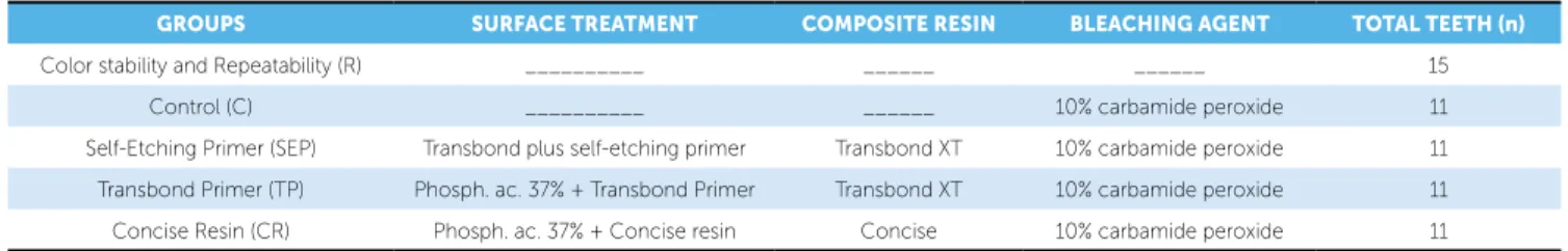
Forty-four incisors previously kept in a 0.1% timol solution were assessed colorimetrically and fifteen were used for the method repeatability test and color stability test. The teeth had their roots cut off with a double-face diamond disc (# 7020, KG Sorensen, Ba-rueri, SP, Brazil) under refrigeration and in low ro-tation. Dental pulps were removed and crowns were brushed using Robinson brushes and pumice-based toothpaste and water in low rotation. The central re-gion — the most flat surface — on the buccal side of each tooth was outlined and dental crowns were included in transparent self-cured acrylic resin blocks (JET, Artigos Odontológicos Clássico Ltda, São Pau-lo, SP, Brazil). The specimens were randomly divid-ed into five groups, as shown in Table 1.
The bracket placed on the lattened surface was that of a maxillary lateral incisor ref. 1030209 (Mo-relli, São Paulo, SP, Brazil), manipulated with the aid of bracket bonding tool (Morelli). The specimens of all groups were placed in the positioning device, their exposed enamels etched with 37% phosphoric acid for 1 minute and underwent bracket-bonding procedures according the instructions provided by the manufac-turers of the diferent adhesive primer systems applied. Before debracketing, the specimens were stored in
distilled water for 24 hours at 37 oC.7,8
Debonding was carried out mechanically with a straight plier How (Unitek), following the technique
proposed by Zachrisson.9 The remaining composite
GROUPS SURFACE TREATMENT COMPOSITE RESIN BLEACHING AGENT TOTAL TEETH (n)
Color stability and Repeatability (R) __________ ______ ______ 15
Control (C) __________ ______ 10% carbamide peroxide 11
Self-Etching Primer (SEP) Transbond plus self-etching primer Transbond XT 10% carbamide peroxide 11
Transbond Primer (TP) Phosph. ac. 37% + Transbond Primer Transbond XT 10% carbamide peroxide 11
Concise Resin (CR) Phosph. ac. 37% + Concise resin Concise 10% carbamide peroxide 11
was removed with a multilaminated bur (K282K – Komet-Brasseler, Lemgo, Germany) in low rotation. The total removal of composite from enamel was verified in a stereoscope microscope (25x).
After bracket debonding, the specimens of groups C, SEP, TP and CR were submerged in a container with aqueous solution containing 250 ml of black tea, 250 ml of coffee, 250 ml of red wine, 250 ml of tobacco solution, 250 ml of coca-cola and 250 ml of
artificial saliva, at 37 oC for 96 hours.
After pigmentation, specimens were subjected to the at-home whitening technique, using 10% carb-amide peroxide (7.82 pH) (Whiteness Perfect 10% – FGM, Joinville, SC, Brazil).
During bleaching, a standardized 0.2 ml whitening substance was applied over the exposed tooth surface, which was kept there for 6 hours a day, during 2 con-secutive weeks. The specimens remained under 100%
relative humidity at 37 oC throughout the experiment.10
Specimens were photographed using position-ing devices, developed with the goal of assessposition-ing the enamel color always at the same spot at the different assessment intervals, at the following time points:
T0 = initial, before bracket bonding; T1 = after
de-bonding and surface polishing; T2 = after
pigmenta-tion; T3 = after the first day of bleaching; T4 = after
the first week of bleaching; T5 = after two weeks of
bleaching.Specimens in the control group were
pol-ished and photographed again at T1 in compliance
with the method applied for experimental groups.
Determining method reliability: Repeatability test
Before starting the experiment, a repeatability test of the photograph shooting and the resulting color measurements was performed on fifteen specimens to ensure the reliability of the method applied. The repeatability test consisted of an assessment of tooth color variation across three photography sessions.
Color stability test on teeth stored in distilled water
This test consisted in the observation of possi-ble color alterations on teeth stored in distilled
wa-ter at 37o C across the experimental period. Fifteen
specimens had their photographic images assessed
at T0 (initial) and T5 (final) intervals. This so-called
group R did not undergo any kind of treatment on their exposed enamel surfaces.
Color measurement of sample
Measurements of L*, a*, b* color dimensions were carried out at the aforementioned time periods
(T0 to T5) on all experimental specimens by using the
histogram function of the Adobe Photoshop 7.0 sot-ware.
The study adopted the CIELAB color system in which colors are deined according to three dimen-sions: L* (lightness), a* and b* (redness/greenness
and yellowness/blueness).11 The numeric values of L*,
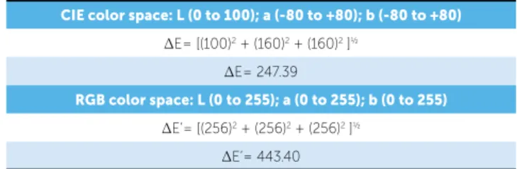
a*, b* obtained from colorimeters and spectropho-tometers vary from 0 to 160. In this study, L*, a*, b* dimensions vary from 0 to 255 because the color as-sessment relied upon the computer-based readings of digital photographs that are obtained in the RGB color space, with 256 diferent hues. Therefore, we needed to adjust the numeric value, which suggests clinical signiicance (DE) (Table 2). So we agreed to name the color diference calculated in this study DE’, to pre-vent wrong comparisons based on DE calculated from L*a*b* color parameters obtained with colorimeters.
Data assessment
The values of L* a* b*resulting from the colori-metric assessment of photographs were statistically evaluated by the MINITAB package.
The ANOVA test (analysis of variance) was per-formed for repeated data, to check for the statistically signiicant diferences at the intervals and among all groups in the study. Once a positive result was observed for ANOVA, we applied the Tukey parametric statis-tics to determine the minimum signiicant diferences among groups at each interval and among diferent in-tervals within each group, at the 1% signiicance level.
Table 2 - ΔE CIELAB color variation (0 – 160 scale) and ΔE’ RGB color varia-tion (0 – 255 scale).
CIE color space: L (0 to 100); a (-80 to +80); b (-80 to +80)
ΔE= [(100)2 + (160)2 + (160)2 ]½
ΔE= 247.39
RGB color space: L (0 to 255); a (0 to 255); b (0 to 255)
ΔE’= [(256)2 + (256)2 + (256)2 ]½
RESULTS
Color stability and repeatability test
The degree of repeatability of the sample was found to be highly reliable, as confirmed by the Analysis of Variance (ANOVA), since no significant difference (p > 0.01) was observed among the values of L* (F = 0.58/ p = 0.57), a* (F = 0.71/ p = 0.8) and b* (F = 0.3/ p = 0.74) for each specimen at the three intervals.
With regards to the color stability test on speci-mens kept in distilled water, the difference in the
val-ues of L*, a*, b* at time points T0 and T5 did not
re-sult in statistically significant differences throughout the sample storage period (Table 3).
Colorimetric assessment results
Value of L*
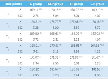
The measurement of luminosity values L*, at time
points T0 to T5, for Control (C), Transbond
Prim-er (TP), Self-Etching Primer (SEP) and Concise Res-in (CR) groups are displayed on Table 4 and Figure 1. Considering diferences in lightness among speci-mens across intervals, it was noted that ater bonding
and debonding of orthodontic brackets (T1) teeth in
groups SEP and CR displayed statistically signiicant diferences (p < 0.01) when compared to teeth in group C (Table 4 and Fig 1). The value of L* decreased in all
groups (C, TP, SEP and CR) between T1 and T2, thus
demonstrating the darkening of the sample ater the pigmentation of the specimens (Table 4 and Fig 1).
A signiicant increase of L* between intervals T0
-T5 and intervals T2-T5 (ΔL’) was observed (Table 4).
Between T2-T3 the diference in L* was not signiicant
for the CR group, indicating a delay in the whitening
efect over this group compared to the others. From T3
to T4 the variation in L* values was statistically
signii-cant for the Control group only. The diference in the
measurement of L* between T4 and T5 was signiicant
(p < 0.01) for all groups. At T4 and T5 the diferences
among the four groups were not statistically signii-cant, hence demonstrating the groups’ homogenous behavior ater two weeks of whitening (Fig 1).
Value of ∆E’
The value of DE’, which expresses the total color difference, was determined from the measurement of L*, a* and b*. There was an increase in the value
Table 3 - Descriptive values of L*a*b* color dimensions related to Group R.
X = mean, S.D. = standard deviation p < 0.01.
Time points L* a* b*
T0 X 159.31 128.22 139.11
S.D. 3.27 0.427 0.480
T5 X 160.17 128.41 139.91
S.D. 3.20 0.433 0.583
Table 4 - Mean, standard deviation and the value of L* comparing control
and experimental groups at time points T0, T1, T2, T3, T4 and T5.
X = mean, S.D. = standard deviation p < 0.01.
A, B, C, D, E show diferences within the same groups at the diferent intervals (columns)
a, b show diferences among diferent groups at the same intervals (rows).
Time points C group SEP group TP group CR group
T0 X 169.11
ABa 170.15 Aa 168.97 Aba 169.12 Aa
S.D. 2.75 3.04 3.01 4.07
T1 X 170.71
Aa 175.72 Bb 173.95 Cab 176.18 BDb
S.D. 3.25 3.00 3.39 3.70
T2
X 159.80 Ca 165.61 Cb 165.09 Ab 163.02 Cab
S.D. 3.72 2.31 3.23 4.07
T3 X 165.01
Ba 170.51 Ab 169.81 Bb 167.62 ACab
S.D. 3.61 2.19 2.92 4.26
T4 X 171.07
Aa 171.38 Aa 171.86 BCa 171.67 ABa
S.D. 2.34 2.55 3.01 3.80
T5 X 180.12
Ea 181.67 Da 180.89 Da 181.16 Da
S.D. 2.95 3.20 3.44 4.06
Figure 1 - Graphic representation of the mean values of L* and their respec-tive conidence intervals, considering Control and Experimental groups at the diferent time intervals (p < 0.01).
190
185
180
175
170
165
160
V
alues of L
*
155
TO T1 T2 T3 T4 T5
C
SEP TP
must be reliable, user-friendly and capable of allowing for a retrospective evaluation of results. In addition, using relatively low-cost and widely available equip-ment like a digital camera is extremely interesting.
The CIELAB color variation system is widely used in dentistry to measure the pigmentation of
com-posites,15,16 assess the color of dental ceramics12 and
teeth,17 the chromatic alteration after dental
bleach-ing5,11,13,14 and after debonding of orthodontic
acces-sories.18 The advantage of the CIELAB color system
is that color differences can be expressed in units (DE), which can be related to the visual perception
and the clinical significance.19 In the literature, the
color value difference (DE) which can be clinically noticeable, i.e., which suggests clinical significance, is
controversial and shows variations from 2.2 to 4.4.17
However, Dozic et al20 found noticeable color
differ-ences under clinical conditions only when DE was greater than 3.0 units. As a result, the calculation of DE’ will be based on a 3.0-unit DE. Therefore, we applied a common rule-of-three equation to calculate the color value difference (DE’) which determines the clinical significance in this study. Because the varia-tion of the mean value of DE is 0 to 247.39 and DE’
is 0 to 443.40, when DE equals 3.0 units,20 DE’ will
be 5.37 units. Thus, the clinical significance in this study was established for values of DE’ > 5.37 units.
Bonding and debonding of orthodontic brackets cause the color on dental surface to change as veriied by the increase in the value of L*, statistically
signii-cant between T0 and T1 for groups SEP, TP and CR,
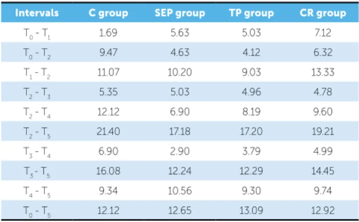
and also clinically signiicant (p < 0.01) for SEP and CR (DE’ = 5.63 and 7.12 respectively) (Tables 4 and 5).
of DE’ from T0 to T1 between group C and
experi-mental groups SEP, TP and CR indicating that the processes of bonding and debonding of orthodontic brackets altered the total color of teeth. Such altera-tion was greater in group CR, which demonstrated
a higher value of DE’ at T1 (Table 5) when compared
against group C.
From T0 to T1 groups SEP and CR displayed
signii-cant color diference ater the debonding of orthodontic
brackets (Table 5). From T1 to T2 all groups showed
sig-niicant color alterations. Groups CR and C displayed
the highest values of DE’ from T1 to T2 (Table 5).
Ater one day of whitening no signiicant color
diference was detected in any group from T2 to T3.
However, ater one week of whitening (T2-T4)
signii-cant color diference was displayed in all groups; con-trol group displayed the greatest diference (Table 5).
Upon assessing the difference among groups at a fixed interval, no significant difference was observed
among the groups at T0. Noticeable color difference
between C and SEP, and between C and CR was
ob-served after the debonding of brackets (T1). After one
and two weeks of whitening there was no significant color difference among the groups (Table 6).
DISCUSSION
The use of speciic colorimetric devices allows a faster and more consistent color assessment than visual
assessments alone.12 The color assessment method
ap-plied in this study was the computer-based evaluation of photographic images, supported by Gerlach, Barker,
Sagel;13 Gerlach, Gibb, Sagel;14 Bentley et al.11 These
authors state that an ideal color assessment method
p < 0.01.
p < 0.01.
Table 5 - Descriptive values of ΔE’ at the diferent evaluated time points.
Table 6 - Descriptive values of ΔE’ according to the experimental and control groups.
Intervals C group SEP group TP group CR group
T0 - T1 1.69 5.63 5.03 7.12
T0 - T2 9.47 4.63 4.12 6.32
T1 - T2 11.07 10.20 9.03 13.33
T2 - T3 5.35 5.03 4.96 4.78
T2 - T4 12.12 6.90 8.19 9.60
T2 - T5 21.40 17.18 17.20 19.21
T3 - T4 6.90 2.90 3.79 4.99
T3 - T5 16.08 12.24 12.29 14.45
T4 - T5 9.34 10.56 9.30 9.74
T0 - T5 12.12 12.65 13.09 12.92
Groups T0 T1 T2 T3 T4 T5
C - SEP 1.22 5.67 5.86 5.54 0.71 1.62
C - TP 0.60 3.88 5.32 4.83 0.84 0.81
C - CR 0.17 6.10 3.25 2.64 0.74 1.08
SEP - TP 1.20 1.79 0.78 0.73 0.61 0.81
SEP - CR 1.21 0.86 2.61 2.91 0.37 0.56
The presence of resin tags in the enamel, illing the “pores” created by the acidic condition of the
enam-el, indicates that it is the driver of those changes.23
The difference among the experimental groups is probably due to the different adhesive systems that
were utilized.23 The increase in the value of L* in the
Concise Resin group was also found in the Osório23
experiment. The presence of resins within the enam-el porosity may have modified a luminous reflection from the dental surface.
Hintz, Bradley and Eliades5 state that there are
three possible variations in bracket debonding proce-dures that can be responsible for the differences ob-served among the experimental surfaces: Quantity, quality and depth of resin tags, meaning the change in the morphology of the enamel as a result of the bonding and debonding procedures, and the amount of enamel lost during debonding. Considering these three possibilities, changes observed in the specimens of this study are probably due to the different resin tags left inside the dental substrate because the dif-ferences are observed among the experimental groups and between those and the control group. The oth-er aforementioned variables act individually on the specimens, regardless of the group, possibly causing high intragroup variance, which was not observed in this study (Tables 4 and 5).
The decrease in the value of L* from T1 to T2 did
not occur homogeneously in all groups (C, TP, SEP and CR) (Table 4 and Fig 1). Pigment absorption was greater in groups C and CR. In case of the control group it was due to lack of tags on the dental surface, which favors the difusion of bleaching agent’s
mole-cules 5. In case of group CR it was due to the improved
color stability of light-cured composites as compared to chemically cured composites (Fig 1). The presence of amines and benzoyl peroxide in chemically cured composite products contributes to a greater
pigmenta-tion when compared to light-cured ones.24,25
After one day of whitening we detected a statis-tically significant increased value of L* for groups C, SEP and TP. This whitening observed in some groups after six hours of exposure to carbamide per-oxide was possibly the result of the pigment depos-ited on the surface of the specimens. Authors report color alteration on dental surface related to the use of 10% carbamide peroxide, observed clinically after
one week of application.26,27 Therefore, the whitening
observed at this interval (T3) was more related to the
superficial removal of the just-deposited than to the bleaching effect itself.
After one week of whitening only the Control group displayed a significant increase in the value of L*, indicating a delayed whitening in the
experimen-tal groups as compared to Control at T3 and T4. These
results are according to those of Hintz, Bradley and
Eliades5 demonstrating the absence of initial response
to whitening in the experimental group. These au-thors report that the experimental group did not re-spond to whitening for the first two weeks. After two weeks, however, the whitening in the experimental group was faster than that in the control group.
At T5, after 14 days of whitening, all groups (C,
SEP, TP and CR) displayed a significant increase (p < 0.01) in the value of L*, which confirms the specimens lighter appearance. It is notable that the teeth became lighter than their initial color (DE’ from
T0 to T5) (Table 5) before pigmentation, confirming
the effectiveness of the bleaching agent.
In the end of the whitening process, at T5, the
val-ues of DE’ among the groups do not demonstrate any clinically significant color difference (Table 6), hence showing that the bonding and debonding of orth-odontic brackets did not interfere on the whitening
of the specimens. Hintz, Bradley and Eliades5 found
similar data by the end of the whitening process, according to which either control or experimental groups displayed significant color alterations. There-fore, the first research hypothesis was rejected since the presence of resin tags did not altered the final re-sult of the at-home whitening process carried out for 14 days, thus demonstrating that the quantity of resin remaining within the dental enamel was not enough to prevent the diffusion of the whitening agent inside the dental structure.
Nevertheless, more time in the application of the whitening agent is required to grant the same degree of color modiication between the control group and the experimental groups as indicated by existing
diferenc-es in the value of L* between T3 and T4, in control and
diferent values of L*. The control groups displayed a darker color than the experimental groups, with difer-ences reaching up to 6 units of L* between C and SE at
T2 (Table 4). Therefore, although the color diferences
among groups C, SEP, TP and CR at T4 (ater 7 days
of whitening) are not signiicant (p > 0.01) (Fig 1), only the control group displayed a statistically
signii-cant color change (T3 and T4). This inding indicates
that the control group changed color more rapidly than the experimental groups, when exposed to bleaching procedures (Table 4 and Fig 1). These indings match
those of Hintz, Bradley and Eliades,5 which state that,
initially, tags do make it more diicult for the whit-ening agent to penetrate the tooth but with a longer exposure to the whitening substance the inal result of the whitening process is not compromised.
In the end, the lack of difference among the exper-imental groups will lead to the rejection of the second research hypothesis, which suggested different group behaviors due to the role played by their specific resin remaining. However, during the experiment, signifi-cant color differences were verified for the different adhesive primer systems applied, especially after the debonding of orthodontic brackets and before dental
whitening (T2 and T3).
Although CIELAB coordinates values are im-portant for an objective color assessment, clinical significance (DE) plays a fundamental role in the
de-termination of what is actually perceived under the social and clinical stand points. Consequently, tak-ing this clinical significance into consideration, the results of this study demonstrate that the bonding and debonding of orthodontic brackets will not prevent the achievement of homogenous dental whitening. Nevertheless, a differentiated clinical protocol should be observed to allow an increased treatment time for the whitening of teeth that have endured bonding and debonding of orthodontic brackets. Despite the similar behavior of human and bovine teeth during
staining and bleaching28 procedures, further studies
using human dental substrates and assessing different intervals are required.
CONCLUSION
In light of the findings in this study, it could be concluded that:
1. Ater 14 days of at-home dental whitening there was no statistically signiicant color diference among the groups that were subjected to bond-ing and debondbond-ing of orthodontic brackets and the control group.
1. Feinman RAB, Madray G, Yarborough D. Chemical, optical and physiologic mechanisms of bleaching products: a review. Pract Periodontics Aesthet Dent. 1991;3(2):32-6.
2. Diedrich P. Enamel alterations from bracket bonding and debonding: a study with the scanning electron microscope. Am J Orthod. 1981;79(5):500-22. 3. Menezes LFS, Chevitarese O. Sealant and resin viscosity and their influence on
formation of resin tags. Angle Orthod. 1995;64(5):383-8.
4. Zachrisson BU, Artun J. Enamel surface appearance after various debonding techniques. Am J Orthod. 1979;75:121-37.
5. Hintz JK, Bradley TG, Eliades T. Enamel colour changes following whitening with 10 per cent carbamide peroxide: a comparison of orthodontically-bonded/ debonded and untreated teeth. Eur J Orthod. 2001;23(4):411-5.
6. Villalta P, Lu H, Okte Z, Garcia-Godoy F, Powers JM. Effects of staining and bleaching on color change of dental composite resin. J Prosthet Dent. 2006;95(2):137-42.
7. Eliades GC, Vougiouklakis GJ, Caputo AA. Degree of double bond conversion in light-cured composites. Dent Mater. 1987;3(1):19-25.
8. Chamda RA, Stein E. Time-related bond strengths of light-cured and chemically cured bonding systems: an in vitro study. Am J Orthod Dentofacial Orthop. 1996;110(4):378-82.
9. Zachrisson BU. Bonding in orthodontics. In: Graber TM, Vanarsdall RL. Orthodontics: current principles and techniques. 3rd ed. St. Louis: Mosby; 2000. 1040 p.
10. Alves EA, Alves FKA, Campos EJ, Mathias P. Susceptibility to caries-like lesions after dental bleaching with different techniques. Quintessence Int. 2007;38(7):e404-9.
11. Bentley C, Leonard RH, Nelson CF, Bentley SA. Quantitation of vital bleaching by computer analysis of photographic images. J Am Dental Assoc. 1999;130(6):809-16.
12. Seghi RR, Johnston WM, O’Brien WJ. Performance assessment of colorimetric devices on dental porcelains. J Dent Res. 1989;68(12):1755-9.
13. Gerlach RW, Barker ML, Sagel PA. Objective and subjective whitening response of two self-directed bleaching systems. Am J Dent. 2000;21:22-8.
14. Gerlach RW, Gibb RD, Sagel PA. A randomized clinical trial comparing a novel 5.3% hydrogen peroxide bleaching strip to 10%, 15% and 20% carbamide peroxide tray-based bleaching systems. Compend Contin Educ Dent. 2002;15:7-12.
REFEREnCEs
15. Scotti R, Mascellani SC, Fornit F. The in vitro color stability of acrylic resins for provisional restorations. Int J Prosthodont. 1997;10(2):164-8.
16. Dietschi D, Campanile G, Holz J, Meyer, JM. Comparison of the color stability of ten new generation composites: an in vitro study. Dent Mater. 1994;10(6):353-62.
17. Dozic A, Kleverlaan CJ, Aartman IHA, Feilzer AJ. Relation in color of three regions of vital human incisors. Dent Mater. 2004;20(9):832-8.
18. Eliades T, Kakaboura A, Eliades G, Bradley TG. Comparison of enamel colour changes associated with orthodontic bonding using two different adhesives. Eur J Orthod. 2001;23(1):85-90.
19. O’Brien WJ, Hemmendinger H, Boenke KM, Linger JB, Groh, CL. Color distribution of three regions of extracted human teeth. Dent Mater. 1997;13(3):179-85.
20. Dozic A, Kleverlaan CJ, Aartman IHA, Feilzer AJ. Relation in color among maxillary incisors and canines. Dent Mater. 2005;21(3):187-91.
21. Ruyter IE, Nilner K, Moller B. Color stability of dental composite resin materials for crown and bridge veneers. Dent Mater. 1987;3(5):246-51.
22. Johnston WN, Kao EC. Assessment of appearance match by visual observation and clinical colorimetry. J Dent Res. 1998;68(5):819-22.
23. Osório LB. Alterações cromáticas e micromorfológicas do esmalte submetido ao procedimento de clareamento pós-descolagem [thesis]. Rio de Janeiro (RJ): Federal University of Rio de Janeiro; 2000.
24. Asmussen E. Factors affecting the color stability of restorative resins. Acta Odontol Scand. 1983;41(1):11-8.
25. Tyas MJ. Colour stability of composite resins: a clinical comparison. Aust Dent J. 1992;37(2):88-90.
26. Kihn PW, Barnes DM, Romberg E, Peterson BS. A clinical evaluation of 10 percent vs. 15 percent carbamide peroxide tooth-whitening agents. J Am Dent Assoc. 2000;131(10):1478-84.
27. Jones HA, Diaz-Arnold MA, Vargas AM, Cobb SD. Colorimetric assessment of laser and home bleaching techniques. J Esthet Dent. 1999;11(2):87-94. 28. Attia ML, Aguiar FHB, Mathias P, Ambrosano GMB, Fontes CM, Liproni PC. The