Dental transposition of canine and lateral incisor and
impacted central incisor treatment: A case report
Tarcisio Jacinto Gebert1, Vinícius Canavarros Palma2, Alvaro Henrique Borges3, Luiz Evaristo Ricci Volpato4
How to cite this article: Gebert TJ, Palma VC, Borges AH, Volpato LER. Dental transposition of canine and lateral incisor and impacted central incisor treatment: A case report. Dental Press J Orthod. 2014 Jan-Feb;19(1):106-12. doi: http://dx.doi.org/10.1590/2176-9451.19.1.106-112.oar
Submitted: January 24, 2012 - Revised and accepted: August 30, 2012
» The authors report no commercial, proprietary or financial interest in the products or companies described in this article.
Contact address: Tarcisio Jacinto Gebert
Av. Campo Grande, 1259 – Primavera do Leste/MT – Brazil CEP: 78.850-000
E-mail: [email protected]
» Patients displayed in this article previously approved the use of their facial and in-traoral photographs.
1 Masters student, Integrated Dental Sciences Program, University of
Cuiabá (UNIC).
2 PhD in Dentistry, State University of São Paulo (UNESP). Professor,
Integrated Dental Sciences Masters Program, UNIC.
3 PhD in Dentistry, University of Ribeirão Preto (UNAERP). Professor,
Integrated Dental Sciences Masters Program, UNIC.
4 PhD in Dentistry, School of Dentistry — USP/ Bauru. Professor, Integrated
Dental Sciences Masters Program, UNIC.
Introduction:Dental transposition and impaction are disorders related to ectopic eruption or failure in tooth eruption, which can affect child physical, mental and social development and may be difficult to be clinically solved. Methods: We describe a case of transposition between the upper left canine and lateral incisor associated with impaction of the central incisor on the same side, in a 12-year-old patient. Conservative treatment involving surgical-orthodontic correction of transposed teeth and traction of the central incisor was conducted. Conclusion: The option of correcting transposition and orthodontic traction by means of the segmented arch technique with devices such as cantilever and TMA rectangular wire loops, although a complex alternative, was proved to be esthetically and functionally effective.
Keywords:Impacted tooth. Ectopic tooth eruption. Corrective orthodontics.
Introdução: transposição e impacção dentárias são distúrbios relacionados à erupção ectópica ou à falha na erupção den-tária, que podem afetar o desenvolvimento físico, psíquico e social da criança, e que podem ser de difícil resolução clínica.
Métodos: é descrito um caso clínico de transposição entre o canino e o incisivo lateral superior esquerdo, associado à impacção do incisivo superior, do mesmo lado, em uma paciente de 12 anos de idade. Optou-se pela realização de tratamento ortodôntico-cirúrgico conservador, envolvendo a correção ortodôntica dos dentes transpostos e o traciona-mento do incisivo central impactado. Conclusão: a opção pela correção da transposição e tracionamento ortodôntico com a utilização da técnica do arco segmentado com uso de dispositivos como cantiléver e alças em fios TMA retan-gulares, apesar de ser uma alternativa de execução complexa, mostrou-se efetiva do ponto de vista estético e funcional.
INTRODUCTION
Impaction is a condition in which complete tooth eruption is hampered by contact with another tooth.1 It is characterized by dental absence in the arch ater its usual period of eruption,2 and its etiology may be relat-ed to general or local factors.3 General factors include endocrine disorders, febrile diseases, radiation, heredity and development factors that may alter the eruption tra-jectory of the tooth germ.4 Local causes include lack of space in the arch, trauma, blocking by supernumerary tooth and lack of coordination between the formation of permanent teeth and deciduous exfoliation.5
Impaction is twice as common in females6 and can occur with any teeth, but the most afected are the lower third molars, upper canines and upper third molars, up-per and lower second premolars and upup-per central inci-sor.7 When the upper permanent incisors are impacted, there may be impairment in physical, psychological and social development of the child.8 Ater diagnosis, the therapeutic decision should prioritize tooth eruption.9
Conversely, tooth transposition, reported since the early nineteenth century, is described not only as a reversal of position between two teeth in the same quadrant of the dental arch, especially in relation to their roots; but also as the development or erup-tion of a tooth in a posierup-tion normally occupied by a non-adjacent element.10 It is considered real or com-plete when the tooth is in fully exchanged position in the dental arch and its roots are parallel to the other teeth; and incomplete when the teeth involved are not in fully exchanged position.11,12,13 It is also more prev-alent in females, in the upper arch, and of unilateral type.11,12,14 The upper permanent canine is the most involved tooth, transposing with the irst premolar in 80% of cases and with the lateral incisor in 20%.12 The causes of tooth transposition also involves general or local factors such as genetic factors with multifacto-rial causes of inheritance, dental anomalies (congen-ital absence of the lateral incisor, cone-shaped lateral incisor, rotations and hypodontia), migration of the developing tooth from its normal eruption path, root dilacerations, dental trauma and intervention in the development of the dental lamina.15
Early diagnosis of transposition in tooth develop-ment and impaction is essential, and greatly inluences the prognosis.16 Depending on the exact position of the impacted incisor, orthodontic movement and positioning
in the dental arch can vary widely. During orthodontic mechanics, occlusal interference should be avoided and root resorption should be controlled by periapical radi-ography so that bone loss does not occur, specially in the buccal bone plate.15 Incisors that are horizontally in-clined, in severe ectopic position or in real or complete tooth transposition are more diicult to treat. In cases of late diagnosis, orthodontic planning interferes not only in the decision of extracting impacted or transposed teeth, but also in the correction of the order of tooth position.11,12,15 Mechanical traction is preferred in the upper central incisors, but potential esthetic and peri-odontal problems may arise.17
Imaging tests are important aids in the diagnosis of these complications. They include the use of periapical, occlusal and panoramic radiographs or cephalograms,18 and, most recently, cone beam tomography.23
The objective of this paper is to report the treatment of a patient with tooth transposition between canine and lateral incisor of the upper let side associated with impaction of the upper central incisor on the same side.
CASE REPORT
A 12-year-old, female, Caucasian patient arrived for evaluation accompanied by her guardian, with her initial documentation at hand and orthodontic appli-ance installed. She had been undergoing treatment for 2 years and 7 months. Her chief complaint was that her front tooth was not born and two teeth had been born in exchanged position. The person responsible for the patient presented a transfer letter in which the orthodontist suggested clinical crown recontouring of the upper let lateral incisor into canine and upper let canine into lateral incisor, in addition to extraction of the retained central incisor.
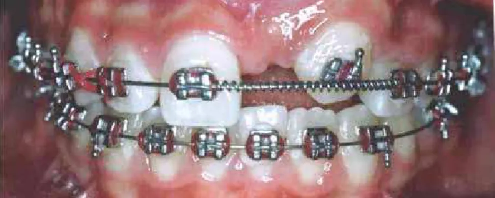
Clinical examination revealed that the patient had permanent dentition with Angle Class II malocclusion on the let side due to migration of the posterior supe-rior segment to a more antesupe-rior position; Class I mal-occlusion on the right side; upper midline deviation to the let; absence of tooth #21 with bulging in the ves-tibular region just below the anterior nasal spine (ANS), lack of space for its eruption, and transposition between teeth #22 and 23 (Fig 1). New radiographic documen-tation was, then, requested.
Its root was fully formed (Fig 2) and had the coronal long axis in a more horizontal position between the middle and apical third of the adjacent teeth roots (Fig 3). In-complete transposition between teeth #22 and 23 and image overlap of tooth #22 root apex and tooth #21 root were conirmed (Fig 4).
Once diagnosis had been completed, orthodon-tic-surgical conservative treatment was planned, al-though impaction of tooth #21 and transposition of teeth #22 and 23 represented unfavorable factors. Treat-ment plan included an attempt to correct transposition of teeth #22 and 23 and traction of tooth #21, which was accepted by the patient and her guardians.
The orthodontic procedures began by removing the appliance previously installed on both arches of the patient, since it was an appliance aimed at a diferent purpose. Ater removal and prophylaxis, new bands and weld of upper triple tube and lower double tube were prepared, in addition to cementing them on the up-per and lower irst molars (Fig 5). The upup-per arch was shaped with impression material for the fabrication of Hilgers auxiliary appliance. Thereater, the ixed metal orthodontic appliance was set with 0.018 x 0.030-in slot in both arches, semi-arches of stabilization with blue 0.016 x 0.016-in Elgiloy wire on the lower right and let sides as well as on the upper right side, an utility arch (UA) in the lower arch, and, on the upper let side, a semi-arch with helicals for intrusion of tooth #13 with TMA 0.017 x 0.025-in wire, with intrusive force of 45 grams to attempt dental transposition (Fig 6). Af-ter two days, the Hilgers appliance was installed for up-per let molar distalization and anchorage of the upup-per premolars on the same side, thus, avoiding extrusion (Fig 7). Ater eight months, the necessary upper mo-lar distalization and canine intrusion were obtained. The Hilgers appliance was removed and a quad-helix appliance (QH) was installed with 0.90 mm stainless steel wire to anchor and maintain upper let molar dis-talization (Fig 8). Additionally, 0.014-in round nick-el-titanium alloy (NiTi) archwire was installed for teeth alignment and leveling without inclusion of tooth #23.
Cortical anchorage was performed in the lower arch with UA, raise of bite with compound resin in the oc-clusal region of the lower molars and, ater alignment and leveling, a 0.014-in stainless steel wire was installed. Then, mesialization of the upper let lateral incisor be-gan to transpose it with the upper let canine by means
of an open nickel-titanium (NiTi) spring in the stainless steel wire between the upper let irst premolar and up-per let lateral incisor. A closed nickel-titanium spring 50g/f was placed from the upper let second premolar to the upper let canine for canine distalization. A semi-arch with rectangular 0.017 x 0.025-in stainless steel wire with 3/16-in medium elastic was also installed (Fig 9). Ater transposition was completed, alignment and leveling were performed with 0.014-in NiTi wire (Fig 10) up to the 0.016 x 0.016-in square stainless steel wire for better stabilization. An open steel spring was used between teeth #11 and 22 to keep the recovered space that would be occupied by tooth #21 (Fig 11). Aterwards, surgical exposure and bonding of the trac-tion accessory were performed.
Closed lap was the surgical technique chosen for traction. Ater 10 days, the sutures were removed and traction of tooth #21 began with 0.014-in NiTi wire superimposed to the square stainless steel archwire with open spring, by applying low magnitude forces in order to prevent unwanted movement of the adjacent teeth (Fig 12). Ater eruption of the retained tooth, the but-ton that was placed during surgery was removed and the bracket was positioned. New alignment and leveling were performed by inserting the tooth in the arch, start-ing with 0.014-in NiTi wire up to the 0.016 x 0.016-in steel square wire during a period of 9 months.
Ater thirty-nine months of active orthodontic treat-ment, the ixed orthodontic appliance was removed and the retainer was installed. For the upper dental arch, a ixed retainer was fabricated with 0.015-in Twist-lex wire from the let canine to the right central inci-sor, associated with a removable plate with continuous arch adapted to the buccal surfaces, from second molar to second molar, without occlusal interferences. As for the lower dental arch, a ixed lingual arch fabricated with 0.80 mm stainless steel wire was adhered to the lingual surfaces of lower canines, with light-cured resin (Fig 13). The case has been inalized and it is currently under clin-ical (Fig 14) and radiographic follow-up (Fig 15).
DISCUSSION
Figure 1 - Patient’s initial photograph showing fixed appliance set with transposition between the upper left canine and lateral incisor, and the absence of upper central incisor on the same side.
Figure 2 - Occlusal radiograph revealing impac-tion of tooth #21 with complete root formaimpac-tion.
Figure 3 - Lateral cephalogram revealing impac-tion of tooth #21 with coronal long axis horizon-tally positioned between the middle and apical third of adjacent teeth roots.
Figure 4 - Panoramic radiograph revealing impaction of tooth #21, #22 and #23 with incomplete trans-position.
Figure 5 - Intraoral photographs with fixed appliance removed and new bands with triple tubes properly cemented. A) Right lateral view. B) Frontal view. C) Left lateral view.
A B C
Figure 6 - Intraoral photograph showing fixed appliance: A) Right lateral view. B) Frontal view and C) Left lateral view with TMA semiarch for #23 intrusion.
Figure 7 - Hilgers appliance properly installed. Figure 8 - Quad-helix appliance installed to main-tain distalization of #26.
Figure 10 - Upper arch alignment after transposi-tion.
Figure 12 - Traction of tooth #21 carried out with 0.014” NiTi wire superimposed to 0.016 x 0.016-in square stainless steel archwire with open spring between #11 and #22.
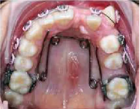
Figure 13 - Final intraoral photographs: A) Frontal view with Hawley retainer. B) Occlusal view of upper arch with fixed retainer from tooth #11 to 23. C) Lower occlusal view with fixed lingual arch.
Figure 9 - Distalization of tooth #23 with closed NiTi spring and mesialization of #22 with open NiTi spring.
Figure 11 - Upper arch with open steel spring used to keep the space left for traction of re-tained tooth #21.
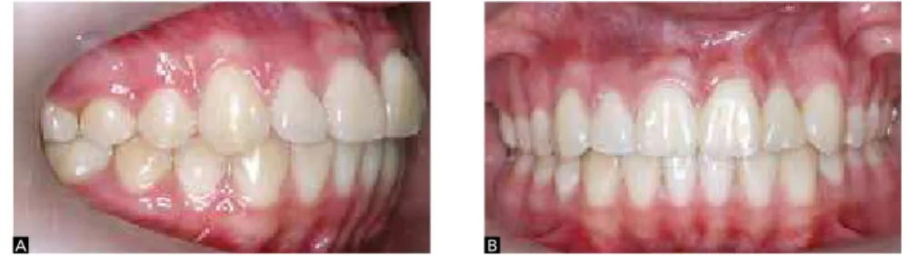
Figure 14 - Final intraoral photographs: A) Right lateral view. B) Frontal view. C) Left lateral view.
Figure 15 - Final panoramic radiograph.
The basal bone most oten afected is the maxilla, with higher incidence of unilateral transposition, which, in this case, the let side prevailed.12-24 The teeth mostly afected by transposition are canine and irst premolar12 as well as canine and lateral incisor.25 The case reported herein presented incomplete transposition between the canine and lateral incisor on the let side, associated with impacted upper let central incisor.
In the presence of an impacted tooth, a frequent complication of traction is the possibility of the tooth not moving due to ankylosis.26 Moving an impacted tooth involves risks of devitalization, discoloration, external root resorption, injury to adjacent teeth, al-veolar bone loss, gingival recession, increase in clini-cal crown length and tooth sensitivity, which can lead to esthetic problems or tooth loss.27 In the reported case, alveolar bone resorption existed prior to treat-ment. Gingival recession and increased clinical crown were also observed.
Traction and conservation of retained anterior teeth, both esthetically and functionally, it is the alternative therapy with the most favorable outcomes.28 There is a great demand for satisfactory esthetic outcomes in the anterior region, and no prosthetic material is superior to the tooth itself. Occlusal problems also decrease, since
there is no tooth loss and the arch remains symmetrical and complete.8 Another important aspect is that the vol-ume of alveolar bone loss resulting from extraction of the incisor is avoided, a frequent situation which is dif-icult to be solved.28 Thus, in the case reported herein, traction of tooth #21 was the treatment of choice, yield-ing satisfactory esthetic and functional results. The sur-gical technique used was the closed technique, in which the tooth is tractioned inside the mucosa and alveolar bone. This technique presents stability, periodontal anatomy and inal esthetic results that are more favor-able than apical reposition of the lap with immediate exposure of the crown ater surgery.28
The technique as well as the orthodontic appliances used in the traction of impacted teeth or in transpo-sition correction will depend on a correct diagnosis and treatment plan.1,3,7,15 When adjacent teeth require individualized and controlled movements, ixed ap-pliances are indicated8. The mechanics of choice must be carefully planned.3,7,13,15 In this case, the segmented arch technique was performed with the use of devices such as cantilever and loop in rectangular wires, which allowed the professional to work with a control system of strength with regard to movement of the central in-cisor, lateral incisor and canine as well as anchorage
units performed individually.3,7,10 However, the treat-ment involves risks, requiring extremely controlled mechanics, care and accurate application to overcome the possibility of failure.
Maintaining the central incisor, lateral incisor and canine in their usual position was essential to achieve balanced occlusion, periodontal health, facial harmony and for establishing patient’s esthetics. Canine guidance was another very positive aspect of the case. As a con-sequence, protrusive and lateral movements were prop-erly maintained, constituting an element of protection of the stomatognathic system, as well as the molar ratio of Class I Angle and correct overjet and overbite, thus allowing occlusal stability and proper dental esthetics.3,7
In view of a case involving tooth transposition between canine and lateral incisor associated with impaction of the
central incisor, the orthodontist must be committed to positioning these teeth correctly, leveling and aligning them in the dental arch within the biological principles that guide the integrity of adjacent tissue structures, thus resulting in a successful treatment.8,10,12 The technique of traction and tooth transposition proved to be highly satisfactory, restoring patient’s esthetics and harmonious occlusal relationships.
CONCLUSION
In the reported case, the choice of transposition and orthodontic traction carried out by means of the seg-mented arch technique performed with devices such as cantilever and loops in rectangular wires, despite being a more diicult alternative, proved to be efective from an esthetic and functional standpoint.
1. Hitchin AD. The impacted maxillary canine. Dent Pract Dent Rec. 1951;2(4):100-3.
2. Lindauer SJ, Rubenstein LK, Hang WM, Andersen WC, Isaacson RJ. Canine impaction identified early with panoramic radiographs. J Am Dent Assoc. 1992;123(3):91-2, 95-7.
3. Moyers RE. Handbook of orthodontics. 4th ed. Chicago: Year Book; 1988. 4. Shapira Y, Kuftinec MM. Treatment of impacted cuspids the azard lasso.
Angle Orthod. 1981;51(3):203-7.
5. Becker A, Smith P, Behar R. The incidence of anomalous maxillary lateral incisors in relation to palatally displaced cuspid. Angle Orthod. 1991;51(1):24-9.
6. Ericson S, Kurol J. Early treatment of palatally erupting maxillary canines by extraction of the primary canines. Eur J Orthod. 1988;10(4):283-95. 7. Moyers RE. Tratamento precoce. In: Ortodontia. Rio Janeiro: Guanabara
Koogan; 1991.
8. Daudt FB, Baraldi CE, Puricelli E. Tratamento orto-cirúrgico de incisive central retido dilacerado e canino retido: relato de caso. J Bras Ortodon Ortop Facial. 2002;7(38):110-6.
9. Lelvesley WD. Minimizing the problem of impacted and ectopic canines. ASDC J Dent Child. 1984;51(5):367-70.
10. Peck S, Peck L, Kataja M. Mandibular lateral incisor canine transposition, concomitant dental anomaliens, and genetic control. Angle Orthod. 1998;68(5):455-66.
11. Neto OJP, Caldas SGFR, Medeiros AM. Transposição dentária: um desafio na clínica ortodôntica – relato de caso. Rev Clin Ortodon Dental Press. 2006;5(4):75-84.
12. Maia FA, Maia NG. Transposição de canino com o incisivo lateral inferior: uma revisão ortodôntica. Rev Dental Press Ortod Ortop Facial. 2000;5(6):79-88. 13. Maia FA, Maia NG. Unusual orthodontic correction of bilateral maxillary
canine-frist pemolar transposition. Angle Orthod. 2005;75(2):266-76. 14. Capelozza Filho L, Cardoso MA, An TL, Bertoz FA. Maxillary canine-first
premolar transposition. Angle Orthod. 2007;77(1):167-75.
15. Capelozza Filho L, Cardoso MA, Cardoso Neto J. Tratamento da transposição de canino e pré-molar superior unilateral: abordagem por meio de mecânica segmentada. Rev Clin Ortod Dental Press. 2007;6(3):73-85.
REFERENCES
16. Shimizu RH, Geraldi Jr GR, Trojan LC, Shimizu IA, Melo ACM. Transposição dentária: um relato de caso. Orthod Sci Pract. 2010;3(12):364-73. 17. Shapira Y, Kuftinec MM, Stom D. Maxillary canine-lateral incisor
transposition: orthodontic management. Am J Orthod Dentofacial Orthop. 1989;95(5):439-44.
18. Caplan D. Transposition of the maxillary canine and the lateral incisor. Dent Pract Dent Rec. 1972;22(8):307.
19. Maia FA, Maia NG. Correção da transposição de canino com primeiro pré-molar na maxila: um desafio ortodôntico possível. Rev Clin Ortod Dental Press. 2006;5(5):79-103.
20. Maia FA. Orthodontic correction of a transposed maxillary canine and lateral incisor. Angle Orthod. 2000;70(4):339-48.
21. Crawford LB. Impacted maxillary central incisor in mixed dentition treatment. Am J Orthod Dentofacial Orthop. 1997;112(7):1-7.
22. Bishara SE. Impacted maxillary canines: a review. Am J Orthod Dentofacial Orthop. 1992;101(2):159-71.
23. Nakajima A, Sameshima GT, Arai Y, Homme Y, Shimizu N, Dougherty H. Two and three-dimensional orthodontic imaging using limited cone beam: computed tomography. Angle Orthod. 2005;75(6):895-903.
24. Peck S, Peck L. Classification of maxillary tooth transpositions. Am J Orthod Dentofacial Orthop. 1995;107(5):505-17.
25. Celikoglu M, Miloglu O, Oztek O. Investigation of tooth transposition in a non-syndromic turkish anatolian population: characteristic features and associated dental anomalies. Med Oral Patol Oral Cir Bucal. 2010;15(5):716-20.
26. Kajiyama K, Kai H. Esthetic management of an unerupted maxillary central incisor with a closed eruption technique. Am J Orthod Dentofacial Orthop. 2000;118(2):224-8.
27. Wreakes G, Cooke MS. The transplantation of canines using direct bonded orthodontic bracket fixation: an improved technique. Br J Orthod. 1979;6(1):5-9.