REVIEW
Glucocorticoid-induced osteoporosis in rheumatic
diseases
Rosa Maria Rodrigues Pereira,
IJoze´lio Freire de Carvalho,
IErnesto Canalis
IIIRheumatology Division, Faculdade de Medicina, Universidade de Sa˜o Paulo, Sa˜o Paulo, Brazil.IIDepartment of Research, Saint Francis Hospital and Medical Center, Hartford, CT, USA; and The University of Connecticut School of Medicine, Farmington, CT, USA.
The aim of this article is to review rheumatological diseases that are associated with glucocorticoid-induced
osteoporosis or fractures and to perform a critical analysis of the current guidelines and treatment regimens. The
electronic database MEDLINE was searched using the date range of July 1986 to June 2009 and the following search
terms: osteoporosis, bone mineral density, fractures, systemic lupus erythematosus, rheumatoid arthritis, systemic
sclerosis, vasculitis, juvenile rheumatoid arthritis, juvenile idiopathic arthritis and juvenile dermatomyositis.
Osteopenia and osteoporosis respectively account for 1.4 to 68.7% and 5.0 to 61.9% of adult rheumatological
diseases. Among juvenile rheumatological disorders, the frequency of low bone mass ranges from 38.7 to 70%. In
general, fracture rates vary from 0 to 25%. Although glucocorticoid-induced osteoporosis has a high rate of
prevalence among rheumatic diseases, a relatively low number of patients on continuous glucocorticoid treatment
receive adequate diagnostic evaluation or preventive therapy. This deficit in patient care may result from a lack of
clear understanding of the attributed risks by the patients and physicians, the high complexity of the treatment
guidelines and poor patient compliance.
KEYWORDS:
Osteoporosis; Glucocorticoids; Bone mineral density; Fractures, Rheumatic diseases.
Pereira RMR, Carvalho JF, Canalis E. Glucocorticoid-induced osteoporosis in rheumatic diseases. Clinics. 2010;65(11):1197-1205. Received for publication onJuly 10, 2010;First review completed onAugust 11, 2010;Accepted for publication onAugust 11, 2010 E-mail: [email protected]
Tel.: 55 11 3061-7490
INTRODUCTION
Glucocorticoids (GC) are frequently used for the
manage-ment of patients with rheumatological diseases. The use of
GC, however, is associated with a variety of adverse effects,
1including the development of osteoporosis and fractures. In
patients who have received GCs for longer than six months,
the estimated glucocorticoid-induced osteoporosis (GIO)
frequency is 50%.
2One-third to one-half of long-term GC
users may develop fractures. Furthermore, the risk of
fractures strongly correlates with the daily and cumulative
dose of GC and does not seem to correlate with the specific
underlying disease.
3The underlying diseases for which GCs
are prescribed, however, usually carry a risk of
osteoporo-sis. The objective of the present study was to review
rheumatic diseases in which GIO fractures have been
described and to perform a critical analysis of the diagnostic
criteria of osteoporosis and low bone mass. In addition, the
current guidelines and treatment barriers for the
manage-ment of GIO will be discussed.
Pathophysiology
The pathogenesis of GIO is multifaceted. Glucocorticoids
have indirect effects on osteoporosis by inhibiting calcium
absorption from the gastrointestinal track and decreasing
the renal tubular reabsorption of calcium and consequentely
secondary hyperparathyroidism. Nevertheless,
hyperpar-athyroidism does not play a central role in the pathogenesis
of GIO, snce the most of patients using chronic GC present
normal levels of serum parathromone. GCs reduce growth
hormone (GH) secretion and may alter the GH/insulin-like
growth factor (IGF)-I axis; however, the serum levels of
IGF-I are normal during osteoporosis, suggesting that alterations
in the GH/IGF-I axis play a minor role in this skeletal
disease. A more important role may be played by skeletal
IGF-I because GCs inhibit IGF-I transcription in osteoblasts.
In addition, GCs inhibit the release of gonadotrophins and
the resulting hypogonadism may contribute to skeletal
disease.
3Glucocorticoids have direct effects on bone cells. Bone
histomorphometric analyses of biopsies obtained from
patients with GIO reveal decreased bone turnover with a
disproportionate reduction in bone formation. GCs reduce
the replication, differentiation and function of osteoblasts
4and increase the apoptosis rates of mature cells, thereby
depleting the osteoblastic cell population and inhibiting the
function of mature cells.
3Furthermore, in the presence of
GCs, bone marrow stromal cells do not differentiate into
osteoblasts; instead, these cells differentiate toward an
adipocyte cell lineage. The underlying mechanism for this
change in cell fate appears to be related to an induction of
CCAAT enhancer binding proteins and possibly by
inhibit-ing Winhibit-ingless (Wnt)/
b
-catenin signaling.
3Moreover, GCs
induce apoptosis in osteocytes and affect the functioning of
Copyrightß2010CLINICS– This is an Open Access article distributed under the terms of the Creative Commons Attribution Non-Commercial License (http:// creativecommons.org/licenses/by-nc/3.0/) which permits unrestricted non-commercial use, distribution, and reproduction in any medium, provided the original work is properly cited.
colony stimulating factor (M-CSF) and receptor activator of
Nuclear fator kappa beta ( NF-kB) ligand (RANK-L). In
addition, GCs decrease the expression of osteoprotegerin in
stromal and osteoblastic cells. Through these mechanisms,
GCs can induce the formation of osteoclasts and favor bone
resorption. GCs also reduce the rate of apoptosis among
mature osteoclasts.
3GIO in Rheumatic Diseases
The electronic database MEDLINE was searched using
the date range of July 1986 to June 2009 and the following
search terms: osteoporosis, bone mineral density, fractures,
systemic lupus erythematosus, rheumatoid arthritis,
sys-temic sclerosis, juvenile rheumatoid arthritis, juvenile
idiopathic arthritis and juvenile dermatomyositis. A total
of 17 studies were found regarding systemic lupus
erythematosus, 16 about rheumatoid arthritis and 13 about
juvenile rheumatic diseases. All of these studies included
patients on GC (Tables 1, 2 and 3).
5–50The definition of
osteoporosis as determined by bone mineral density
(T-score
,
-2.5) and osteopenia (T-score
,
-1.0 to -2.5) should be
the definition used for postmenopausal women;
51however,
it is not applicable to GIO because patients on GCs can
fracture at T-scores in the normal or osteopenic range.
Along these lines, the International Society of Clinical
Densitometry recommends the following definition: ‘‘below
the expected range for age’’ for Z-scores lower than -2.0 and
‘‘within the expected range for age’’ for Z-scores above -2.0
for premenopausal women, children and adolescents
instead of osteoporosis/osteopenia for GIO.
52Never-theless, published reports use different definitions for
diagnosing GIO, thereby limiting the effectiveness of study
comparisons. For example, the term osteoporosis may not
be used for patients with fractures and a bone mineral
density greater than -2.5.
53Systemic Lupus Erythematosus
Studies on systemic lupus erythematosus demonstrate
a frequency of osteoporosis from 4.0 to 48.8% and of
osteo-penia from 1.4 to 68.7%.
5–21Fractures were
evalu-ated in four of these reports, with a frequency of 5.0 to
21.4%.
7,11–13A negative association between bone mass and
glucocorticoid use was documented in
,
60% of these
studies.
5–7,13,15–17,20,21Other possible associations with low
bone mass in subjects with lupus were the chronicity of the
disease, disease duration, low body mass index and weight,
increased age, habitual drinking, positive serum markers
of inflammation, renal dysfunction, menopause and
phy-sical dysfunction (Table 1).
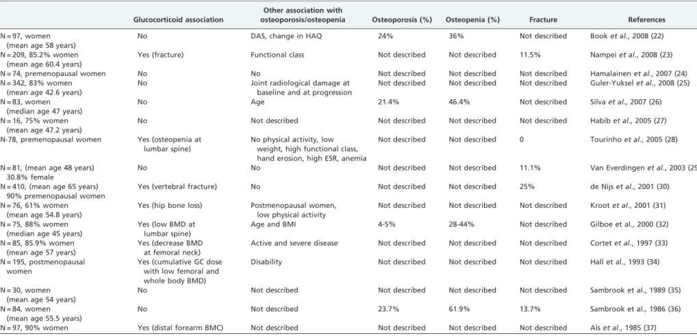
6–8,10–14,17–18,20Rheumatoid Arthritis
The frequency of osteoporosis in patients with
rheuma-toid arthritis (RA) ranges from 4 to 24% and the frequency
of osteopenia ranges from 28 to 61.9%.
22,26,32,36Fractures
were evaluated in four studies out of 16, which showed a
prevalence of osteoporosis of 0 to 25% in patients with
rheumatoid arthritis;
23,28,30,36however, only two of these
patients exhibited an association with GC use.
23–30De Nijs
et al
., showed that each 1 mg prednisone equivalent increase
in the daily dose of GC increased the risk of vertebral
deformities and symptomatic vertebral fractures in
pati-ents with RA.
30Glucocorticoid use was associated with
decreased bone mass in 56.2% of subjects with RA.
28,31–35,37change in the Health Assessment Questionnaire,
radiologi-cal joint damage, age, postmenopausal state, low physiradiologi-cal
activity, body mass index, disability, functional class and
anemia (Table 2).
22,23,25,26,28,31–34Juvenile Rheumatic Diseases
Some studies have addressed GIO in juvenile idiopathic
arthritis (JIA), which includes juvenile rheumatoid arthritis
(JRA), juvenile chronic arthritis (JCA), juvenile systemic
lupus erythematosus and juvenile dermatomyositis.
38–50The prevalence of low bone mineral density in children
with rheumatic diseases is difficult to assess because various
studies have used different cut-off points for Z-scores (e.g.,
,
1.0,
,
2.0).
41,44–47,50Recently, the International Society of
Clinical Densitometry defined low bone mineral density as a
Z-score below –2.0 in children and adolescents in an attempt
to standardize clinical data.
52,54In JIA, an association
between glucocorticoid use and low bone mass was
observed in four out of five studies.
39–41Santiago
et al.
,
found a relationship between pulse therapy with
methyl-prednisolone (30 mg/kg per day for at least three days) and
low bone mass in juvenile dermatomyositis.
43An
associa-tion between glucocorticoid use and low bone mass was
also observed in two studies that evaluated patients with
juvenile systemic lupus erythematosus
47,48and in a study
that evaluated several juvenile rheumatic diseases.
49Seven
studies evaluated the prevalence of fractures and only one
42demonstrated
an
association
with
this
complication
(Table 3).
38,42,44–47,50Systemic Sclerosis
Several studies have assessed bone mass in patients with
systemic sclerosis;
55–58however, only two reports included
patients on glucocorticoid therapy.
57,58These authors did
not find an association between osteoporosis and
glucocor-ticoid use in this disease.
57,58Systemic Vasculitis
Few studies have addressed GIO in systemic vasculitis
and only polymyalgia rheumatica and giant cell arteritis
have been described.
59–62The frequency of osteoporosis has
been shown to vary from 14.9 to 85%.
60,61Vertebral fractures
were analyzed in a study that compared placebo with
calcitonin and found an incidence of fractures between 11–
14%.
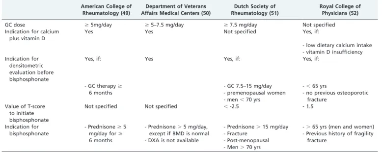
59Guidelines for GIO
There are a number of guidelines regarding the
manage-ment of GIO in patients who are receiving glucocorticoid
treatment or that will be starting this therapy. We have
reviewed the guidelines established by the American
College of Rheumatology (ACR),
63the Department of
Veterans Affairs Medical Centers (VMACs),
64the Dutch
Society of Rheumatology (DSR)
65and the Royal College of
Physicians (RCP).
66Various similarities among these four
Table 1 -
GIO in systemic lupus erythematosus.
Number (N) Population Glucocorticoid association
Other associations with osteoporosis and osteopenia
Osteoporosis (%)
Osteopenia
(%) Fracture References
N = 98, premenopausal women
Yes (cumulative GC dose with low lumbar spine BMD, and GC duration with low hip BMD)
No 6.1% 41.9% Not described Yeapet al., 2009 (5)
N = 100, premenopausal women
Yes (low BMD at hip) Chronic disease damage, low BMI 5.0% 40% Not described Mendoza-Pintoet al., 2009 (6)
N = 40, men
(mean age 42.6 years)
Yes (low BMD at lumbar spine) Increased age, habitual drinking, low BMI
Not described 30% 5% Moket al., 2008 (7)
N = 163, women (median age 47 years)
No Age, low weight, inflammatory markers, renal dysfunction, high chronic damage
23% Not described Not described Almehedet al., 2007 (8)
N = 60, premenopausal women
No No 6.6% 46.7% Not described Chonget al., 2007 (9)
N = 307, 65%
premenopausal women
No Disease damage Not described Not described Not described Leeet al., 2006 (10)
N = 70, premenopausal women (mean age 31.8 years)
No BMD Not described Not described 21.4% Borba et al, 2005 (11)
N = 107, 93% women (mean age 41.1 years)
Yes (vertebral fracture with intravenous methylprednisolone)
Low BMI 4% 39% 20% Bultink et al., 2005 (12)
Menopause Vitamin D deficiency N = 242, 95.4% women
(median age 39.9 years)
Yes (low BMD) Age, menopause 10.3% 50.8% 9.1% Yee et al., 2005 (13)
N = 205 patients No Age and damage index 48.8% 18% Not described Pineauet al., 2004 (14) N = 118, premenopausal
women
Yes (decreased BMD at lumbar spine and trochanter)
No Not described 1.4% Not described Uaratanawang et al., 2003 (15)
N = 32, women (mean age 43.2 years)
Yes (decreased BMD at lateral spine and total hip)
Not described Not described 68.7% Not described Boyanov et al., 2003 (16)
N = 79, women (mean age 49 years)
Yes (daily and cumulative dose) High functional class 23.7% 61.9% Not described Bhattoaet al., 2002 (17)
N = 64 patients No Body weight, disease duration, and damage index
10.9% Not described Not described Beckeret al., 2001 (18)
N = 23, men
(mean age 45.6 years)
No No 17.4% Not described Not described Bhattoaet al., 2001 (19)
N = 75, 88% women (median age 45 years)
Yes (low BMD at lumbar spine) Age, BMI 9% 41% Not described Gilboeet al., 2000 (20)
N = 97, women (mean age 44.2 years)
Yes (low BMD at lumbar spine) Not described 13.4% 44.3% Not described Kipen et al., 1997 (21)
BMD, bone mineral density; BMI, body mass index; GC, Glucocorticoid; Osteopenia and osteoporosis defined using WHO classification.
CLINICS
2010;65(11
):1197-1205
GIO
in
rheumatic
disease
Pereira
RMR
Table 2 -
GIO in rheumatoid arthritis.
Glucocorticoid association
Other association with
osteoporosis/osteopenia Osteoporosis (%) Osteopenia (%) Fracture References
N = 97, women (mean age 58 years)
No DAS, change in HAQ 24% 36% Not described Booket al., 2008 (22)
N = 209, 85.2% women (mean age 60.4 years)
Yes (fracture) Functional class Not described Not described 11.5% Nampeiet al., 2008 (23)
N = 74, premenopausal women No No Not described Not described Not described Hamalainenet al., 2007 (24) N = 342, 83% women
(mean age 42.6 years)
No Joint radiological damage at baseline and at progression
Not described Not described Not described Guler-Yukselet al., 2008 (25)
N = 83, women (median age 47 years)
No Age 21.4% 46.4% Not described Silvaet al., 2007 (26)
N = 16, 75% women (mean age 47.2 years)
No Not described Not described Not described Not described Habibet al., 2005 (27)
N-78, premenopausal women Yes (osteopenia at lumbar spine)
No physical activity, low weight, high functional class, hand erosion, high ESR, anemia
Not described Not described 0 Tourinhoet al., 2005 (28)
N = 81, (mean age 48 years) 30.8% female
No No Not described Not described 11.1% Van Everdingenet al., 2003 (29)
N = 410, (mean age 65 years) 90% premenopausal women
Yes (vertebral fracture) No Not described Not described 25% de Nijset al., 2001 (30)
N = 76, 61% women (mean age 54.8 years)
Yes (hip bone loss) Postmenopausal women, low physical activity
Not described Not described Not described Krootet al., 2001 (31)
N = 75, 88% women (median age 45 years)
Yes (low BMD at lumbar spine)
Age and BMI 4-5% 28-44% Not described Gilboe et al., 2000 (32)
N = 85, 85.9% women (mean age 57 years)
Yes (decrease BMD at femoral neck)
Active and severe disease Not described Not described Not described Cortetet al., 1997 (33)
N = 195, postmenopausal women
Yes (cumulative GC dose with low femoral and whole body BMD)
Disability Not described Not described Not described Hall et al., 1993 (34)
N = 30, women (mean age 54 years)
No Not described Not described Not described Not described Sambrook et al., 1989 (35)
N = 84, women (mean age 55.5 years)
No Not described 23.7% 61.9% 13.7% Sambrook et al., 1986 (36)
N = 97, 90% women Yes (distal forearm BMC) Not described Not described Not described Not described Alset al., 1985 (37) BMD, bone mineral density; BMI, body mass index; DAS, Disease Activity Score; HAQ, Health Assessment Questionnaire; ERS, erythrosedimentation rate
disease
et
al.
CLINICS
2010;65
(11):1197-12
Table 3 -
GIO in juvenile rheumatic diseases.
Disease Number (n) Population Glucocorticoid association
Other associations with low bone mass
Low bone
mass (%) Fracture References
JIA/JCA/JRA N = 62, 69.4% girls (median age 11.4 years)
No No Not described 10% Valtaet al., 2007 (38)
JIA/JCA/JRA N = 28, 57.1% girls (mean age 11 years)
Yes (low BMD at lumbar spine)
Age and age of disease onset
Not described Not described Celikeret al.,2003 (39)
JIA/JCA/JRA N = 18, 38.9% girls (mean age 11 years)
Yes (low BMD at lumbar spine)
No Not described Not described Cetinet al., 1998 (40)
JIA/JCA/JRA N = 62, 58.1% girls (5–18 years) Yes (low BMD at distal radius and lumbar spine)
Long disease duration 50–60% Not described Pereiraet al., 1998 (41)
JIA/JCA/JRA N = 46 Yes (fracture) Not described Not described 50% Varonoset al.,1987 (42) Juvenile dermatomyositis N = 20 girls (mean age 13.4 years) Yes (GC pulse therapy with
low BMD in hip)
Lean mass Not described Not described Santiagoet al., 2008 (43)
Juvenile dermatomyositis N = 10 girls (mean age 11.8 years) No Weight 70% 0 Castroet al., 2005 (44) Juvenile dermatomyositis N = 15, 60% girls
(mean age 7.9 years)
No Disease duration 66.7% (Z,-1) 33.3% Stewartet al., 2003 (45)
Juvenile systemic lupus erythematosus
N = 36 girls (mean age 17.7) No Disease 38.7% 22.6% Regioet al., 2008 (46)
Juvenile systemic lupus erythematosus
N = 70, 65% girls (mean age 26.4 years)
Yes (low bone mineral content)
Male gender 41% 6% Lilleby et al., 2005 (47)
Juvenile systemic lupus erythematosus
N = 20, 90% girls (mean age 14.5 years)
Yes (BMD loss at lumbar spine)
No Not described Not described Trapani et al., 1998 (48)
Juvenile systemic lupus erythematosus, JIA
N = 20, 65% girls (mean age 13.1 years)
No No Not described Not described Kashef et al., 2007 (49)
Juvenile dermatomyositis, juvenile systemic lupus erythematosus, vasculitis
N = 36, 91.6% girls (mean age 11.4 years)
Yes (lower BMD at lumbar spine, hip, total body)
Younger and prepubertal
40% JSLE and 27% JDM/vasculitis
0 Alsufyani et al., 2005 (50)
JIA, juvenile idiopathic arthritis; JCA, juvenile chronic arthritis; JRA, juvenile rheumatoid arthritis; DAS, Disease Activity Score; HAQ, Health Assessment Questionnaire
CLINICS
2010;65(11
):1197-1205
GIO
in
rheumatic
disease
Pereira
RMR
The Royal College of Physicians’ guidelines also reinforce
the necessity to use the lowest GC dose possible.
66Barriers in the Management of GIO
Although numerous guidelines for GIO management
have been published, previous studies suggest that a
relatively low percentage of patients who receive
contin-uous glucocorticoid treatment are evaluated or
adminis-tered preventive treatment for GIO. Saag
et al
., studied more
than 3,000 adult men and women who had undergone
long-term glucocorticoid therapy. These authors found that bone
mineral density testing was performed in 19% of
post-menopausal women and in 6% of women under the age of
50 years.
67The use of antiosteoporotic medication was most
common among postmenopausal women and its use
approached 50% in this group. In addition, the medical
specialty of the physician providing care influenced both
testing and treatment regimens. Notably, testing rates were
3 to 4 times greater in rheumatology practices than in
internal medicine or family medicine practices.
67,68There
are several possible explanations for these low compliance
rates with current guidelines. For example, published
guidelines are inconsistent regarding who should be
treated. In addition, the dose and duration of glucocorticoid
therapy are not standardized. Guidelines are also difficult to
implement due to the physician’s focus on the underlying
disease. In select cases, there may be limited access to
densitometry. Furthermore, patients and physicians may
not have a clear perception of the risk of GIO and patients
may not accept treatment. Our findings suggest that
unification of guidelines regarding the glucocorticoid dose
that would require treatment (such as prednisone
equiva-lents
$
5 mg/day for at least 3 months), requirement of
densitometric evaluation (premenopausal women and
patients on GCs) and indication of bisphosphonates for
prevention (postmenopausal women and men) and
treat-ment (T-score
,
-1.0 or previous fragility fracture in
postmenopausal women and men) could be of value to
medical practioners.
69Treatment
Because GCs induce an overall negative calcium balance,
adequate calcium and vitamin D supplementation is
important. A Cochrane Database Meta-Analysis concluded
that calcium and vitamin D supplementation should be
started in all patients who are administered glucocorticoids
because of their low toxicity, low cost and the possible
benefit in terms of fracture risk.
70Vitamin D is a hormone
that increases intestinal calcium absorption and increases its
reabsorption in distal renal tubules. Serum levels of at least
30 ng/mL (82 nmol/L), and optimally of 40–60 ng/mL, of
25-hydroxyvitamin D should be the target treatment
regi-men for GIO manageregi-ment. To achieve these levels, 1,000 to
2,000 IU of oral vitamin D daily may be necessary.
71 American College ofRheumatology (49)
Department of Veterans Affairs Medical Centers (50)
Dutch Society of Rheumatology (51)
Royal College of Physicians (52)
GC dose $5mg/day $5–7.5 mg/day $7.5 mg/day Not specified
Indication for calcium plus vitamin D
Yes Yes Not specified Yes, if:
- low dietary calcium intake - vitamin D insufficiency Indication for
densitometric evaluation before bisphosphonate
Yes, if: Yes Yes, if: Yes, if:
- GC therapy$ 6 months
- GC 7.5–15 mg/day - premenopausal women - men,70 yrs
-,65 yrs
- no previous osteoporotic fracture
Value of T-score to initiate bisphosphonate
Not specified Not specified ,-2.5 - 1.5
Indication for bisphosphonate
- Prednisone$5 mg/day for$ 6 months
- Prednisone.5 mg/day, except if BMD is normal - DXA is not available
- Prednisone.15 mg/day - Fracture
- Post-menopausal - Men.70 yrs
-.65 yrs (men and women) - Previous history of fragility
fracture
GC, Glucocorticoid
Table 5 -
Guidelines for the treatment of GIO.
American College of Rheumatology (49)
Department of Veterans Affairs Medical Centers (50)
Dutch Society of Rheumatology (51)
Royal College of Physicians (52)
GC dose $5mg/day Not specified Not specified Not specified
Indication for calcium plus vitamin D Yes Yes Not specified Yes
Indication for densitometric evaluation before bisphosphonates
Yes Yes Not specified Yes
Indication for bisphosphonate BMD,-1.0 - low BMD
- history of fracture
Bisphosphonates are indicated for the prevention and
treatment of GIO and most guidelines recommend the use
of these drugs.
63–66The prevention and treatment goals of
bisphosphonate use are stabilized or increased bone mineral
density, as well as reduced frequency of fractures. A study
using risedronate showed a decrease in vertebral fractures
after one year of treatment.
72Currently, alendronate (70 mg/
week or 10 mg/day) and risedronate (35 mg/week or 5 mg/
day) are the only oral antiresorptive drugs that are
recommended in GIO. Recently, zoledronic acid was
approved for the prevention and treatment of GIO. In a
multicenter, double-blind, double-dummy, randomized
controlled trial that included 833 patients, a single 5 mg
intravenous infusion of zoledronic caused a greater increase
in bone mineral density than oral risedronate at 5 mg
daily.
73Bisphosphonate treatment is recommended while
patients are on glucocorticoids; however, in subjects with
significant bone loss, therapy may need to be continued
following the discontinuation of glucocorticoids.
Caution needs to be exercised when considering the use
of bisphosphonates in women of childbearing age with
GIO,
63,74,75given that bisphosphonates have an extended
half-life and may cross the placenta with potentially
unfavorable effects on fetal skeletal development. A recent
review of 51 human cases examining exposure to
bispho-sphonates before or during pregnancy did not demonstrate
skeletal abnormalities or other congenital malformations in
the infants.
75Similarly, a related case-controlled study
suggested that preconceptional and first-trimester use of
bisphosphonates may pose limited fetal risk.
76Nevertheless,
these studies included a small number of subjects and the
safety of bisphosphonates in women of childbearing age,
during pregnancy or while lactating is unknown. Therefore,
physicians should carefully weigh the risks and benefits of
bisphosphonate therapy in premenopausal women.
Although the guidelines do not address the use of
anabolic therapies in GIO, this approach appears to be ideal
because glucocorticoids reduce bone formation. Saag
et al.,
published a randomized multicenter trial to compare use of
oral alendronate (10 mg/day) and subcutaneous
teripara-tide (20
m
g/day) over 18 months in patients with established
GIO. The study showed that among patients with
osteo-porosis with a high risk for fracture, the bone mineral
density increase in patients receiving teriparatide was
greater than in those receiving alendronate.
77The study
did not possess enough statistical power to detect
differ-ences in the incidence of fractures, although the number of
vertebral fractures was significantly lower in the
teripara-tide arm than in the alendronate arm. Subsequently, the
study was extended to 36 months and these results
confirmed a higher increase in bone mineral density and
fewer new vertebral fractures in subjects treated with
teriparatide as compared to alendronate.
78CONCLUSION
After an extensive review of the literature, it was
observed that the frequency of GIO varies due to different
study designs and the lack of a uniform definition of GIO.
Similarly, currently available guidelines use different
recommendations for the prevention and treatment of
GIO, thereby creating practical difficulties. Consequently,
patients who are frequently exposed to GCs are not assessed
or treated.
ACKNOWLEDGMENTS
This work was supported by grants from National Council for Scientific and Technological Development (305691/2006-6 to R.M.R.P. and 300665/2009-1 to J.F.C.) and Federico Foundation (J.F.C. and R.M.R.P.).
REFERENCES
1. Cutolo M, Seriolo B, Pizzorni C, Secchi ME, Soldano S, Paolino S, et al. Use of glucocorticoids and risk of infections. Autoimmun Rev. 2008;8:153-5, doi: 10.1016/j.autrev.2008.07.010.
2. van Staa TP, Leufkens HG, Cooper C. The epidemiology of corticoster-oid-induced osteoporosis: a meta-analysis. Osteoporos Int. 2002;13:777-87, doi: 10.1007/s001980200108.
3. Canalis E, Mazziotti G, Giustina A, Bilezikian JP. Glucocorticoid-induced osteoporosis: pathophysiology and therapy. Osteoporos Int 2007;18:1319-28, doi: 10.1007/s00198-007-0394-0.
4. Pereira RM, Delany AM, Canalis E. Cortisol inhibits the differentiation and apoptosis of osteoblasts in culture. Bone 2001;28:484-90, doi: 10. 1016/S8756-3282(01)00422-7.
5. Yeap S, Fauzi A, Kong N, Halim A, Soehardy Z, Rahimah S, et al. Influences on bone mineral density in Malaysian premenopausal systemic lupus erythematosus patients on corticosteroids. Lupus 2009;18:178-81, doi: 10.1177/0961203308094995.
6. Mendoza-Pinto C, Garcı´a-Carrasco M, Sandoval-Cruz H, Esca´rcega RO, Jime´nez-Herna´ndez M, Etchegaray-Morales I, et al. Risks factors for low bone mineral density in pre-menopausal Mexican women with systemic lupus erythematosus. Clin Rheumatol. 2009;28:65-70, doi: 10.1007/ s10067-008-0984-z.
7. Mok CC, Cheung MY, Ho LY, Yu KL, To CH. Risk and predictors of work disability in Chinese patients with systemic lupus erythematosus. Lupus. 2008;17:1103-7, doi: 10.1177/0961203308094280.
8. Almehed K, Forsblad d’Elia H, Kvist G, Ohlsson C, Carlsten H. Prevalence and risk factors of osteoporosis in female SLE patients-extended report. Rheumatology Oxford. 2007;46:1185-90, doi: 10.1093/ rheumatology/kem105.
9. Chong HC, Chee SS, Goh EM, Chow SK, Yeap SS. Dietary calcium and bone mineral density in premenopausal women with systemic lupus erythematosus. Clin Rheumatol. 2007;26:182-5, doi: 10.1007/s10067-006-0258-6.
10. Lee C, Almagor O, Dunlop DD, Manzi S, Spies S, Chadha AB, et al. Disease damage and low bone mineral density: an analysis of women with systemic lupus erythematosus ever and never receiving corticosteroids. Rheumatology. (Oxford) 2006;45:53-60, doi: 10.1093/rheumatology/ kei079.
11. Borba VZ, Matos PG, da Silva Viana PR, Fernandes A, Sato EI, Lazaretti-Castro M. High prevalence of vertebral deformity in premenopausal systemic lu´pus erythematosus patients. Lupus. 2005;14:529-33, doi: 10. 1191/0961203305lu2154oa.
12. Bultink IE, Lems WF, Kostense PJ, Dijkmans BA, Voskuyl AE. Prevalence of and risk factors for low bone mineral density and vertebral fractures in patients with systemic lupus erythematosus. Arthritis Rheum. 2005;52:2044-50, doi: 10.1002/art.21110.
13. Yee CS, Crabtree N, Skan J, Amft N, Bowman S, Situnayake D, et al. Prevalence and predictors of fragility fractures in systemic lupus erythematosus. Ann Rheum Dis. 2005;64:111-3, doi: 10.1136/ard.2003. 018127.
14. Pineau CA, Urowitz MB, Fortin PJ, Ibanez D, Gladman DD. Osteoporosis in systemic lupus erythematosus: factors associated with referral for bone mineral density studies, prevalence of osteoporosis and factors associated with reduced bone density. Lupus. 2004;13:436-41, doi: 10. 1191/0961203303lu1036oa.
15. Uaratanawong S, Deesomchoke U, Lertmaharit S, Uaratanawong S. Bone mineral density in premenopausal women with systemic lupus erythematosus. J Rheumatol 2003;30:2365-8.
16. Boyanov M, Robeva R, Popivanov P. Bone mineral density changes in women with systemic lupus erythematosus. Clin Rheumatol. 2003;22:318-23, doi: 10.1007/s10067-003-0743-0.
17. Bhattoa HP, Bettembuk P, Balogh A, Szegedi G, Kiss E. Bone mineral density in women with systemic lupus erythematosus. Clin Rheumatol. 2002;21:135-41, doi: 10.1007/s10067-002-8272-9.
18. Becker A, Fischer R, Scherbaum WA, Schneider M. Osteoporosis screening in systemic lupus erythematosus: impact of disease dura-tion and organ damage. Lupus. 2001;10:809-14, doi: 10.1177/ 096120330101001108.
19. Bhattoa HP, Kiss E, Bettembuk P, Balogh A. Bone mineral density, biochemical markers of bone turnover, and hormonal status in men with systemic lupus erythematosus. Rheumatol Int. 2001;21:97-102, doi: 10. 1007/s00296-001-0149-8.
20. Gilboe IM, Kvien TK, Haugeberg G, Husby G. Bone mineral density in systemic lupus erythematosus: comparison with rheumatoid arthritis and healthy controls. Ann Rheum Dis. 2000;59:110-5, doi: 10.1136/ard.59. 2.110.
CLINICS 2010;65(11):1197-1205 GIO in rheumatic disease
erythematosus and the role of steroids. J Rheumatol. 1997;24:1922-9. 22. Book C, Karlsson M, Akesson K, Jacobsson L. Disease activity and
disability but probably not glucocorticoid treatment predicts loss in bone mineral density in women with early rheumatoid arthritis. Scand J Rheumatol. 2008;37:248-54, doi: 10.1080/03009740801998747. 23. Nampei A, Hashimoto J, Koyanagi J, Ono T, Hashimoto H, Tsumaki N,
et al. Characteristics of fracture and related factors in patients with rheumatoid arthritis. Mod Rheumatol. 2008;18:170-6, doi: 10.1007/ s10165-008-0032-5.
24. Ha¨ma¨la¨inen H, Kaarela K, Kro¨ger H, Kauppi M, Ja¨rvenpa¨a¨ S, Hakala M, et al. Changes in bone mineral density in premenopausal women with rheumatoid arthritis during a two-year follow-up. Joint Bone Spine. 2007;74:482-7, doi: 10.1016/j.jbspin.2007.01.021.
25. Gu¨ler-Yu¨ksel M, Bijsterbosch J, Goekoop-Ruiterman YP, de Vries-Bouwstra JK, Hulsmans HM, de Beus WM, et al. Changes in bone mineral density in patients with recent onset, active rheumatoid arthritis. Ann Rheum Dis. 2008;67:823-8, doi: 10.1136/ard.2007.073817. 26. Silva RG, Pippa MG, Zerbini CA. Evaluation of body composition and
bone mineral density in women with rheumatoid arthritis. Rev Assoc Med Bras. 2007;53:135-41, doi: 10.1590/S0104-42302007000200018. 27. Habib GS, Haj S. Bone mineral density in patients with early rheumatoid
arthritis treated with corticosteroids. Clin Rheumatol. 2005;24:129-33, doi: 10.1007/s10067-004-0989-1.
28. Tourinho TF, Stein A, Castro JA, Brenol JC. Rheumatoid arthritis: evidence for bone loss in premenopausal women. J Rheumatol. 2005;32:1020-5.
29. van Everdingen AA, Siewertsz van Reesema DR, Jacobs JW, Bijlsma JW. Low-dose glucocorticoids in early rheumatoid arthritis: discordant effects on bone mineral density and fractures? Clin Exp Rheumatol. 2003;21:155-60.
30. de Nijs RN, Jacobs JW, Bijlsma JW, Lems WF, Laan RF, Houben HH, et al. Osteoporosis Working Group, Dutch Society for Rheumaology. Prevalence of vertebral deformities and symptomatic vertebral fractures in corticosteroid treated patients with rheumatoid arthritis. Rheumatology. (Oxford) 2001;40:1375-83, doi: 10.1093/rheumatology/ 40.12.1375.
31. Kroot EJ, Nieuwenhuizen MG, de Waal Malefijt MC, van Riel PL, Pasker-de Jong PC, Laan RF. Change in bone mineral Pasker-density in patients with rheumatoid arthritis during the first decade of the disease. Arthritis Rheum. 2001;44:1254-60, doi: 10.1002/1529-0131(200106)44:6, 1254::AID-ART216.3.0.CO;2-G.
32. Gilboe IM, Kvien TK, Haugeberg G, Husby G. Bone mineral density in systemic lupus erythematosus: comparison with rheumatoid arthritis and healthy controls. Ann Rheum Dis. 2000;59:110-5, doi: 10.1136/ard.59. 2.110.
33. Cortet B, Flipo RM, Blanckaert F, Duquesnoy B, Marchandise X, Delcambre B. Evaluation of bone mineral density in patients with rheumatoid arthritis. Influence of disease activity and glucocorticoid therapy. Rev Rhum Engl Ed. 1997;64:451-8.
34. Hall GM, Spector TD, Griffin AJ, Jawad AS, Hall ML, Doyle DV. The effect of rheumatoid arthritis and steroid therapy on bone density in postmenopausal women. Arthritis Rheum. 1993;36:1510-6, doi: 10.1002/ art.1780361105.
35. Sambrook PN, Cohen ML, Eisman JA, Pocock NA, Champion GD, Yeates MG. Effects of low dose corticosteroids on bone mass in rheumatoid arthritis: a longitudinal study. Ann Rheum Dis. 1989;48:535-8, doi: 10. 1136/ard.48.7.535.
36. Sambrook PN, Eisman JA, Yeates MG, Pocock NA, Eberl S, Champion GD. Osteoporosis in rheumatoid arthritis: safety of low dose corticoster-oids. Ann Rheum Dis. 1986;45:950-3, doi: 10.1136/ard.45.11.950. 37. Als OS, Gotfredsen A, Christiansen C. The effect of glucocorticoids on
bone mass in rheumatoid arthritis patients. Influence of menopausal state. Arthritis Rheum. 1985;28:369-75, doi: 10.1002/art.1780280403. 38. Valta H, Lahdenne P, Jalanko H, Aalto K, Ma¨kitie O. Bone health and
growth in glucocorticoid-treated patients with juvenile idiopathic arthritis. J Rheumatol 2007;34:831-6.
39. Celiker R, Bal S, Bakkalog˘lu A, Ozaydin E, Coskun T, Cetin A, et al. Factors playing a role in the development of decreased bone mineral density in juvenile chronic arthritis. Rheumatol Int. 2003;23:127-9. 40. Cetin A, Celiker R, Dinc¸er F, Ariyu¨rek M. Bone mineral density in
children with juvenile chronic arthritis. Clin Rheumatol. 1998;17:551-3, doi: 10.1007/BF01451301.
41. Pereira RM, Corrente JE, Chahade WH, Yoshinari NH. Evaluation by dual X-ray absorptiometry (DXA) of bone mineral density in children with juvenile chronic arthritis. Clin Exp Rheumatol. 1998;16:495-501. 42. Varonos S, Ansell BM, Reeve J. Vertebral collapse in juvenile chronic
arthritis: its relationship with glucocorticoid therapy. Calcif Tissue Int. 1987;41:75-8, doi: 10.1007/BF02555248.
43. Santiago RA, Silva CA, Caparbo VF, Sallum AM, Pereira RM. Bone mineral apparent density in juvenile dermatomyositis: the role of lean body mass and glucocorticoid use. Scand J Rheumatol. 2008;37:40-7, doi: 10.1080/03009740701687226.
Braz J Med Biol Res. 2005;38:309-13.
45. Stewart WA, Acott PD, Salisbury SR, Lang BA. Bone mineral density in juvenile dermatomyositis: assessment using dual x-ray absorptiometry. Arthritis Rheum. 2003;48:2294-8, doi: 10.1002/art.11211.
46. Regio P, Bonfa´ E, Takayama L, Pereira RM. The influence of lean mass in trabecular and cortical bone in juvenile onset systemic lupus erythema-tosus. Lupus. 2008;17:787-92, doi: 10.1177/0961203308089446.
47. Lilleby V, Lien G, Frey Frøslie K, Haugen M, Flatø B, et al. Frequency of osteopenia in children and young adults with childhood-onset systemic lupus erythematosus. Arthritis Rheum. 2005;52:2051-9, doi: 10.1002/art. 21115.
48. Trapani S, Civinini R, Ermini M, Paci E, Falcini F. Osteoporosis in juvenile systemic lupus erythematosus: a longitudinal study on the effect of steroids on bone mineral density. Rheumatol Int. 1998;18:45-9, doi: 10. 1007/s002960050056.
49. Kashef S, Saki F, Karamizadeh Z, Kashef MA. Bone mineral density in children wth systemic lupus erythematosus and juvenile rheumatoid arthritis. Ann Saudi Med. 2007;27:427-31, doi: 10.4103/0256-4947.51458. 50. Alsufyani KA, Ortiz-Alvarez O, Cabral DA, Tucker LB, Petty RE, et al.
Bone mineral density in children and adolescents with systemic lupus erythematosus, juvenile dermatomyositis, and systemic vasculitis: relationship to disease duration, cumulative corticosteroid dose, calcium intake, and exercise. J Rheumatol. 2005;32:729-33.
51. (1994) Assessment of fracture risk and its application to screening for postmenopausal osteoporosis. Geneva: World Health Organization; 1994. Technical Report Series, No. 843.
52. Official positions of the International Society for Clinical Densitometry Updated 2007. Available at www.iscd.org/Visitors/positions/ OfficialPositionsText.cfm Accessed July 5, 2009.
53. Langsetmo L, Goltzman D, Kovacs CS, Adachi JD, Hanley DA, Kreiger N, et al. Repeat Low-Trauma Fractures Occur Frequently Among Men and Women who have Osteopenic Bone Mineral Density. J Bone Miner Res. 2009;24:1515-22, doi: 10.1359/jbmr.090319.
54. Khan AA, Bachrach L, Brown JP, Hanley DA, Josse RG, Kendler DL, et al. Standards and guidelines for performing central dual-energy X-ray absorptiometry in premenopausal women, men, and children. J Clin Densitom. 2004;51-63, doi: 10.1385/JCD:7:1:51.
55. Souza RB, Borges CT, Takayama L, Aldrighi JM, Pereira RM. Systemic sclerosis and bone loss: the role of the disease and body composition. Scand J Rheumatol. 2006;35:384-7, doi: 10.1080/03009740600704296. 56. Frediani B, Baldi F, Falsetti P, Acciai C, Filippou G, Spreafico A, et al.
Clinical determinants of bone mass and bone ultrasonometry in patients with systemic sclerosis. Clin Exp Rheumatol. 2004;22:313-8.
57. Yuen SY, Rochwerg B, Ouimet J, Pope JE. Patients with scleroderma may have increased risk of osteoporosis. A comparison to rheumatoid arthritis and noninflammatory musculoskeletal conditions. J Rheumatol. 2008;35:1073-8.
58. Sampaio-Barros PD, Costa-Paiva L, Filardi S, Sachetto Z, Samara AM, Marques-Neto JF. Prognostic factors of low bone mineral density in systemic sclerosis. Clin Exp Rheumatol. 2005;23:180-4.
59. Healey JH, Paget SA, Williams-Russo P, Szatrowski TP, Schneider R, Spiera H, et al. A randomized controlled trial of salmon calcitonin to prevent bone loss in corticosteroid-treated temporal arteritis and polymyalgia rheumatica. Calcif Tissue Int. 1996;58:73-80, doi: 10.1007/ BF02529727.
60. Andersson R, Rundgren A, Rosengren K, Bengtsson BA, Malmvall BE, Mellstro¨m D. Osteoporosis after long-term corticosteroid treatment of giant cell arteritis. J Intern Med. 1990;227:391-5, doi: 10.1111/j.1365-2796. 1990.tb00177.x.
61. Hatz HJ, Helmke K. Polymyalgia rheumatica and giant cell arteritis; diagnosis and side effects of low-dose long-term glucocorticoid therapy. Z Rheumatol 1992;51:213-21.
62. Pearce G, Ryan PF, Delmas PD, Tabensky DA, Seeman E. The deleterious effects of low-dose corticosteroids on bone density in patients with polymyalgia rheumatica. Br J Rheumatol. 1998;37:292-9, doi: 10.1093/ rheumatology/37.3.292.
63. (2001) American College of Rheumatology Ad Hoc Committee on Glucocorticoid-Induced Osteoporosis Recommendations for the preven-tion and treatment of glucocorticoid-induced osteoporosis: 2001 update. Arthritis Rheum. 2001;44:1496-503, doi: 10.1002/1529-0131(200107)44: 7,1496::AID-ART271.3.0.CO;2-5.
64. Adler RA, Hochberg MC. Suggested guidelines for evaluation and treatment of glucocorticoid-induced osteoporosis for the Department of Veterans Affairs. Arch Intern Med. 2003;163:2619-24, doi: 10.1001/ archinte.163.21.2619.
65. Geusens PP, de Nijs RN, Lems WF, Laan RF, Struijs A, van Staa TP, et al. Prevention of glucocorticoid osteoporosis: a consensus document of the Dutch Society for Rheumatology. Ann Rheum Dis. 2004; 63:324-5, doi: 10. 1136/ard.2003.008060.
Osteoporosis: guidelines for prevention and treatment. London, Royal College of Physicians. 2002.
67. Saag KG, Gehlbach SH, Curtis JR, Youket TE, Worley K, Lange JL. Trends in prevention of glucocorticoid-induced osteoporosis. J Rheumatol. 2006;33:1651-7.
68. Guzman-Clark JR, Fang MA, Sehl ME, Traylor L, Hahn TJ. Barriers in the management of glucocorticoid-induced osteoporosis. Arthritis Rheum. 2007;57:140-6, doi: 10.1002/art.22462.
69. Solomon DH, Morris C, Cheng H, Cabral D, Katz JN, Finkelstein JS, et al. Medication use patterns for osteoporosis: an assessment of guidelines, treatment rates, and quality improvement interventions. Mayo Clin Proc. 2005;80:194-202, doi: 10.4065/80.2.194.
70. Berris KK, Repp AL, Kleerekoper M. Glucocorticoid-induced osteoporo-sis. Curr Opin Endocrinol Diabetes Obes. 2007;14:446-50.
71. Heaney RP. The vitamin D requirement in health and disease. J Steroid Biochem Mol Biol. 2005;97:13-9, doi: 10.1016/j.jsbmb.2005.06.020. 72. Wallach S, Cohen S, Reid DM, Hughes RA, Hosking DJ, Laan RF, et al.
Effects of risedronate treatment on bone density and vertebral fracture in patients on corticosteroid therapy. Calcif Tissue Int. 2000;67:277-85, doi: 10.1007/s002230001146.
73. Reid DM, Devogelaer JP, Saag K, Roux C, Lau CS, Reginster JY, et al. Zoledronic acid and risedronate in the prevention and treatment of glucocorticoid-induced osteoporosis (HORIZON): a multicentre, double-blind, double-dummy, randomised controlled trial. Lancet. 2009;373:1253-63, doi: 10.1016/S0140-6736(09)60250-6
74. Franchimont N, Canalis E. Management of glucocorticoid induced osteoporosis in premenopausal women with autoimmune disease. Autoimmun Rev. 2003;2:224-8, doi: 10.1016/S1568-9972(03)00056-9. 75. Djokanovic N, Klieger-Grossmann C, Koren G. Does treatment with
bisphosphonates endanger the human pregnancy? J Obstet Gynaecol Can. 2008;30:1146-8.
76. Levy S, Fayez I, Taguchi N, Han JY, Aiello J, Matsui D, et al. Pregnancy outcome following in utero exposure to bisphosphonates. Bone. 2009;44:428-30, doi: 10.1016/j.bone.2008.11.001.
77. Saag KG, Shane E, Boonen S, Marı´n F, Donley DW, Taylor KA, et al. Teriparatide or alendronate in glucocorticoid-induced osteoporosis. N Engl J Med. 2007;357:2028-39, doi: 10.1056/NEJMoa071408. 78. Saag KG, Zanchetta JR, Devogelaer JP, Adler RA, Eastell R, See K, et al.
Effects of teriparatide versus alendronate for treating glucocorticoid-induced osteoporosis: thirty-six-month results of a randomized, double-blind, controlled trial. Arthritis Rheum. 2009;60:3346-55, doi: 10.1002/art.24879.
CLINICS 2010;65(11):1197-1205 GIO in rheumatic disease