Hydroxyethyl Starch (HES 130/0.4) Impairs
Intestinal Barrier Integrity and Metabolic
Function: Findings from a Mouse Model of
the Isolated Perfused Small Intestine
Yuk Lung Wong1,2☯, Ingmar Lautenschläger1,2☯, Heike Dombrowsky2, Karina Zitta1,
Berthold Bein1, Thorsten Krause3, Torsten Goldmann4, Inez Frerichs1, Markus Steinfath1, Norbert Weiler1, Martin Albrecht1*
1Department of Anesthesiology and Intensive Care Medicine, University Medical Center Schleswig-Holstein, Kiel, Germany,2Division of Barrier Integrity, Research Center Borstel, Leibniz-Center for Medicine and Biosciences, Borstel, Germany,3Division of Mucosal Immunology and Diagnostics, Research Center Borstel, Leibniz-Center for Medicine and Biosciences, Borstel, Germany,4Division of Clinical and Experimental Pathology, Research Center Borstel, Leibniz-Center for Medicine and Biosciences, Borstel, Germany
☯These authors contributed equally to this work. *[email protected]
Abstract
Background
The application of hydroxyethyl starch (HES) for volume resuscitation is controversially dis-cussed and clinical studies have suggested adverse effects of HES substitution, leading to increased patient mortality. Although, the intestine is of high clinical relevance and plays a crucial role in sepsis and inflammation, information about the effects of HES on intestinal function and barrier integrity is very scarce. We therefore evaluated the effects of clinically relevant concentrations of HES on intestinal function and barrier integrity employing an iso-lated perfused model of the mouse small intestine.
Methods
An isolated perfused model of the mouse small intestine was established and intestines were vascularly perfused with a modified Krebs-Henseleit buffer containing 3% Albumin (N=7) or 3% HES (130/0.4; N=7). Intestinal metabolic function (galactose uptake, lactate-to-pyruvate ratio), edema formation (wet-to-dry weight ratio), morphology (histological and electron microscopical analysis), fluid shifts within the vascular, lymphatic and luminal com-partments, as well as endothelial and epithelial barrier permeability (FITC-dextran translo-cation) were evaluated in both groups.
Results
Compared to the Albumin group, HES perfusion did not significantly change the wet-to-dry weight ratio and lactate-to-pyruvate ratio. However, perfusing the small intestine with 3%
OPEN ACCESS
Citation:Wong YL, Lautenschläger I, Dombrowsky H, Zitta K, Bein B, Krause T, et al. (2015)
Hydroxyethyl Starch (HES 130/0.4) Impairs Intestinal Barrier Integrity and Metabolic Function: Findings from a Mouse Model of the Isolated Perfused Small Intestine. PLoS ONE 10(3): e0121497. doi:10.1371/ journal.pone.0121497
Academic Editor:James F. Collins, University of Florida, UNITED STATES
Received:August 28, 2014
Accepted:February 2, 2015
Published:March 23, 2015
Copyright:© 2015 Wong et al. This is an open access article distributed under the terms of the Creative Commons Attribution License, which permits unrestricted use, distribution, and reproduction in any medium, provided the original author and source are credited.
Data Availability Statement:All relevant data are within the paper and its Supporting Information files.
Funding:The authors have no support or funding to report.
Competing Interests:The authors have declared
HES resulted in a significant loss of vascular fluid (p<0.01), an increased fluid accumulation in the intestinal lumen (p<0.001), an enhanced translocation of FITC-dextran from the vas-cular to the luminal compartment (p<0.001) and a significantly impaired intestinal galactose uptake (p<0.001). Morphologically, these findings were associated with an aggregation of intracellular vacuoles within the intestinal epithelial cells and enlarged intercellular spaces.
Conclusion
A vascular perfusion with 3% HES impairs the endothelial and epithelial barrier integrity as well as metabolic function of the small intestine.
Introduction
A common therapy for the treatment of hypovolemia is the application of crystalloid and col-loidal solutions for fluid resuscitation [1]. Among others, hydroxyethyl starch (HES), a synthet-ic nonionsynthet-ic starch derivate whsynthet-ich is available in various molecular weight and substitution forms, is frequently applied in the clinic [1,2]. However, several recently published studies have suggested a negative benefit-risk ratio of HES, showing an increased mortality after fluid resuscitation with HES [1,3,4].
The intestine is a typical barrier organ with a large inner surface area and one of its major function is to maintain a selective barrier between the organism and the environment [5]. Under physiological conditions the intestinal endo- and epithelia preserve the fluid homeosta-sis and barrier function between the vascular, interstitial and luminal compartments. This im-portant function is for example impaired during inflammatory processes and microbial sepsis, which induce an increased endothelial and epithelial permeability leading to intestinal
edema formation and passage of bacterial toxins as well as pathogens into the systemic circula-tion [6–10]. In spite of the central role of the intestine in metabolism, inflammation and sepsis, information about the effects of HES solutions on intestinal function and barrier integrity is still very scarce [8,11,12].
To gain insight into the possible effects of HES on intestinal function and barrier integrity, we evaluated the HES mediated cellular effects employing a newly established isolated perfused model of the mouse small intestine showing that the vascular perfusion with clinically relevant concentrations of HES impairs the endothelial and epithelial barrier integrity as well as meta-bolic function of the intestine.
Materials and Methods
Animals
Female C57/BL6 mice (15–25g; Charles River, Sulzfeld, Germany) were used for all experi-ments. Animals were housed with standard diet and waterad libitumfor at least 24 hours
Experimental protocol
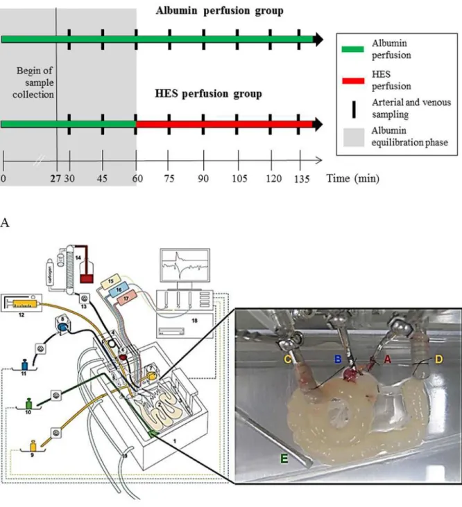
To evaluate the intestinal effects of a vascularly perfused HES solution, two experimental set-tings were established. The first group (“Albumin perfusion group”, control, N = 7) received a 135 min vascular perfusion with Albumin (3%) containing buffer. The second group (“HES perfusion group”, HES, N = 7) received a vascular Albumin (3%) perfusion for 60 min (equili-bration phase) followed by a HES 130/0.4 (3%; Fresenius Kabi, Bad Homburg, Germany) per-fusion for 75 min. Perper-fusions were continuously applied without intermittent stops. When establishing the mouse model of the isolated perfused intestine, various control experiments were performed to investigate the physiological and metabolic stability of the perfused intes-tines during the ex-vivo experiment. Employing the described experimental setup, mouse in-testines are physiologically and metabolically stable for up to 135 minutes. This was the main reason, why all experiments were performed for a maximum time period of 135 minutes in-cluding an equilibration phase of 60 minutes. To exclude influences of the perfusion time on the observed effects, the results obtained at a respective time point in the HES perfusion group were compared to the respective time point in the Albumin perfusion group (inter group com-parison) as well as to the“internal”HES control which consisted of 60 min equilibration with Albumin prior to the HES perfusion (intra group comparison;Fig. 1A).
Preparation of the small intestine, cannulation and perfusion
Mice were anesthetized by inhalation of 1–3% sevoflurane and an additional intraperitoneal in-jection of ketamine up to a maximum dose of 40 mg/kg. After opening the abdomen by a mid-line incision, further preparation steps were performed under a binocular microscope. Parts of the duodenum, the jejunum and ileum were isolated for the perfusion as described in detail for the rat [13]. All animals were killed under narcosis by cervical dislocation. The cannulation sys-tem used for perfusing the mouse intestine was based on our recently published rat model [14]. Minor modifications and adaptions were necessary due to animal size and anatomical differ-ences between the rat and mouse system (Fig. 1B). Briefly, we used a custom made perfusion system from Hugo Sachs Elektronik-Harvard Apparatus (March-Hugstetten, Germany) with modified cannulas and weight sensors. For perfusion, the aorta (in close proximity to the supe-rior mesenteric artery), the hepatic portal vein as well as the proximal and distal small intestine were cannulated (see inset inFig. 1B). For the vascular perfusion a modified Krebs-Henseleit solution containing 2 mM lactobionic acid, 7.4 mM glucose, 30 mM mannitol, 0.8 mM gluta-mine, 122μg/l norepinephrine hydrochloride, 12.6 mM HEPES, and 3% Albumin (BSA,
Fig 1. Experimental setting and basic components of the perfusion model.A) Experimental setting and time frame B) isolated perfused mouse small intestine apparatus. Using a custom made, heated chamber (1) an isolated small intestine (2) is perfused (vascular system: red, blue; luminal system: yellow; lymphatic system: green) while placed on a built-in microbalance (3). A moveable cannulating block (4) carries the tubings, heat exchanger cannula holders (5), and bubble trap (6). Height-adjustable reservoirs (7, 8) allow clamping both afterloads to zero. For online analysis of fluid homeostasis all emanating liquids are quantified by use of three balances (9 to 11). Constant flow perfusion is applied by a syringe pump (12) and a roller pump (13). The vascular perfusate is pH equilibrated, oxygenated and prewarmed with a tempered hollow fiber dialyzer flushed with carbogen gas (14). Pressure transducers (15–17) allow online detection of the luminal (yellow), venous (blue) and arterial (red) pressures. All data are recorded on a personal computer (18). To secure constant temperature, the chamber and cannulating block are water-jacketed and warmed by a water bath (19). The inset shows a representative photograph of a perfused small intestine. A, arterial cannula; B, venous cannula; C, oral intestinal lumen cannula; D, aboral intestinal lumen cannula; E, lymphatic suction needle; modified from [14]. A representative video presentation of the perfused small intestine is available as online supporting information (S1 Movie).
luminal perfusion pressures were recorded continuously. A blood gas analyser (ABL700, Radi-ometer Copenhagen, Bønshøj, Denmark) was used to measure O2and CO2partial pressures, pH, electrolytes, glucose and lactate of the arterial inflow and venous outflow every 15 minutes
(Fig. 1B). A representative perfused small intestine with typical peristaltic movements can be
seen in the supporting information provided online (S1 Movie).
Pre- and post- perfusion tissue preparation
One 3 cm long proximal portion of the small intestine was obtained before as well as after per-fusion and the mesentery was removed. The wet weight was determined immediately after de-pletion of intestinal liquid while for the evaluation of the dry weight, the same sample was dehumidified for 96 h at 55°C. Moreover, at the end of the perfusion experiment an approxi-mately 3 cm long portion of the intestine was fixed in 4% formaldehyde for
histological examination.
Evaluation of the vascular lactate-to-pyruvate ratio
The lactate-to-pyruvate ratio was employed as a parameter for anaerobic and aerobic metabo-lism. Pyruvate was determined in the venous outflow by a quantitative enzymatic photometric method (Sigma-Aldrich, Munich, Germany). Additionally, the vascular lactate concentration was evaluated using a blood gas analyser (ABL700, Radiometer Copenhagen, Bønshøj, Den-mark) and the lactate-to-pyruvate ratio was calculated.
Quantification of vascular, lymphatic and luminal FITC-dextran
FITC-dextran (150 kDa; Sigma-Aldrich, Munich, Germany) was added into the vascular per-fusate in a concentration of 40 mg/l to determine the endothelial and epithelial permeability for macromolecules. Under physiological conditions the endothelial and also the epithelial bar-rier is impermeable for the 150 kDa FITC-dextran [15]. Samples of venous, lymphatic, and lu-minal outflow were collected every 15 minutes and analysed for the FITC-dextran content using a fluorescence ELISA reader (excitation 485 nm, emission 530 nm; FL 600 microplate fluorescence Reader, MWG-Biotech, Ebersberg, Germany).
Determination of vascular galactose
In order to evaluate the resorptive capacity of the intestine, 30 mM of lactose were supplied with the luminal perfusion buffer and vascular galactose (derived from the luminal lactose) was determined by a commercially available assay kit (Raffinose/D-Galactose Assay Kit, Mega-zyme, Bray, Ireland). Due to initially high variations in galactose uptake during the equilibra-tion phase, statistical analyses were only performed with samples from time points 60 min and beyond.
Histological examination
Statistical analysis
Statistical analyses were performed using GraphPad Prism 5 (GraphPad Software, San Diego, USA). Data are presented as mean values with standard deviations (SD). Statistical compari-sons were performed using Student’s t-tests, one-way ANOVA (for intra group comparisons) and two-way ANOVA (for inter group comparisons) with Bonferroni post-tests. Differences were considered to be statistically significant if p was less than 0.05. Non-parametric data were analysed by Kruskal-Wallis test and Dunns post-test. For the two-way ANOVA data were transformed using [Y = log(Y)].
Results
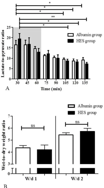
Quantification of the vascular lactate-to-pyruvate ratio
As a parameter for the metabolic status the vascular lactate-to-pyruvate ratio (LPR) was deter-mined and all values were within the physiological range of aerobic metabolism. There were no statistically significant differences between the time response characteristics of the LPR in the HES perfusion group and the time response characteristics of LPR in the Albumin perfusion group (p>0.05; inter group comparison;Fig. 2A). However, due to the equilibration of the
system, a time dependent reduction of LPR was observed in the HES and Albumin perfusion group [HESt30(19.36 ± 6.94) vs. HESt105(8.74 ± 2.21); HESt120(8.23 ± 2.75); HESt135(7.28 ± 1.99); p<0.05 for HESt105and HESt120, p<0.01 for HESt135; intra group comparison.
Albu-mint30(17.34 ± 4.94) vs. Albumint105(9.42 ± 1.42); Albumint120(8.74 ± 2.41); Albumint135 (9.11 ± 2.46); p<0.05 for all; intra group comparison].
Evaluation of the tissue wet-to-dry weight ratio
The wet-to-dry-weight ratio (W/d) was evaluated before (W/d1) and after (W/d2) the perfu-sion with HES and Albumin, respectively. No statistically significant differences were detected between the HES perfusion group and the Albumin perfusion group at the start or end of per-fusion (p>0.05;Fig. 2B).
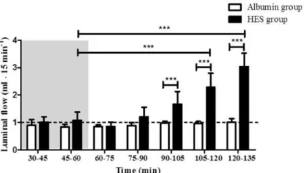
Vascular perfusion with HES leads to a fluid shift to the luminal
compartment
Vascular HES perfusion resulted in an alteration of fluid distribution and significantly increased luminal flow [HESt45–60(1.08 ± 0.29 ml15 min-1) vs. HESt105–120(2.28 ± 0.51 ml15 min-1); HESt120–135(3.04 ± 0.48 ml15 min-1); p<0.001 for all; intra group comparison;Fig. 3]. The
luminal flow in the Albumin perfusion group showed no changes during perfusion (p>0.05;
intra group comparison;Fig. 3). The luminal flow between the Albumin perfusion group and the HES perfusion group differed significantly [Albumint90–105(0.98 ± 0,06 ml15 min-1);
Al-bumint105–120(0.96 ± 0.08 ml15 min-1); Albumint120–135(1.01 ± 0.13 ml15 min-1) vs. HES
t90–105(1.66 ± 0.47 ml15 min-1); HESt105–120(2.28 ± 0.51 ml15 min-1); HESt120–135(3.04 ± 0.48 ml15 min-1); p<0.001 for all; inter group comparison;Fig. 3]. Control experiments with
Vascular perfusion with HES increases the permeability of the
endothelial and epithelial barrier
The endothelial and epithelial barrier integrity was estimated by determining the translocation of vascularly applied FITC-dextran into the luminal compartment. No changes in the luminal Fig 2. Lactate-to-pyruvate ratio and wet-to-dry weight ratio.A) Lactate-to-pyruvate ratio in the Albumin perfusion and HES perfusion group, B) wet-to-dry weight ratio before (W/d1) and after (W/d2) perfusion, in the Albumin perfusion and HES perfusion group. Areas shaded in grey indicate the equilibration phase. Error bars denote the mean±SD. Albumin (N = 6), HES (N = 7).*, p<0.05;**, p<0.01; ns, not significant.
FITC-dextran concentration were detected in the Albumin perfusion group (p>0.05; intra
group comparison;Fig. 4). Vascular perfusion with HES time-dependently increased the luminal FITC-dextran concentration [HESt45(2.04 ± 1.73 03BCgml-1); HESt60(1.57 ± 1.37μgml-1);
HESt75(1.14 ± 1.41μgml-1); HESt90(2.70 ± 3.39μgml-1) vs. HESt135(16.71 ± 5.17μgml-1); p<0.05 for t45 and t90, p<0.01 for t60, p<0.001 for t75; intra group comparison;Fig. 4].
Furthermore, compared to the Albumin perfusion group the luminal FITC-dextran concentra-tion was significantly increased in the HES perfusion group [Albumint105(0.49 ± 0.26μgml-1);
Albumint120(0.47 ± 0.27μgml-1); Albumint135(0.60 ± 0.37μgml-1) vs. HESt105(7.03 ± 6.41μgml-1); HESt120(12.24 ± 5.91μgml-1); HESt135(16.71 ± 5.17μgml-1); p<0.001 for all;
Fig 3. Luminal flow.Luminal effluent flow was measured in 15 minutes intervals. Areas shaded in grey indicate the equilibration phase. The default luminal flow rate is represented by a dashed line. Error bars denote the mean±SD. Albumin (N = 7), HES (N = 7).***, p<0.001.
doi:10.1371/journal.pone.0121497.g003
Fig 4. Luminal FITC-dextran concentrations.Luminal FITC-dextran concentrations were evaluated every 15 minutes and sufficient amounts of effluents for analyses were obtained at time point 45 min and beyond. Areas shaded in grey indicate the equilibration phase. Error bars denote the mean±SD. Albumin (N = 7), HES (N = 7).*, p<0.05;**, p<0.01;***, p<0.001.
inter group comparison;Fig. 4]. Control experiments with prolonged HES application (without equilibration phase) showed that the luminal FITC-dextran concentration reached the saturation point after 105 minutes of HES perfusion (data not shown). In addition, measurements of the lymphatic FITC-dextran concentrations also showed a significant increase during perfusion with HES (S1 Fig.). Note: The differences in the FITC-dextran concentrations between the HES and Albumin group during the equilibration period (Fig. 4; HES/Albumint45, HES/Albumint60) can be explained by inter-individual variations of the intestines used in the respective perfusion ex-periments. The respective intestines were however not excluded from the study as all other physi-ologic and metabolic parameters were unaltered and within the expected limits.
Vascular perfusion with HES alters the morphology of intestinal epithelial
cells
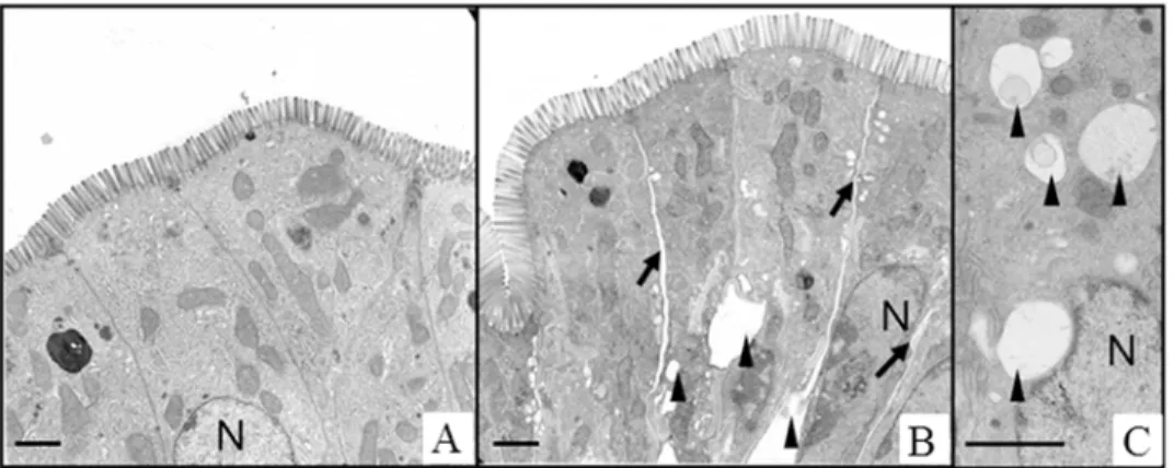
The histomorphological damage of the intestinal epithelium was analysed by employing a histological stability score [14]. No significant differences were detected between the HES perfusion group and the Albumin perfusion group (HES 0.59 ± 0.09 vs. Albumin 0.61 ± 0.08; p>0.05; data not shown). However, electron microscopic analyses suggested distinct cellular
and subcellular changes in the small intestine after HES perfusion. In the HES perfusion group expanded intercellular spaces were found within the intestinal epithelium and epithelial cells contained numerous intracellular vacuoles. These changes were not observed after Albumin perfusion (Fig. 5).
Vascular perfusion with HES impairs the resorptive capacity of the small
intestine
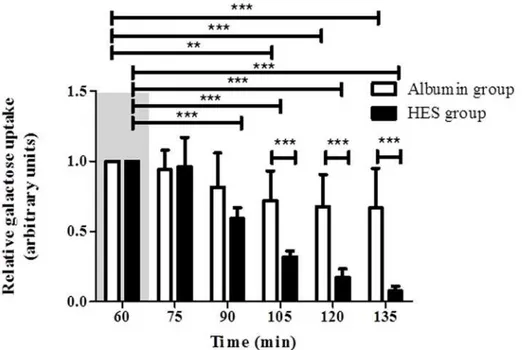
To validate the resorptive capacity and metabolic function of the isolated intestine during HES and Albumin perfusion, the luminal galactose uptake was measured. (LPR) Vascular perfusion with Albumin led to a time-dependent reduction of galactose uptake [Albumint60(set to 1) vs. Albumint105(0.72 ± 0.21); Albumint120(0.68 ± 0.23); Albumint135(0.67 ± 0.28); p<0.01 for t105, p<0.001 for t120 and t135; intra group comparison;Fig. 6]. A similar observation was
found for the perfusion with HES which caused an even faster reduction of luminal galactose uptake [HESt60(set to 1) vs. HESt75(0.96 ± 0.21); HESt90(0.59 ± 0.076); HESt105(0.32 ±
Fig 5. Ultrastructure of HES and Albumin perfused intestinal tissue.A) Albumin perfusion group, B and C) HES perfusion group. HES perfusion leads to widened intercellular spaces and appearance of intracellular vacuoles. Scale bars denote approximately 2μm. Black arrows, intercellular gaps; black arrowheads, vacuoles; N, nucleus.
0.04); HESt120(0.17 ± 0.06); HESt135(0.08 ± 0.03); p<0.001 for all; intra group comparison;
Fig. 6]. With increasing perfusion time, the galactose uptake differed significantly between
the Albumin perfusion group and the HES perfusion group [Albumint105(0.72 ± 0.21); Albumint120(0.68 ± 0.23); Albumint135(0.67 ± 0.28) vs. HESt105(0.32 ± 0.04); HESt120 (0.17 ± 0.06); HESt135(0.08 ± 0.03); p<0.001 for all; inter group comparison;Fig. 6].
Discussion
The application of hydroxyethyl starch (HES) for volume resuscitation in critically ill patients is controversially discussed and several recent clinical studies have suggested adverse effects of HES substitution, leading to increased patient mortality [1,3,18]. Although of high clinical relevance, up to now the effects of HES on intestinal function and barrier integrity are mainly unknown.
Ex-vivo model of the perfused intestine
Using a model of the isolated perfused mouse small intestine in combination with Krebs-Hen-seleit based perfusion media, we analysed the intestinal effects of a vascular perfusion with 3% HES in relation to a control perfusion with 3% Albumin. The employed system is a further de-velopment of our previously described model of the perfused rat intestine [14] and was de-signed to permit a detailed analysis of edema formation and permeability changes in the mouse intestine by monitoring fluid distributions and FITC-dextran transport from the vasculature to the interstitial space, the lymph and the lumen.
Effects of HES on intestinal barrier function
One of our main results is a significantly altered fluid shift from the intestinal vasculature into the lumen of the small intestine during HES perfusion, indicative of an increased endothelial Fig 6. Intestinal galactose uptake.The relative galactose uptake (t60= 1) was measured every 15 minutes.
Areas shaded in grey indicate the equilibration phase. Error bars denote the mean±SD. Albumin (N = 6), HES (N = 7).**, p<0.01;***, p<0.001.
and epithelial barrier permeability. Our flow-rate data showed an increased luminal flow while the venous flow was reduced under perfusion with HES, suggesting a loss of vascular fluid into the intestinal lumen due to negative effects of HES on endothelial and epithelial barrier integri-ty. These results were confirmed by our FITC-dextran measurements, where FITC-dextran of 150 kDa was supplemented to the vascular perfusion buffer. While in the Albumin perfusion group only very low concentrations of FITC-dextran appeared in the luminal effluents, HES perfusion resulted in a dramatic increase of luminal FITC-dextran levels, confirming the hy-pothesis that HES impairs the endothelial and epithelial barrier function in the small intestine. Our results are also in accordance with data published by Gao et al. who employed a rat model of hemorrhagic shock and detected an initially higher intestinal bacterial translocation to the liver after HES resuscitation compared to Ringer’s solution [19]. A study by Li et al investigated the effects of different HES concentrations on ischemia-reperfusion injury of rat intestinal trans-plantations showing that high-concentration HES solutions (>3%) resulted in
histomorpholo-gical damage and were not appropriate for intestinal preservation [20]. Employing an isolated perfused guinea pig heart model, Jacob et al. demonstrated a post-ischemic transient increase in vascular leak with Krebs-Henseleit buffer containing 6% HES 130/0.4 but not with 5% Albumin [21]. Interestingly, the authors also showed that augmenting the perfusion buffer with human Albumin improved the endothelial integrity after ischemia by protecting the endothelial glyco-calyx [22]. In contrary to the above mentioned studies that suggest negative effects of HES on endothelial and epithelial barrier function, using a rat model of hemorrhagic shock Wang et al. demonstrated that HES resuscitation is able to reduce intestinal permeability [23] and that LPS-and sepsis induced capillary permeability is decreased after vascular HES perfusion [24,25] pointing towards possible protective effects of a perfusion with HES under hemorrhagic and/or septic conditions, in which endothelial permeability may already be increased.
Effects of HES on intestinal morphology
Although the clinical effects of a perfusion with HES are well described, the detailed mecha-nisms of HES on epithelial cells are still mainly unknown. Our electron microscopic experi-ments revealed numerous of intracellular vacuoles and enlarged intercellular spaces in the HES perfusion group, while these signs were absent after Albumin perfusion. Several authors tected similar structures in cells and classified them as HES containing vesicles that might be de-rived from lysosomes [26–28]. Others showed that the amount of vacuoles increased
concomitantly with the dosage of HES used for perfusion [29]. Our findings of enlarged inter-cellular spaces after HES perfusion might be explained by an accumulation of interstitial fluids. HES molecules are able to overcome the endothelial barrier and reside in the interstitial com-partment of the respective tissue, where they are stable for several years [28,29]. As the HES molecule is osmotically highly active and is able to bind large amounts of water [30], the fluid shift from the vascular to the interstitial compartment might be increased, leading to the development of a transient interstitial edema. As a consequence of the increased tissue pressure, intercellular spaces in the intestinal epithelium expand and damage of tight junctions with in-creased fluid shift into the intestinal lumen may result. A similar morphological observation was also reported by Jacob et al. employing an isolated perfused heart model. Interstitial spaces with-in the cardiac tissue were more dense with-in the Albumwith-in perfusion group than after perfusion with HES, pointing towards an interstitial fluid accumulation in the HES group [21].
Effects of HES on the resorptive capacity of the intestine
membrane is achieved by an uniporter (GLUT2) [31]. Our galactose uptake measurements in which 30 mM of lactose were supplied with the luminal perfusion buffer suggest several direct or indirect HES mediated mechanisms, which could function alone or in combination to im-pair galactose absorption in the intestine: (i) HES attenuates the digestion of lactose into galac-tose by inhibiting intestinal lactase activity and/or by reducing lacgalac-tose concentration due to the increased fluid shift into the lumen (ii) HES impairs the uptake of galactose by intestinal epi-thelial cells via an interaction with SGLT1 (iii) HES negatively influences the transfer of galac-tose to the vascular compartment by inhibition of GLUT2. As we do not have sufficient biochemical data that support one the above mentioned mechanisms of HES action we cannot exclude the possibility that the attenuation of intestinal galactose uptake and metabolic func-tion is rather and simply a consequence of the HES mediated deranged barrier integrity and disturbed tissue homeostasis than a direct effect of HES on the responsible transporter mole-cules and enzymes.
Limitations of the study
The employed model of the isolated perfused small intestine offers the unique possibility to in-vestigate organ-specific effects of a vascular perfusion with HES, while at the same time inter-fering systemical effects of HES are excluded. However, the following points should be
considered when interpreting the results of our study. (I) A single pass application of a cell free vascular perfusate was performed in our study. Therefore, the model does not account for pos-sible effects of systemic immune cells and humoral factors on the described HES and Albumin mediated mechanisms. (II) The perfusate used in our system was deficient in plasma proteins including coagulation factors, which may have an influence on the endothelial barrier function. (III) Possible effects mediated by the splanchnic nerves are not reflected in the perfused model of the small intestine, as the innervation was disrupted during organ preparation. However, the autonomously working enteric nervous system is still functioning as can be seen by the regular peristaltic movements of the perfused intestine in our model (S1 Movie).
Conclusion
Taken together, we show that a vascular perfusion with clinically relevant concentrations of HES impairs the endothelial and epithelial barrier integrity as well as metabolic function of the small intestine. Our ex-vivo experiments extend recent studies that suggested negative effects of HES during fluid resuscitation and we propose the small intestine as a potential target for HES mediated adverse effects.
Supporting Information
S1 Fig. Relative venous flow.Venous flow was measured in 15 minutes intervals (t30–45 = 1). Areas shaded in grey indicate the equilibration phase. Bars denote the mean ± SD. Albumin (N = 7), HES (N = 7)., p<0.01;, p<0.001.
(TIF)
S2 Fig. FITC-dextran concentration.Lymphatic FITC-dextran concentrations were evaluated every 15 minutes. Areas shaded in grey indicate the equilibration phase. Bars denote the mean ± SD. Albumin (N = 7), HES (N = 7)., p<0.05.
(TIF)
Acknowledgments
The authors are indebted to K. Masuhr, C. Schildhauer and J. Sarau for technical assistance as well as N. Speck for language proofreading of the manuscript.
Author Contributions
Conceived and designed the experiments: YLW IL MA NW IF TK HD. Performed the experi-ments: YLW IL TG TK. Analyzed the data: YLW IL KZ MA. Wrote the paper: YLW IL KZ BB IF MS NW MA.
References
1. Myburgh JA, Finfer S, Bellomo R, Billot L, Cass A, Gattas D, et al. Hydroxyethyl starch or saline for fluid resuscitation in intensive care. N Engl J Med. 2012; 367(20):1901–11. Epub 2012/10/19. doi:10.1056/ NEJMoa1209759PMID:23075127
2. Finfer S, Liu B, Taylor C, Bellomo R, Billot L, Cook D, et al. Resuscitation fluid use in critically ill adults: an international cross-sectional study in 391 intensive care units. Crit Care. 2010; 14(5):R185. Epub 2010/10/19. doi:10.1186/cc9293PMID:20950434
3. Perner A, Haase N, Guttormsen AB, Tenhunen J, Klemenzson G, Aneman A, et al. Hydroxyethyl starch 130/0.42 versus Ringer's acetate in severe sepsis. N Engl J Med. 2012; 367(2):124–34. Epub 2012/06/ 29. doi:10.1056/NEJMoa1204242PMID:22738085
4. Rochwerg B, Alhazzani W, Sindi A, Heels-Ansdell D, Thabane L, Fox-Robichaud A, et al. Fluid Resus-citation in Sepsis: A Systematic Review and Network Meta-analysis. Annals of internal medicine. 2014. Epub 2014/07/23.
5. Podolsky DK. Mucosal immunity and inflammation. V. Innate mechanisms of mucosal defense and re-pair: the best offense is a good defense. Am J Physiol. 1999; 277(3 Pt 1):G495–9. Epub 1999/09/14. PMID:10484372
6. Feng X, Hu Y, Ding J, Ge Y, Song J, Ai Q, et al. Early treatment with hydroxyethyl starch 130/0.4 causes greater inhibition of pulmonary capillary leakage and inflammatory response than treatment instituted later in sepsis induced by cecal ligation and puncture in rats. Ann Clin Lab Sci. 2007; 37(1):49–56. PMID:17311869
7. Sun Z, Wang X, Andersson R. Role of intestinal permeability in monitoring mucosal barrier function. History, methodology, and significance of pathophysiology. Dig Surg. 1998; 15(5):386–97. Epub 1998/ 12/09. PMID:9845620
8. Feng X, Liu J, Yu M, Zhu S, Xu J. Protective roles of hydroxyethyl starch 130/0.4 in intestinal inflamma-tory response and survival in rats challenged with polymicrobial sepsis. Clin Chim Acta. 2007; 376(1– 2):60–7. Epub 2006/09/01.
9. Sun ZW, Wang XD, Deng XM, Wallen R, Gefors L, Hallberg E, et al. The influence of circulatory and gut luminal challenges on bidirectional intestinal barrier permeability in rats. Scand J Gastroenterol. 1997; 32(10):995–1004. Epub 1997/11/15. PMID:9361172
10. Blikslager AT, Roberts MC, Young KM, Rhoads JM, Argenzio RA. Genistein augments prostaglandin-induced recovery of barrier function in ischemia-injured porcine ileum. Am J Physiol Gastrointest Liver Physiol. 2000; 278(2):G207–16. Epub 2000/02/09. PMID:10666044
11. Kimberger O, Arnberger M, Brandt S, Plock J, Sigurdsson GH, Kurz A, et al. Goal-directed colloid ad-ministration improves the microcirculation of healthy and perianastomotic colon. Anesthesiology. 2009; 110(3):496–504. Epub 2009/02/20. doi:10.1097/ALN.0b013e31819841f6PMID:19225390
12. Lobo SM, Orrico SR, Queiroz MM, Contrim LM, Cury PM. Comparison of the effects of lactated Ringer solution with and without hydroxyethyl starch fluid resuscitation on gut edema during severe splanchnic ischemia. Braz J Med Biol Res. 2008; 41(7):634–9. Epub 2008/08/23. PMID:18719746
13. Monchik GJ, Russell PS. Transplantation of small bowel in the rat: technical and immunological consid-erations. Surgery. 1971; 70(5):693–702. PMID:4399244
14. Lautenschlager I, Dombrowsky H, Frerichs I, Kuchenbecker SC, Bade S, Schultz H, et al. A model of the isolated perfused rat small intestine. Am J Physiol Gastrointest Liver Physiol. 2010; 298(2):G304–13. doi:10.1152/ajpgi.00313.2009PMID:19910525
15. Hulstrom D, Svensjo E. Intravital and electron microscopic study of bradykinin-induced vascular perme-ability changes using FITC-dextran as a tracer. J Pathol. 1979; 129(3):125–33. PMID:529011
17. Bye WA, Allan CH, Trier JS. Structure, distribution, and origin of M cells in Peyer's patches of mouse ileum. Gastroenterology. 1984; 86(5 Pt 1):789–801. Epub 1984/05/01. PMID:6706062
18. Haase N, Perner A. Is hydroxyethyl starch 130/0.4 safe? Crit Care. 2012; 16(2):116. Epub 2012/03/13. doi:10.1186/cc11200PMID:22405319
19. Gao XY, Zhou Q, Wu CY, Pang QF, Cao JL, Zeng YM. [Comparison of effects of two kinds of fluid for re-suscitation on bacterial translocation and inflammation of small intestine in rats with hemorrhagic shock]. Zhongguo Wei Zhong Bing Ji Jiu Yi Xue. 2006; 18(3):146–9. Epub 2006/03/10. PMID: 16524504
20. Li XL, Zou X, Nie G, Song ML, Li G, Cui W. Role of hydroxyethyl starch in ischemia-reperfusion injury in rat intestinal transplantation. Transplantation proceedings. 2013; 45(6):2491–6. Epub 2013/08/21. doi: 10.1016/j.transproceed.2013.02.133PMID:23953568
21. Jacob M, Bruegger D, Rehm M, Welsch U, Conzen P, Becker BF. Contrasting effects of colloid and crys-talloid resuscitation fluids on cardiac vascular permeability. Anesthesiology. 2006; 104(6):1223–31. PMID:16732094
22. Jacob M, Paul O, Mehringer L, Chappell D, Rehm M, Welsch U, et al. Albumin augmentation improves condition of guinea pig hearts after 4 hr of cold ischemia. Transplantation. 2009; 87(7):956–65. Epub 2009/04/09. doi:10.1097/TP.0b013e31819c83b5PMID:19352113
23. Wang P, Li Y, Li J. Protective roles of hydroxyethyl starch 130/0.4 in intestinal inflammatory response and oxidative stress after hemorrhagic shock and resuscitation in rats. Inflammation. 2009; 32(2):71–82. Epub 2009/02/25. doi:10.1007/s10753-009-9105-7PMID:19238531
24. Tian J, Lin X, Guan R, Xu JG. The effects of hydroxyethyl starch on lung capillary permeability in en-dotoxic rats and possible mechanisms. Anesth Analg. 2004; 98(3):768–74, table of contents. PMID: 14980935
25. Lv R, Zhou W, Chu C, Xu J. Mechanism of the effect of hydroxyethyl starch on reducing pulmonary cap-illary permeability in a rat model of sepsis. Ann Clin Lab Sci. 2005; 35(2):174–83. PMID:15943182 26. Parth E, Jurecka W, Szepfalusi Z, Schimetta W, Gebhart W, Scheiner O, et al. Histological and
immu-nohistochemical investigations of hydroxyethyl-starch deposits in rat tissues. Eur Surg Res. 1992; 24 (1):13–21. PMID:1375158
27. Sirtl C, Laubenthal H, Zumtobel V, Kraft D, Jurecka W. Tissue deposits of hydroxyethyl starch (HES): dose-dependent and time-related. Br J Anaesth. 1999; 82(4):510–5. PMID:10472213
28. Jurecka W, Szepfalusi Z, Parth E, Schimetta W, Gebhart W, Scheiner O, et al. Hydroxyethylstarch de-posits in human skin—a model for pruritus? Arch Dermatol Res. 1993; 285(1–2):13–9. Epub 1993/01/ 01.
29. Stander S, Szepfalusi Z, Bohle B, Stander H, Kraft D, Luger TA, et al. Differential storage of hydro-xyethyl starch (HES) in the skin: an immunoelectron-microscopical long-term study. Cell Tissue Res. 2001; 304(2):261–9. PMID:11396719
30. Wiedermann CJ, Joannidis M. Accumulation of hydroxyethyl starch in human and animal tissues: a sys-tematic review. Intensive Care Med. 2014; 40(2):160–70. Epub 2013/11/22. doi: 10.1007/s00134-013-3156-9PMID:24257970