Arq Neuropsiquiatr 2008;66(3-A):563-565
563
Clinical / Scientiic note
INTERNAL CAROTID ARTERY BLOOD
BLISTER-LIKE ANEURYSM
Joacil Carlos da Silva
1, Igor Vilela Faquini
2, Matheus Augusto Pinto Kitamura
2,
Hildo Rocha Cirne de Azevedo-Filho
1ANEURISMA DA CARóTIDA INTERNA NãO RELACIONADA A RAMO pERfURANTE
Restauração Hospital, Recife PE, Brazil: 1Neurosurgeon, 2Neurosurgery Resident.
Received 13 February 2008, received in inal form 17 Abril 2008. Accepted 17 May 2008.
Dr. Joacil Carlos da Silva – Rua Agenor Lopes 424/701 - 51021-110 Recife PE - Brasil. E-mail: [email protected] Aneurysms located at nonbranching sites in the
supra-clinoid internal carotid artery (ICA) were originally charac-terized, in 1986, as aneurysms protruding from the dorsal wall of the ICA1, but they are also known as blood blister-like aneurysms (BBLA)2 or ICA anterior wall aneurysms3. They are rare, comprising 0.9 to 6.5% of all ICA aneurysms3.
These lesions are fairly small, sometimes appearing as just a protrusion of the vessel wall (expressed as being “blood blister-like”). They grow rapidly in a short time and easily rupture, especially during surgery. Eventually they cannot be eliminated easily by the ordinary clipping pro-cedure requiring wrapping or encircling methods4-6.
Curiously these aneurysms are more often reported by oriental authors1-5, 7-9. We will present a 37-year male Bra-zilian case successfully treated with clip application.
CASE
A 37-year-old man presented with sudden onset of severe headache, vomiting and briely consciousness disturbance. Ex-amination revealed mild meningismus, but no focal neurological deicits. A CT scan of the brain revealed thick and diffuse sub-arachnoid hemorrhage (SAH) involving the carotid cistern and left sylvian issure. The angiogram revealed a small irregularity of the ventral left supraclinoid artery suspicious for a BBLA.(Fig 1)
We performed a left pterional craniotomy, with micro dis-section and exposure of the optic nerve and the ICA. The lateral issure was widely opened allowing visualization of ICA bifurca-tion, middle and anterior cerebral arteries. There were signs of hemorrhage in the carotid cistern and sylvian issure. The clot was carefully dissected away from the optic nerve and ICA in order to approach the involved segment. The aneurysm resem-bled a focal dissection, with no emerging vessels in neighbor-hood. Parallel application of a titanium straight angle clip was performed without temporary clipping of ICA. Care was taken to extend the clip beyond the aneurysm margins with minimal ICA stenosis (Figs 2 and 3).
The post-operative period was uneventful and the patient was discharged after control angiogram which revealed com-pletely exclusion of the aneurysm and ICA patency (Fig 4).
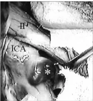
Fig 2. Microphotograph. Microdissection showing the blood blister-like aneurysm located at a nonbranching region of ICA. Note the close relationship with optical nerve. II, left optical nerve; ICA, left internal carotid artery; *aneurysm.
Arq Neuropsiquiatr 2008;66(3-A)
564
Internal carotid artery Silva et al.
DISCUSSION
BBLA have some unique characteristics that make dis-tinction from the common saccular or berry aneurysms1-4,6-8. Rhoton introduced three rules related to the anatomy of saccular aneurysms that should be considered when planning the operative approach to these lesions. First, these aneurysms arise at a branching site on the parent artery. Second, saccular aneurysms arise at a turn or curve in the artery. Saccular aneurysms arise on the convex, not concave, side of the curve. Third, saccular aneurysms point in the direction that the blood would have gone if the curve at the aneurysm site were not present10.
BBLA contradict the irst Rhoton’s rule as they are fre-quently located at the dorsal or anterior wall of ICA, a nonbranching region3.
The histological characteristics of BBLA include fo-cal wall defects covered with clot and ibrous tissue. The focal wall defects may be the result of laceration of the ICA wall caused by ulceration and penetration into the internal elastica lamina, resulting from arteriosclerosis.
The walls of BBLA are composed of only normal
adven-titia, in an abrupt transformation from the sclerotic ICA wall. In contrast, ordinary berry aneurysms are formed by thickened intima and/or adventitia, with rich collagen and inlammatory cells, that is continuous with the three-lay-ered arterial wall2,3.
Therefore, arteriosclerosis alone may not explain the causative mechanism for BBLA. Associated hypertension, ulceration resulting from arteriosclerosis, arterial dissec-tion and hemodynamic stress may also inluence the oc-currence of these aneurysms1,3.
Dissection of the ICA was often observed in patients with BBLA but not in patients with saccular-type aneu-rysms, suggesting the presence of speciic pathological features in BBLA. Hemodynamic stress is thought to initi-ate berry aneurysms and may also inluence the occur-rence of BBLA1-4.
The treatment options include direct lesion clipping6, wrapping or encircling techniques4,8 and endovascular oblit-eration9 of the ICA with or without bypass procedures.3 The main surgical problem is the extremely fragile na-ture of the walls and the usually large defect in the artery. The direct clip application could lead to avulsion of the an-eurysm or to obliteration of the parent vessel. Reconstruc-tion techniques may involve direct suture5, Sundt clip appli-cation6, vascular titanium clamps and bypass procedures3. Parallel clipping of the ICA (including the normal ar-terial wall beyond the lesion), under decreased pressure produced by temporary clamping of the cervical ICA, is recommended, to avoid breakage of the transitional zone
Fig 3. Microphotograph. Parallel clipping of the ICA was possible ex-tending the blades of the clip beyond arterial wall defect. II, left op-tical nerve; ICA, left internal carotid artery.
Fig 4. Post operative carotid angiogram. Note absence of ICA steno-sis. ICA, left internal carotid artery; A1, left anterior cerebral artery; M1, left middle cerebral artery.
between the normal ICA and the aneurysm wall3. Another
clip option is the Sundt encircling clip developed for di-rect reconstruction of ICA6.
Arq Neuropsiquiatr 2008;66(3-A)
565 Internal carotid artery Silva et al.
While avoiding these dangerous lesions, proximal ICA occlusion by endovascular or surgical means may be a treatment option, but this does not eliminate rebleeding risk posed by retrograde low1,3.
Technically, the endovascular occlusion of focal ICA segments is not easy9. Unlike aneurysm clips, the precise and stable placement of endovascular devices such as coils and balloons is dificult. For compact and precise trapping, balloons may be preferred to coils, but ICA BBLA are very fragile and usually do not have uniform conigurations that accommodate balloons. In a small series of six patients endovascular treated, there was regrowth in all cases7,9.
In conclusion, the blood blister-like aneurysms are a rare but important cause of subarachnoid hemorrhage. The early recognition of these lesions is crucial to adequate treatment planning. Neurosurgeons should attempt to the fragility and extension of arterial wall defect as well to the rather elevated risks of regrowth and rebleeding.
Nowadays, microsurgery with arterial wall reconstruc-tion (direct clip or encircling techniques) is preferable to endovascular treatment.
REfERENCES
1. Nakagawa F, Kobayashi S, Takemae T, Sugita K. Aneurysms protrud-ing from the dorsal wall of the internal carotid artery. J Neurosurg 1986; 65:303-308.
2. Ishikawa T, Nakamura N, Houkin K, Nomura M. Pathological consid-eration of a “blister-like” aneurysm at the superior wall of the internal carotid artery: case report. Neurosurgery 1997;40:403-406.
3. Ogawa A, Suzuki M, Ogasawara K. Aneurysms at nonbranching sites in the supraclinoid portion of the internal carotid artery: internal ca-rotid artery trunk aneurysms. Neurosurgery 2000;47:578-586. 4. Kubo Y, Ogasawara K, Tomitsuka N, Otawara Y, Watanabe M, Ogawa A.
Wrap-clipping with polytetraluoroethylene for ruptured blisterlike an
-eurysms of the internal carotid artery. J Neurosurg 2006;105:785-787. 5. Joo S, Kim T, Moon K, et al. Arterial suturing followed by clip
reinforce-ment with circumferential wrapping for blister-like aneurysms of the internal carotid artery. Surg Neurol 2006;66:424-429.
6. Sekula R, Cohen D, Quigley M, Jannetta P. Primary treatment of a blis-ter-like aneurysm with an encircling clip graft: technical case report. Neurosurgery 2006; 59[ONS Suppl 1]:ONS-168.
7. Tanoue S, Kyosue H, Matsumoto S, Yamashita M, Nagatomi H, Mori H. Ruptured “blisterlike” aneurysm with a pseudoaneurysm forma-tion requiring delayed intervenforma-tion with endovascular coil emboliza-tion. J Neurosurg 2004;101:159-162.
8. Yanaka K, Meguro K, Nose T. Repair of a tear at the base of a blisterlike aneurysm with suturing and an encircling clip: technical note. Neuro-surgery 2002;50:218-221.