Arq Neuropsiquiatr 2007;65(3-B):845-847
SCHISTOSOMIASIS MANSONI PRESENTING
AS A CEREBELLAR TUMOR
Case report
Joacil Carlos da Silva, Frederico de Melo Tavares de Lima,
Cláudio Henrique Vidal, Hildo Cirne Rocha de Azevedo Filho
ABSTRACT - The Manson’s schistosomiasis tumoral form rarely affects the brain. There are only 12 cases prior related with a mean age of 25 years and a male predominance. We describe a 16-year-old Brazilian Northeastern boy with a cerebellar mass lesion. The radiological aspect was considered compatible with glioma and a gross total resection was performed. Microscopic examination disclosed intraparenchymal granulomas surrounding Schistosoma mansoni eggs. The case is compared with the literature findings and some peculiar aspects of this trematode infection are reviewed.
KEY WORDS: schistosomiasis, tumor, cerebellum.
Esquistossomose mansônica apresentando-se como tumor cerebelar: relato de caso
RESUMO - A forma pseudotumoral da esquistossomose mansônica raramente acomete o encéfalo. Apenas 12 casos foram previamente descritos com idade média de 25 anos e predominância do sexo masculino. Relatamos um caso em paciente do nordeste brasileiro de 16 anos portador de lesão expansiva cerebelar. O aspecto radiológico foi considerado compatível com tumor cerebelar primário e ressecção radical reali-zada. Estudo histopatológico demonstrou granulomas intraparenquimatosos contornando ovos de Schis-tosoma mansoni. O caso é comparado com os achados da literatura e aspectos peculiares dessa neuropa-sitose são discutidos.
PALAVRAS-CHAVE: esquistossomose, tumor, cerebelo.
Department of Neurosurgery, Restauração Hospital, Recife PE, Brazil.
Received 8 February 2007, received in final form 15 May 2007. Accepted 27 June 2007.
Dr. Joacil Carlos da Silva - Rua Agenor Lopes 424 / 701 - 51021-110 Recife PE - Brasil. E-mail: joacil_carlos@hotmail.com
Schistosomiasis is one of the most common para-site infections. Approximately 200 million people are carriers and another 600 million are exposed to the risk of infection in Latin America, Africa and Asia. This metazoal infection is caused by trematodes of the
ge-nus Schistosoma and three species cause significant
disease in humans: Schistosoma mansoni (endemic to countries in South America, central and southern Af-rica and Saudi Arabi); S. haematobium (distributed throughout Africa and Middle East); and S. japonicum
(restricted to Japan, China and southeast Asia). Hu-mans are the definitive host, althought aquatic snails act as the intermediate host. Adult parasites live in the mesenteric (S. mansoni and S. japonicum) or pel-vic (S. haematobium) veins and females of this genus lay hundreds of eggs per day, which are excreted in human urine or feces. The most commom urinary or intestinal symptoms are caused by a localized inflam-matory response surrounding these eggs; other
or-gans such as the lungs, liver, and central neurvous sys-tem (CNS) may be involved by hematogenous dissem-ination of the eggs or by migration of adult worms to ectopic locations1-5. CNS clinical involvement is
un-commom, cerebral lesions most frequently occur in cases of Japanese schistosomiasis6, whereas the
oth-er two types (Manson’s and haematobium) may in-volve the spinal cord5
. The cerebral or cerebellar tu-moral form of schistosomiasis mansoni is extremely rare, there are only 12 cases prior related1-5,7-10
. We describe a curious case in which Manson’s schistosomiasis simulated a posterior fossa tumour.
CASE
We received the informed consent from the patient’s family permitting this publication.
846 Arq Neuropsiquiatr 2007;65(3-B)
and were not associated with visual scintillation, position, movement, nausea or vomiting. They were occuring dai-ly and were not relieved by administration of commom analgesics. The patient’s medical history disclosed no rel-evant findings.
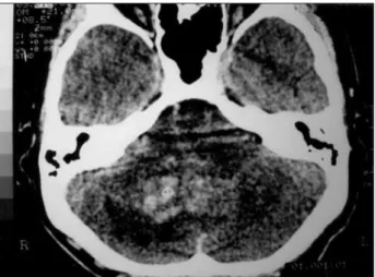
Examination – The patient appeared to be a healthy young boy. General physical examination was normal. Neu-rological examination showed slight gait ataxia and inco-ordination of the movements of the right limbs. Computer tomography (CT) scan showed a heterogeneus, hyperdense area, of irregular borders, enhanced by intravenous con-trast, in the right cerebellar hemisphere (Fig 1).
Surgery – The lesion was considered compatible with glioma and suboccipital craniotomy with gross total resec-tion was performed. The “tumour” infiltrated the cerebel-lar cortex and white matter. It was bloodless and the con-sistency was slightly more firm than normal tissue.
Histological findings – Microscopic analysis showed sec-tions of leptomeninges and of the cerebellum cortex and white matter containing numerous S. mansoni eggs involved in chronic granulomatous inflammatory reaction (Fig 2).
Postoperative evolution – The patient was submitted to chemotherapy with oxamniquine. His symptoms grad-ually improved.
DISCUSSION
Neurological complications arising from schistoso-miasis are uncommom and was initially described in 1899 by Yamagiva. This author performed an autopsy
in a carrier of Katayama disease who suffered focal seizures and identified a brain granuloma11.
Symptomatic brain involvement is most commom in cases of Japanese schistosomiasis. Eventually, en-cephalopathy or encephalitis develop during the acute stage of the infection, referred to as “Kata-yama fever”. In chronic disease, raised intracranial pressure and focal signs or seizures can result from granulomatous inflammation. Neuroschistosomiasis caused by S. haematobium and S. mansoni infections usually involve the spinal cord with multiple granu-lomas or a focal granulomatous mass that cause lum-bosacral transverse myelitis2,3,5
.
Fig 1. CT scan disclosing a cerebellar heterogeneous and ir-regular lesion.
Arq Neuropsiquiatr 2007;65(3-B) 847
It is curious that clinical brain involvement do not develop more frequently in patients with Manson’s schistosomiasis. Some autopsy studies provided his-tological evidence of cerebral and cerebellar ecto-pic eggs location in infected individuals, particularly those with the hepatosplenic form of the disease as-sociated with pulmonary hypertension5,9,10. The lack
of neurological symptoms in such cases may be ex-plained by the small ammount of eggs that are dis-seminated over a wide area and are often associated with only mild inflammation. Symptoms are more likely to occur when a large number of eggs is con-centrated in one area and the associated granuloma-tous inflammation produces a tumorlike mass2. The
tumoral form of cerebellar schistosomiasis mansoni, as it is observed in our case and the twelve other in the literature, showed not to be associated with oth-er sevoth-ere forms of viscoth-eral involvement by S. manso-ni. Probably, we may expect that those patients were carriers of mild intestinal or hepatointestinal forms.
It must be discussed how the S. mansoni ova reach the brain. There are two possibilities: the eggs may be carried into the brain through the arterial system or through the retrograde venous path, or in other way, it is possible an anomalous migration of adult worms and local deposition of ova. The first mech-anism occurs in carriers of the hepatosplenic form of schistosomiasis, in which eggs distributed in the brain and surrounded by inflammatory reaction were found in 26% of autopsy cases. This finding leads to the conclusion that cerebral Manson’s schistosomiasis is quite commom, although it is not clinically relevant in the majority of cases. The hypothesis of arterial embolization supposes the presence of either pre-existing or disease-related pulmonary arteriovenous shunts resulting of organizing thombi in the pulmo-nary circulation causing pulmopulmo-nary arterial hyperten-sion. Another possible path for the arterial embo-lization would be related to the portal-pulmonary anastomoses through the azygos veins facilitade by the presence of portal hypertension. The local depo-sition of eggs after migration of adult worms is sug-gested by two facts: worms ares occasionally found inside the leptomeningeal veins and eggs grouped in the same area are found1,4,5
.
The main region involved in the tumoral form is the vertebro-basilar circulation territory. This fact could be explained by the anomalous migration hy-pothesis: a local deposition of eggs after the adult worm reach the CNS through venous anastomoses
between brain, spinal cord and the portal system by the Batson’s venous vertebral plexus, probably the portal hypertension facilitates this migration2-12
.
The majority of Manson’s schistosomiasis tumoral cases, as in our report, were young adult males (9 males, 3 females, mean age 25 years) who had previ-ously been healthy. These patients typically present-ed within three months of the onset of neurological symptoms, the most commom being headache, focal signs and seizures2,4,5,7-10,12
.
The ideal management is a controversial issue be-cause this is an extremely rare and almost unexpect-ed diagnostic. Patients who were treatunexpect-ed with com-plete surgical resection and antihelmintic medica-tion tended to have a good outcome, whereas those who underwent partial resection or biopsy more of-ten had residual or recurrent symptoms despite an-tihelmintic therapy. It is not predictable if there is a possibility of clinical remission without gross ressec-tion2,4,5,7-10,12.
In conclusion, Manson’s schistosomiasis must be considered in the differential diagnosis when a young male patient from an endemic region pres-ents with a brain mass lesion in the posterior circu-lation territory. There is evidence, from the previous reported cases, of a beneficial effect of a aggressive surgical resection followed by specific chemothera-py with praziquantel or oxamniquine.
REFERENCES
1. Gonçalves E, Fonseca A, Pitella J. Frequency of schistosomiasis man-soni, of its clinicopathological forms and of the ectopic locations of the parasite in autopsies in Belo Horizonte, Brazil. J Trop Med Hyg 1995; 98:289-295.
2. Mackenzie I, Guha A. Manson’s schistosomiasis presenting as a brain tumor. J Neurosurg 1998;89:1052-1054.
3. Pitella J. Neuroschistosomiasis. Brain Pathol 1997;7:649-662. 4. Pitella J, Gusmão S, Carvalho G. Tumoral form of cerebral
schistosomi-asis mansoni: a report of four cases and a review of the literature. Clin Neurol Neurosurg 1996;98:15-20.
5. Pitella J, Lana-Peixoto M. Brain involvement in hepatosplenic schisto-somiasis mansoni. Brain 1981;104:621-631.
6. Greenield J, Pritchard B. Cerebral infection with Schistosoma japoni -cum. Brain 1937;60:361-372.
7. Bambirra E, Andrade J, Cesarini I. The tumoral form of schistosomia-sis: report of a case with cerebellar involvement. Am J Trop Med Hyg 1984;33:76-79.
8. Cabral G, Pitella J. Tumoral form of cerebellar schistosomiasis mansoni: report of a surgically treated case. Acta Neurochir (Wien) 1989;99:148-151.
9. Gjerd I, Mörk S, Larsen J. Cerebral schistosomiasis presentin as a brain
tumour. Eur Neurol 1984;23:229-236.
10. Scrimgeour E, Gajdusek D. Involvement of the central nervous system
in schistosoma mansoni and S. haematobium infection: a review. Brain
1985;108:1023-1038.
11. Faust E. An inquiry into the ectopic lesions in schistosomiasis. Am J Trop Med 1948;28:175-181.
