SYNDROMES RELATED TO SODIUM AND
ARGININE VASOPRESSIN ALTERATIONS
IN POST-OPERATIVE NEUROSURGERY
Ana P.D. Cardoso
1, Desanka Dragosavac
1, Sebastião Araújo
1, Antonio L.E. Falcão
1,
Renato G.G. Terzi
1, Margaret de Castro
2, Fabiana G. Marcondes
1, Taís G. Melo
1,
Rosmari A.R.A. Oliveira
1, Eliana A. Cintra
1ABSTRACT - Background: Cerebral salt wasting syndrome (CSWS), syndrome of inappropriate antid-iuretic hormone secretion (SIADH) and diabetes insipidus (DI) are frequently found in postoperative neurosurgery. Purpose: To identify these syndromes following neurosurgery. Method: The study in-cluded 30 patients who had been submitted to tumor resection and cerebral aneurysm clipping. Sodium levels in serum and urine and urine volume were measured daily up to the 5th day following surgery. Plas-ma arginine vasopressin (AVP) was measured on the first, third and fifth days post-surgery. Results: CSWS was found in 27/30 patients (90%), in 14 (46.7%) of whom it was associated with a reduction in the levels of plasma AVP (mix syndrome). SIADH was found in 3/30 patients (10%). There was no difference between the two groups of patients. Conclusion: CSWS was the most common syndrome found, and in half the cases it was associated with DI. SIADH was the least frequent syndrome found.
KEY WORDS: cerebral salt wasting syndrome, syndrome of inappropriate antidiuretic hormone secretion, diabetes insipidus, mixed syndrome, neurosurgery.
Síndromes relacionadas com alterações de sódio e arginina-vasopressina no pós-operatório de neurocirurgia
RESUMO - Introdução: A síndrome perdedora de sal (SPS), síndrome da secreção inapropriada do hormô-nio antidiurético (SIADH) e diabetes insipidus (DI) são freqüentemente encontradas no pós-operatório de neurocirurgia. Objetivo: Identificar essas síndromes relacionadas à neurocirurgia. Método: Foram estu-dados 30 pacientes submetidos à ressecção de tumor (n=19) e clipagem de aneurisma (n=11) cerebral du-rante os primeiros cinco dias do pós-operatório. Os pacientes foram submetidos a dosagens diárias de só-dio sérico e urinário até o 5º dia pós-operatório, com controle de volume urinário neste período e dosa-gem de arginina-vasopressina (AVP) plasmática no 1º, 3º e 5º dias pós-operatórios. Resultados: A SPS foi encontrada em 27/30 pacientes (90%), em 14/27 (46,7%) associada à diminuição dos níveis de AVP plasmá-tica (síndrome mista). A SIADH foi encontrada em 3/30 pacientes (10%). Não houve diferença entre os dois grupos de pacientes. Conclusão: A SPS foi a síndrome mais freqüente, em metade de casos associada ao DI. A SIADH foi a menos freqüente.
PALAVRAS-CHAVE: síndrome perdedora de sal, síndrome de secreção inapropriada de hormônio antidiu-rético, diabetes insipidus, síndrome mista, neurocirurgia.
1Intensive Care Unit, Department of Surgery, School of Medical Sciences, State University of Campinas, Campinas SP, Brazil
(UNICAMP); 2
Department of Clinical Pathology, Faculty of Medicine of Ribeirão Preto, University of São Paulo, Ribeirão Preto SP, Bra-zil (FMRP/USP). Financial support: Foundation for Research Support of the State of São Paulo (FAPESP), grant number 00/05990-9. Received 30 January 2007, received in final form 18 May 2007. Accepted 25 June 2007.
Dra. Desanka Dragosavac - Rua Olimpio Pattaro 364 - Residencial Barão do Café - 13085-045 Campinas SP - Brasil. E-mail: desanka@ unicamp.br ou [email protected]
Alterations in sodium are frequently found in
neurological and neurosurgical patients1-7
and are associated with alterations in the osmolarity of the extracellular medium and changes in the volume of the cells of the central nervous system (CNS). They
may aggravate the condition of these patients8
. Hyponatremia is the sodium disturbance most fre-quently found in patients with severe cerebral
dam-age occurring in 29-33% of patients following
sub-arachnoid hemorrhage (SAH)9-12
. Initially, hyponatre-mia in patients with intracranial disease was attrib-uted to cerebral salt wasting syndrome (CSWS), as proposed by Peters et al. in 195013. Later, the
syn-drome of inappropriate antidiuretic hormone secre-tion (SIADH), described by Schwartz et al. in 195714,
hypona-tremia in neurological patients with excessive uri-nary sodium loss, and the original concept of CSWS
was abandoned1,8,14-17. Hypernatremia is the sodium
disturbance least frequently found in these patients and its principal cause is neurogenic diabetes insipi-dus. Improved knowledge with respect to the phys-iology of arginine vasopressin (AVP), the possibility
of its measuring by radioimmunoassay2,16-21 and the
report of hypovolemia in the majority of neurologi-cal patients presenting hyponatremia and natriuresis, led investigators to question SIADH in brain disease. Many questions remain unanswered with respect to SIADH since few studies in which AVP was measured have been reported and results have been contradic-tory1,2,7. At the present time, CSWS has again been
proposed as a cause of hyponatremia associated with
neurological disease. The majority of patients diag-nosed with SIADH are hypovolemic and, when treat-ed with fluid restriction, may develop a stroke with consequent worsening of their neurological
condi-tion18, suggesting that CSWS may be the most
proba-ble cause of hyponatremia in these patients1,2,4,5,7,16,19.
Other studies suggest that hyponatremia may be iat-rogenic, secondary to the excessive administration of hypotonic liquids20.
CSWS and SIADH show very similar laboratory characteristics13-15 and are equally associated with
neurological disease. However, they differ with re-spect to volemic states since SIADH is associated with normovolemia or hypervolemia, whereas CSWS is as-sociated with fluid depletion2,4,5,7. Although SIADH
is characterized by fluid retention, the majority of
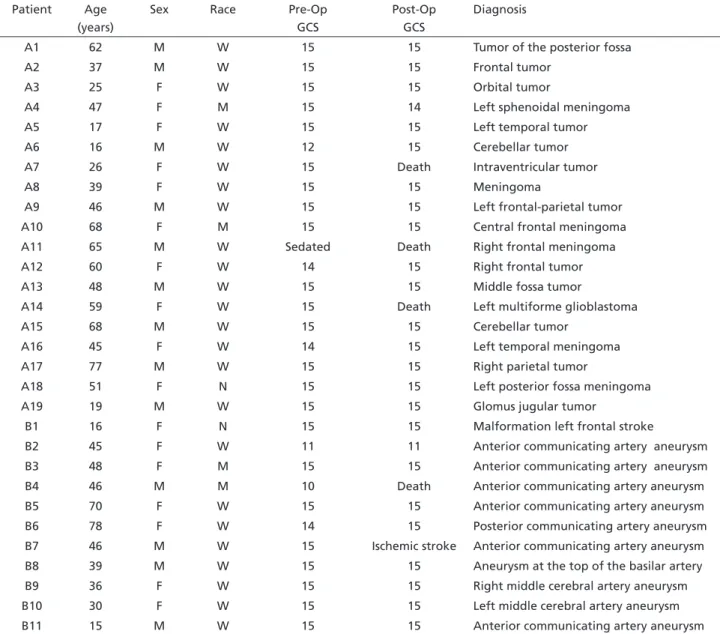
Table 1. Demographic data, pre- and postoperative Glasgow Coma Scale score and neurologic diagnoses in patients of Group A and B. Patient Age
(years)
Sex Race Pre-Op
GCS
Post-Op GCS
Diagnosis
A1 62 M W 15 15 Tumor of the posterior fossa
A2 37 M W 15 15 Frontal tumor
A3 25 F W 15 15 Orbital tumor
A4 47 F M 15 14 Left sphenoidal meningoma
A5 17 F W 15 15 Left temporal tumor
A6 16 M W 12 15 Cerebellar tumor
A7 26 F W 15 Death Intraventricular tumor
A8 39 F W 15 15 Meningoma
A9 46 M W 15 15 Left frontal-parietal tumor
A10 68 F M 15 15 Central frontal meningoma
A11 65 M W Sedated Death Right frontal meningoma
A12 60 F W 14 15 Right frontal tumor
A13 48 M W 15 15 Middle fossa tumor
A14 59 F W 15 Death Left multiforme glioblastoma
A15 68 M W 15 15 Cerebellar tumor
A16 45 F W 14 15 Left temporal meningoma
A17 77 M W 15 15 Right parietal tumor
A18 51 F N 15 15 Left posterior fossa meningoma
A19 19 M W 15 15 Glomus jugular tumor
B1 16 F N 15 15 Malformation left frontal stroke
B2 45 F W 11 11 Anterior communicating artery aneurysm
B3 48 F M 15 15 Anterior communicating artery aneurysm
B4 46 M M 10 Death Anterior communicating artery aneurysm
B5 70 F W 15 15 Anterior communicating artery aneurysm
B6 78 F W 14 15 Posterior communicating artery aneurysm
B7 46 M W 15 Ischemic stroke Anterior communicating artery aneurysm
B8 39 M W 15 15 Aneurysm at the top of the basilar artery
B9 36 F W 15 15 Right middle cerebral artery aneurysm
B10 30 F W 15 15 Left middle cerebral artery aneurysm
B11 15 M W 15 15 Anterior communicating artery aneurysm
Table 2. Alterations used in the differential diagnosis of syndromes responsible for so-dium disturbances found in the postoperative phase of patients in this study.
CSWS SIADH DI
Fluid balance ↓ Normal or ↑ ↓
Volume of urine ↑ Normal or ↓ ↑
Serum sodium ↓ ↓ ↑
Urinary sodium ↑ ↑ Normal
Plasma AVP Normal ↑ ↓
↓decrease; ↑increase.
patients show no signs of hypervolemia since one-third of the remaining fluid is maintained in the ex-tracellular compartment, making diagnosis difficult8.
Measurement of AVP can be useful in the differen-tial diagnosis of these syndromes2,7. However, it can
also be influenced by several factors involved in the postoperative phase of neurosurgical patients, that are able to stimulate the non-osmotic secretion of AVP, such as stress, an increase in intracranial pres-sure (ICP), pain, use of medication (antipsychotics, tri-cyclic antidepressives, carbamazepine, morphine, an-esthetics, metoclopramide), hypovolemia, prolonged recumbency and positive-pressure mechanical venti-lation4,7,16,22-27
. Considering that patients submitted to neurosurgery may frequently suffer sodium and AVP alterations of various causes and since differen-tial diagnosis of these alterations is essendifferen-tial to guar-antee adequate treatment, their accurate diagnosis is of great importance17,18
.
The objective of the present study is therefore to identify the presence of syndromes related to sodium alterations occurring in neurological patients during the postoperative period.
METHOD
The study was carried out at the Teaching Hospital of the State University of Campinas (UNICAMP), Brazil. It was approved by the Internal Review Board under protocol number 142/99. Signed, informed consent was obtained from the patients themselves or from their next-of-kin pri-or to admission to the study.
Thirty consecutive patients (males and females) over 13 years of age, who had been submitted to craniotomy either for resection of a brain tumor (Group A, n=19) or for clipping of an aneurysm of the cerebral artery (Group B, n=11) were enrolled to the study. The groups of patients were consid-ered homogenic with respect to the parameters of age, sex, race, and the pre- and postoperative Glasgow Coma Scale (GCS) scores, (Table 1). Sodium measurements in serum and urine (12 hour, overnight urine) were carried out on post-operative days 1-5 (D1-D5), urinary volume was measured in the 12-hour overnight period and plasma arginine vasopres-sin (AVP) measurements were carried out by radioimmuno-assay on postoperative days 1, 3 and 5 (D1, D3, D5).
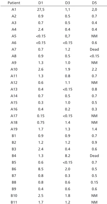
Table 3. Plasma arginine vasopressin values on the 1st
, 2nd
and 3rd
postoperative days in patients of Groups A and B
Patient D1 D3 D5
A1 27,5 1,1 2,0
A2 0.9 0.5 0.7
A3 0.7 0.5 0.4
A4 2.4 0.4 0.4
A5 <0.15 0,7 NM
A6 <0.15 <0.15 0.6
A7 0.7 1.2 Dead
A8 0.9 0.5 <0.15
A9 1.3 1.0 NM
A10 2.6 1.9 2.2
A11 1.3 0.8 0.7
A12 0.6 1.1 NM
A13 0.4 <0.15 0.8
A14 0.7 0.5 0.7
A15 0.3 1.0 0.5
A16 0.4 0.2 0.3
A17 0.15 <0.15 NM
A18 0.75 1.4 NM
A19 1.7 1.3 1.4
B1 0.9 0.9 0.7
B2 1.2 1.2 0.9
B3 2.4 0.4 0.6
B4 1.3 8.2 Dead
B5 0.6 <0.15 0.7
B6 8.5 2.0 0.5
B7 0.8 0.3 0.5
B8 0.8 0.6 0.15
B9 0.4 0.6 0.6
B10 2.5 1.8 NM
B11 1.7 1.2 NM
Normal value of plasma AVP: 0.5-5.0 pg/mL; NM, not measurable.
Exclusion criteria included: age <13 years, pregnan-cy, diagnosis of hypophyseal tumor and previously diag-nosed alterations in cardiac, renal, adrenal, thyroid or he-patic function.
Diagnostic criteria used for related syndromes are shown in Table 2.
The Mann-Whitney test was used in the statistical anal-ysis of the comparison of continuous measurements be-tween the two groups and the Chi-square test or Fisher’s Exact test were used for the comparison of proportions. Significance was established at 5% .
RESULTS
Hydro-electrolytic and plasma AVP alterations
– Plasma AVP levels are shown in Table 3. Table 4
shows the occurrence of alterations in sodium, uri-nary volume and plasma AVP for each patient. These alterations, permitted a differential diagnosis of the syndromes (CSWS, SIADH, DI), based on the diagnos-tic criteria already described in Table 2.
Distribution of syndromes according to group – There were no statistically significant differences in the distribution of syndromes between the two groups (p=0.7737), as shown in Table 5. This allowed us to group them into one single set, showing that of the 30 patients in the study, 27 developed CSWS, of which 13/27 developed CSWS alone and 14/27 devel-oped CSWS associated with DI (mixed syndrome); in
Table 4. Alterations in sodium, natriuresis, urinary volume and plasma AVP found in patients of both groups with the respective diagnoses of the syndromes involved.
Patient Hyponatremia Hypernatremia Natriuresis Polyuria ↑ AVP ↓AVP Diagnosis
A1 X X X X SIADH
A2 X X CSWS
A3 X X X CSWS + DI
A4 X X X X CSWS + DI
A5 X X X CSWS + DI
A6 X X X X CSWS + DI
A7 X X X CSWS
A8 X X X X CSWS + DI
A9 X X X CSWS
A10 X X X CSWS
A11 X X X CSWS
A12 X X CSWS
A13 X X X X CSWS + DI
A14 X X X X CSWS
A15 X X X X CSWS + DI
A16 X X X X CSWS + DI
A17 X X X CSWS + DI
A18 X X X CSWS
A19 X X CSWS
B1 X X CSWS
B2 X X X CSWS
B3 X X X CSWS + DI
B4 X X X SIADH
B5 X X X X CSWS + DI
B6 X X X SIADH
B7 X X X CSWS + DI
B8 X X X X CSWS + DI
B9 X X X CSWS + DI
B10 X X X CSWS
B11 X X X CSWS
this period no patient developed DI on its own and three presented SIADH.
DISCUSSION
Hyponatremia in neurological and neurosurgical patients can occur secondary to cerebral salt wast-ing syndrome10,13,18,28 or syndrome of inappropriate
antidiuretic hormone secretion15
and iatrogenically as a result of a large infusion of hypotonic liquid20. In
this study, no patient received hypotonic liquid in ex-cess, and sodium and fluid replacement were carried out whenever necessary. Therefore, hyponatremia, present in 19/30 patients (63.3%) in the postopera-tive phase, must not have occurred iatrogenically but must have been secondary to CSWS or SIADH.
The most common cause of hypernatremia in neu-rological patients is diabetes insipidus and this con-dition may also occur iatrogenically or secondary to neurological lesions. The most common form of dia-betes insipidus is that wich folows trauma or surgery
to the region of the pituitary and hypothalamus29
. As we excluded patients with pituitary tumor there were no patients with isolated diabetes insipidus. Diabetes insipidus may exibit as three patterns: transient, per-manent and triphasic. The triphasic patern is observed more often clinicaly. First, a polyuric phase occurs and lasts 4-5 days.
Inhibition of ADH causes the polyuric phase. Sec-ond, an antidiuretic phase of 5-6 days occurs, with results from release of stored hormone and the third phase can be permanent DI, when stores of ADH are exausted and the cells that produce more ADH are absent or unable to produce.
In this study, hypernatremia was observed iatro-genically in 2/30 patients (6.7%) due to the adminis-tration of hypertonic liquid and because of the excess correction of hyponatremia. No patient presented hypernatremia associated with low levels of AVP.
Urine sodium loss (natriuresis) can be caused by CSWS or SIADH and most studies show that its most probable cause is CSWS, natriuresis being preceded by hyponatremia and not correlated to AVP. This
sug-gests that CSWS is the principal cause of natriuresis in these patients2,10,30
. The present study also shows that most of the patients who developed hyponatremia and natriuresis presented no increase in plasma AVP levels, suggesting that the probable cause of natri-uresis in these patients is CSWS.
Increased plasma levels of AVP occur due to SI-ADH. However, most studies show that in the imme-diate postoperative period and in the initial stages of SAH, the increase in AVP may be attributed to surgi-cal stress, increased ICP, use of medication (metochlo-pramide, morphine, anticonvulsants), pain and/or loss of blood during surgery, and not to SIADH2,10,20,27,28.
Some authors also suggest the participation of AVP in CSWS. This hypothesis suggests that hypovolemia stimulates AVP secretion in a physiologically correct manner despite the suppressive effect of hyponatre-mia on AVP, and that in SIADH the increase in AVP occurs inappropriately since there is an expansion in the actual vascular volume.
In this study, increased plasma AVP measurements (>5.0 pg/mL) were found in only 3/30 patients (10%), and 3/150 measurements at the first posoperatory day. Considering that none of these patients devel-oped hypovolemia, which could have justified the increase in AVP, the cause may be due to SIADH or to the physiological response to surgical stress.
Decreased plasma levels of AVP (<0.5 pg/mL) were found in 14/30 patients (46.7%), in 23 of 150 mea-surements during the postoperative period. The most common cause of the reduction in plasma secretion of AVP in neurological patients is hyponatremia or secondary to neurological lesions .
In this study, the reduction in the plasma levels of AVP was associated with hyponatremia. However, all patients also presented CSWS concomitantly and re-duction in the plasma levels of AVP thereby prevent the patients from developing higher hyponatremia. It was also observed that, in the postoperative phase, 27/30 patients (90%) developed cerebral salt wast-ing syndrome, 13 of these (43.3%) developwast-ing CSWS alone and 14/27 (46.7%) developing CSWS associated
Table 5. Distribution of syndromes between the groups.
CSWS CSWS + DI DI SIADH
Group A 9 9 0 1
Group B 4 5 0 2
Total 13 14 0 3
% 43.3 46.7 0 10
with diabetes insipidus (mixed syndrome). Three out of thirty patients (10%) suffered an increase in plas-ma AVP levels. There was no statistically significant difference in the distribution of syndromes between the two groups (p=0.7737), which allows us to ana-lyze them as a single set.
The incidence of CSWS among all the patients was greater than that of SIADH, which is in agreement with reports from various other authors, who also observed a greater prevalence of CSWS, compared to SIADH in neurological patients1,2,4,5,10,18,30. The
co-existence of DI and CSWS (mixed syndrome) observed in our patients has also been described by Yamaki
et al.17, who studied two patients postoperatively
following surgery for pituitary tumor and observed the presence of severe natriuresis and hyponatre-mia in a study patient who developed DI (fluid loss >2.36 mL/kg/h), which led the authors to conclude that hyponatremia resulting from CSWS in intracra-nial diseases may develop in patients with DI.
Simi-lar findings were described by Laredo et al.29, who
reported hypernatremia resulting from DI, followed by hyponatremia due to sodium urinary loss compat-ible with CSWS, in the immediate postoperative pe-riod of two neurosurgical patients. However, plasma measurements of AVP were not carried out in either of these studies.
Studies reported in the literature about the altera-tions in hydro-electrolytic control and the syndromes responsible for these disturbances are mostly case re-ports and retrospective studies. These studies include small, diverse populations and analyze few variables using a variety of methods. Few studies have been carried out using a control group and their results are conflicting. In these studies, the differential diag-nosis between CSWS and SIADH is based on volemic state, as measured by indirect, insufficient methods with no comparative studies between the different methods. Also, in the majority of studies on CSWS in a group of patients, examples are cited but there are no group values.
In the present study, difficulty was found in con-trolling the water and sodium balance in patients spontaneously ingesting liquids. Otherwise, addi-tional data could have been available, particularly in the case of those patients who developed CSWS associated with DI, justifying the association of these syndromes. Since the patients with CSWS are hypovo-lemic and vohypovo-lemic state is the most potent stimulus of AVP secretion, levels of the latter should be high. However, in this study, osmotic stimulus apparently prevailed over volemic state, leading to a fall in AVP
in patients with CSWS. The doubt remains regarding whether the decrease in AVP associated with CSWS can be considered a compensatory mechanism or a “protector” of plasma sodium, increasing the loss of free water. This compensatory mechanism may in-crease fluid loss even further, which could be harm-ful to these patients. This fluid loss may indeed be compensatory and “protective” in the attempt to re-duce the cerebral edema and intracranial hyperten-sion that may be caused by the severe hyponatremia. In this case, a cerebral natriuretic factor may be in-volved in the physiopathology of CSWS, as suggested by some authors7,29,30
.
There are no large studies reported in the litera-ture on CSWS alone or in association with DI (mixed syndrome); neither are there studies on the participa-tion of intracranial pressure (ICP) in the hydro-elec-trolytic imbalance of the syndromes involved. Un-fortunately, in our study, ICP was also not routinely monitored. Therefore, additional studies are required to provide more data on the association of cerebral salt wasting syndrome with diabetes insipidus, the in-fluence of intracranial pressure in hydro-electrolytic control, on the role of the atrial natriuretic factor and the possible existence of a brain natriuretic fac-tor, involved in the pathophysiology of cerebral salt wasting syndrome.
In conclusion, in this study cerebral salt wasting syndrome was the syndrome most frequently found, being detected in 90% of cases, and was associated
most often with diabetes insipidus (46.7%) (mixed
syndrome). Diabetes insipidus was not found alone
in any of our patients. Syndrome of inappropriate antidiuretic hormone secretion was found less fre-quently (10%) and may be merely a response to sur-gical stress. There was no difference between the two groups studied (cerebral tumor resection and clipping of cerebral artery aneurysm) in the incidence of the three syndromes. An adequate diagnosis is very important in defining treatment, reducing the complications and mortality related to the inade-quate management of sodium disturbances during the neurological postoperative phase.
REFERENCES
1. Nelson PB, Seif SM, Maroon JC, Robinson AG. Hyponatremia in intra-cranial disease: perhaps not the syndrome of inappropriate secretion of antidiuretic hormone (SIADH). J Neurosurg 1981;55:938-941. 2. Nelson PB, Seif S, Gutai J, Robinson AG. Hyponatremia and
natriure-sis following subarachnoid hemorrhage in a monkey model. J Neuro-surg 1984; 60:233-237.
3. Diringer M, Landenson PW, Borel C, Hart GK, Kirsch JR, Hanley DF. Sodium and water regulation in a patient with cerebral salt wasting. Arch Neurol 1989;46:928-930.
4. Sivakumar V, Rajshekhar V, Chandy MJ. Management of neurosurgical patients with hyponatremia and natriuresis. Neurosurgery 1994;34:269-274.
5. Damaraju SC, Rajshekhar V, Chandy MJ. Validation study of a central venous pressure-based protocol for the management of neurosurgical patients with hyponatremia and natriuresis. Neurosurgery 1997;40:312-317.
6. Bacic A, Gluncic I, Gluncic V. Disturbances in plasma sodium in pa-tients with war head injuries. Mil Medicine 1999;164:214-217. 7. Harrigan MR. Cerebral salt wasting syndrome. Crit Care Clin 2001;17:
125-138.
8. Palmer BF. Hyponatraemia in a neurosurgical patient: syndrome of in-appropriate antidiuretic hormone secretion versus cerebral salt wast-ing. Nephrol Dial Transplant 2000;15:262-268.
9. Wong MF, Chin NM, Lew TW. Diabetes insipidus in neurosurgical pa-tients. Ann Acad Med Singapore 1998; 27:340-343.
10. Wijdicks EF, Vermeulen M, Haaf JA, Hijdra A, Bakker WH, van Gijn J. Volume depletion and natriuresis in patients with a ruptured intracra-nial aneurysm. Ann Neurol 1985;18:211-216.
11. Hasan D, Wijdicks EF, Vermeulen M. Hyponatremia is associated with cerebral ischemia in patients with aneurysmal subarachnoid haemor-rhage. Ann Neurol 1999;27:106-108.
12. Diringer MN, Wu KC, Verbalis JG, Hanley DF. Hypervolemic thera-py prevents volume contraction but not hyponatremia following sub-arachnoid hemorrhage. Ann Neurol 1992;31:543-550.
13. Peters JP, Welt LG, Sims EA, Orloff J, Needham J. A salt-wasting syn-drome associated with cerebral disease. Trans Assoc Am Physicians 1950;63:57-64.
14. Schwartz WB, Bennett W, Curelop S, Bartter FC. A syndrome of renal sodium loss and hyponatremia probably resulting from inappropriate secretion of antidiuretic hormone. Am J Med 1957;23:529-542.
15. Bartter FC, Schwartz WB. The syndrome of inappropriate secretion of antidiuretic hormone. Am J Med 1967;42:790-806.
16. Vingerhoets F, Tribolet N. Hyponatremia hypo-osmolarity in neurosur-gical patients: “appropriate secretion of ADH“ and “cerebral salt wast-ing syndrome“. Acta Neurochir 1988;91:50-54.
17. Yamaki T, Tano-Oka A, Takahashi A, Imaizumi T, Suetake K, Hashi K. Cerebral salt wasting distinct from syndrome of inappropriate secretion of antidiuretic hormone (SIADH). Acta Neurochir 1992;115:156-162. 18. Wijdicks EF, Vermeulen M, Hijdra A, van Gijn J. Hyponatremia and
ce-rebral infarction in patients with ruptured intracranial aneurysms. Is luid restriction harmful? Ann Neurol 1985;17:137-140.
19. Coenraad MJ, Meinders AE, Taal JC, Bolk JH. Hyponatremia in intra-cranial disorders. Netherlands J Med 2001;58:123-127.
20. Chung HM, Kluge R, Schrier RW, Anderson RJ. Postoperative hypona-tremia: a prospective study. Arch Intern Med 1986;146:333-336. 21. Robertson GL, Mahr EA, Athar S, Sinha T. Development and clinical
application of a new method for radioimmunoassay of arginine vaso-pressin in human plasma. J Clin Invest 1973;52:2340-2352.
22. Saborio P, Tipton GA, Chan JC. Diabetes insipidus. Pediatr Rev 2000;21: 122-129.
23. Reeder RF, Harbaugh RE. Administration of intravenous urea and nor-mal saline for the treatment of hyponatremia in neurosurgical patients. J Neurosurg 1989;70:201-206.
24. Rabinstein AA, Wijdicks EF. Hyponatremia in critically ill neurological patients: review. Neurologist 2003;9:290-300.
25. Miller M. Syndrome of excess antidiuretic hormone release. Crit Care Clin 2001;17:11-23.
26. Dickerson RN. Hyponatremia in neurosurgical patients: syndrome of inappropriate antidiuretic hormone or cerebral salt wasting syndrome? Hospital Pharmacy 2002;12:1336-1340.
27. Oh MS, Carroll HJ. Cerebral salt wasting syndrome: we need better proof of its existence. Nephron 1999;82:110-114.
28. Isotani E, Suzuki R, Tomita K, et al. Alterations in plasma concentra-tions of natriuretic peptides and antidiuretic hormone after subarach-noid hemorrhage. Stroke 1994;25:2198-2203.
29. Laredo S, Yuen K, Sonnenberg B, Halperin ML. Coexistence of central diabetes insipidus and salt wasting: the dificulties in diagnosis, chang-es in natremia and treatment. J Am Soc Nephrol 1996;7:2527-2532. 30. Tisdall M, Crocker M, Watkiss J, Smith FRCA. Disturbances of sodium