Tumoral lesions of the spine can represent great challenges for a surgical team. Many techniques have been proposed for surgical training as a learning pro-cess for young surgeons or for the simulation of com-plex procedures. A rapid prototyping technique has been presented recently as an option for these purpos-es1. Among prototyping forms the stereolithography, a technique developed in 1987 for aerospace industry stands out. It is based on models and graphic computa-tion of images and create solid molds in resins, ceramic or metals1,2. As of the end of the 90’s the first adapta-tions were accomplished for application in the biomed-ical area, substituting the drawings of graphic compu-tation with images of a medical scanner such as com-puterized tomography (CT) and magnetic resonance image (MRI), whereby physical models of the human anatomy are built3 . Several techniques of rapid
proto-typing have been developed, and all are based on the construction of anatomical representations in a three-dimensional structure, elaborated layer by layer1,4. The technology is based on the photopolimeryzing of a res-in through an ultra violet laser. A controller positions a vertical elevator, initially on a container full of photo-sensitive resin, so that just a fine liquid layer is on the platform1,5. When the laser reaches the surface of the resin, this is solidified and the first layer of the model is built. After this, the elevator goes down submerg-ing the layer recently solidified. The process of scan-ning laser is repeated and a second layer is elaborated on top of the first. The process of scanning laser is re-peated so that the whole model is built. The purpose of this work is to demonstrate the practical use of the stereolithography an auxiliary method for training and surgical simulation1.
APLICATION OF THE STEREOLITHOGRAPHY
TECHNIQUE IN COMPLEX SPINE SURGERY
Wellingson Silva Paiva
1, Robson Amorim
1,
Douglas Alexandre França Bezerra
1, Marcos Masini
2ABSTRACT - Many techniques have been proposed for surgical training as a learning process for young surgeons or for the simulation of complex procedures. Stereolithograpfy, a rapid prototyping technique, has been presented recently as an option for these purposes. We describe the case of a 12 years old boy, diagnosed with Ewing´s sarcoma in the cervical spine. After a surgical simulation accomplished in the pro-totype, built by stereolithography, the patient was submitted to a C4 corpectomy and to a C4 and C3 lam-inectomy with anterior and posterior fixation, a non intercurrence procedure. This technique is an inno-vative and complementary tool in diagnosis and therapy. As a result, it is easier for the surgeon to under-stand the complexity of the case and plan the approach before any surgical procedure.
KEY WORDS: spine surgery, rapid prototyping, stereolithography.
Aplicação da técnica de estereolitografia em cirurgias complexas da coluna
RESUMO - Lesões tumorais da coluna podem representar um grande desafio para equipe cirúrgica. A es-tereolitografia é uma técnica de construção de peças anatômicas a partir de desenhos gráficos ou exa-mes radiológicos. Apresentamos um paciente de 12 anos, com quadro de cervicobraquialgia à direita com diagnóstico de sarcoma de Ewing. Ao exame neurológico, o paciente apresentava-se com paresia em MSD grau IV e leve hipoestesia em dermátomos de C6 à T1 à direita. RM evidenciou edema intra e extra-ósseo com compressão medular e instabilidade cervical. Realizada simulação cirúrgica no protótipo construído por estereolitografia. Foi submetido a corpectomia de C4 e a laminectomia C4 e C3, com fixação anterior e posterior, procedimento sem intercorrências. A estereolitografia é ferramenta inovadora no auxílio diag-nóstico e terapêutico. Seu uso permite ao cirurgião entender fisicamente o sítio da lesão, estudar a via de acesso e perceber a real complexidade do caso antes do procedimento cirúrgico.
PALAVRAS-CHAVE: cirurgia da coluna, prototipagem rápida, estereolitografia.
Clínica Queops de Neurologia e Neurocirurgia, Brasília DF, Brazil: 1Neurosurgery Resident of Hospital das Clinicas - University of São
Paulo, São Paulo SP, Brazil; 2Professor of Neurosurgery, PhD - University of Planalto Central, Brasília DF, Brazil.
Received 27 June 2006, received in final form 1 December 2006. Accepted 10 February 2007.
Dr. Wellingson Paiva - Rua Ovidio Pires de Campos 171 / 511 - 05403-010 São Paulo SP - Brasil. E-mail: [email protected]
444 Arq Neuropsiquiatr 2007;65(2-B)
METHOD
This is a report of a case of the stereolithography tech-nique application. In this case, the graphic models were used in extension “. stl”. We used a specific device SLA 250 (3D system Inc. Valência, CA, USA) to elaborate the ste-reographic model. The radiologic images were transferred through an optical disk for better graphic interface. In this case a careful planning of the model with its surgery instru-mental. This study was approved by the ethical committee
of the institution and also its publication was authorized by child’s guardians.
Case – We describe the case of 12 years old, white, male, student, complaining about a sudden and intense pain in the neck lasting for 45 days. According to the patient, 38 days after the symptoms had started the pain also reached the right arm. In the neurological examination the patient presented right arm paresis (grade 4) and slight
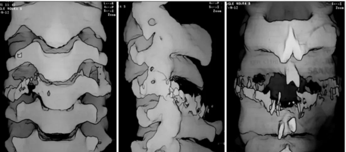
hypoes-Fig 1. Computed tomography image with 3D reconstruction of the case showing osteolytic lesion in cervical spine.
Fig 2. Anatomical representation of the patient’s cervical spine, already in physical mold.
Arq Neuropsiquiatr 2007;65(2-B) 445
tesia in C6 to T1 right dermathomus. In a complementary investigation (Figs 1 and 2) the X-ray of cervical spine in-dicated pathologic fracture of C4 vertebra with C4-C5 an-terolistese (grade 1). The scyntilography of the whole body with Tc - 99 showed tenuous increase of osteogenic activi-ty in C4. CT with tumoral lesion bone committing C4 with soft parts components (suggestive of osteoblastoma) MRI evidenced edema intra and extra-bone with spine compres-sion and cervical instability.
After a surgical simulation accomplished in the proto-type, built by stereolithography, the patient was submitted to a C4 corpectomy and to a C4 and C3 laminectomy with anterior and posterior fixation (Fig 3). The hystopathologic exam and immunohystochemic were compatible with neu-roectodermic sarcoma of Ewing. The patient developed a partial redemption of painful symptoms and the recovery of his muscular force. Subsequently, the patient was sub-mitted to a complementary radiotherapeutic treatment.
DISCUSSION
There were no complications during the proce-dure, which supports the idea that the process of stereolithography can be used in complex spine op-erations. In this case the previous training on ste-reolithographic mold allowed the procedure to be accomplished with an appropriated preparation be-sides evaluating the ideal dimensions of the plate system, screws and cables. This technique presents a great potential application, allowing visualization of tumors and its relationship with neighboring anat-omy. It was described for visualization of maxillo-fa-cial tumors and dental alterations3,5
, stereotatics and brain aneurysm surgeries6, evaluation of dimensions of orthopaedics prostheses4. It is also possible to de-termine the standardization of surgical techniques, when accompanied by special procedures. In the case of surgery for placement of prostheses the
stereo-lithographic model has been used to determine ideal dimensions of the material to be implanted1.
Of all the techniques the stereolithography is the most used, presenting application in the aero-space industry for more than two decades1. The adaptation for medical appliance with the substitution of graph-ic drawings for radiologgraph-ical exams can be done with-out the loss of precision, maintaining a level with variation of 1mm in the graphic interface7
. The limi-tation for regular use of this technique is the high cost involved in prototyping process (the important support to complex procedures).
In conclusion, this technique is an innovative and complementary tool in diagnosis and therapy. As a result, it is easier for the surgeon to understand the complexity of the case and plan the approach be-fore any surgical procedure. Careful planning and previous rehearsal reduce the risk of surprises during an operation. Moreover, it can improve decisional knowledge which represents an active role in a sur-gical procedure.
REFERENCES
1. Petzold R, Zeilhofer HF, Kalender WA. Rapid protyping technology in medicine basics and applications. Comput Med Imaging Graph 2002;23:277-284.
2. Choi JY, Choi JH, Kim NK, et al. Analysis of errors in medical rapid pro-totyping models. Int J Oral Maxillofac Surg 2002;31:23-32.
3. D’Urso PS, Barker TM. Stereolithographic biomodelling in cranio-maxillo-facial surgery: a prospective trial. J Craniomaxillofac Surg 1999;27:30-37. 4. Potamianos P, Amis AA, Forester AJ. Rapid prototyping for
orthopae-dic surgery. Proc Inst Mech Eng 1998;212:383-393.
5. Poukens J, Haex J, Riediger D. The use of rapid prototyping in the pre-operative planning of distraction osteogenesis of the cranio-maxillofa-cial skeleton. Comput Aided Surg 2003;8:146-154.
6. Wurm G, Tomancok B, Pogady P, Holl K, Trenkler J. Cerebrovascular stereolithographic biomodeling for aneurysm surgery: technical note. J Neurosurg 2004;100:139-145.