Approximately half of childhood leukoencepha-lopathies remain unclassified despite extensive inves-tigation1. Recently, some of them have been identi-fied and categorized based on their distinct abnor-malities detected on magnetic resonance imaging (MRI)2-6
. A novel distinct leukoencephalopathy was described in 2003 by Van der Knaap et al.7 Symptoms begin during childhood, with slow progression. Dis-tinctive clinical findings include cerebellar,
pyrami-dal and dorsal column dysfunction. Laboratory inves-tigation is unrevealing. Typical findings on MRI and proton magnetic resonance spectroscopy (1H-MRS) in-clude abnormal cerebral and cerebellar white matter, brainstem and spinal cord tracts and elevated lactate (Lac) within the abnormal white matter. This recent-ly identified disease has been called leukoencepha-lopathy with brainstem and spinal cord involvement and high lactate (LBSL).
LEUKOENCEPHALOPATHY WITH BRAINSTEM
AND SPINAL CORD INVOLVEMENT
AND HIGH BRAIN LACTATE
Report of three Brazilian patients
Daniel Gurgel Fernandes Távora
1, Mauro Nakayama
1, Rômulo Lopes Gama
1,
Thereza Cristina de Lara Alvim
1, Dalton Portugal
2, Enio Alberto Comerlato
3ABSTRACT - A novel leukoencephalopathy was recently identified based on magnetic resonance imaging (MRI) and proton magnetic resonance spectroscopy (1H-MRS) findings. Leukoencephalopathy with
brain-stem and spinal cord involvement and high lactate (LBSL) is an autosomal recessive disorder character-ized by early onset of symptoms and slowly progressive cerebellar, pyramidal and spinal cord dorsal col-umn dysfunction. MRI and 1H-MRS typically show abnormalities within cerebral and cerebellar white
mat-ter, a characteristic involvement of brainstem and spinal cord tracts and elevated lactate in the abnormal white matter. We present three cases with characteristic clinical and neuroimaging findings of this disor-der. Some additional unique findings of our patients are discussed, like distal motor neuropathy and ele-vated creatine kinase in the serum.
KEY WORDS: leukoencephalopathy, brainstem, spinal cord, lactate.
Leucoencefalopatia com envolvimento do tronco cerebral e da medula espinal e elevação do lactato cerebral: relato de três casos brasileiros
RESUMO - Uma nova leucoencefalopatia foi recentemente descrita com base em achados característicos de ressonância magnética e espectroscopia de prótons por ressonância magnética. Leucoencefalopatia com envolvimento do tronco cerebral e da medula espinal e elevação do lactato cerebral é uma doença au-tossômica recessiva de aparecimento precoce e evolução lenta, caracterizada por disfunção cerebelar, pi-ramidal e das colunas dorsais da medula. Ressonância magnética e espectroscopia de prótons tipicamen-te demonstram anormalidades na substância branca cerebral e cerebelar, com envolvimento caractipicamen-terísti- característi-co de tratos no troncaracterísti-co encefálicaracterísti-co e na medula espinhal e aumento de lactato na substância branca cere-bral anormal. Relatamos três casos com achados clínicos e de neuroimagem característicos. Achados adi-cionais peculiares aos nossos pacientes são discutidos, como a elevação da creatina-quinase sérica e a pre-sença de neuropatia motora distal.
PALAVRAS-CHAVE: leucoencefalopatia, tronco cerebral, medula espinal, lactato.
The Sarah Network of Hospitals for Rehabilitation, Fortaleza CE, Brazil: Departments of 1
Radiology, 2
Clinical Neurology and 3
Clini-cal Neurophysiology.
Received 22 September 2006, received in final form 15 December 2006. Accepted 7 March 2007.
We describe three cases with clinical, neurophysi-ologic, laboratory, MRI and 1
H-MRS findings sugges-tive of this new entity.
METHOD
We have studied three patients from two unrelated families. All patients were born in the same geographic area, in the northeast region of Brazil. Patients 1 and 2 are siblings. A careful interview regarding family history was taken and an experienced neurologist performed a detailed clinical evaluation. Assessment of muscle strength was per-formed using the Medical Research Council scale.
Laboratory studies included blood cell count, blood gas-es, glucose, ammonium-ion, uric acid, transaminasgas-es, cre-atine kinase, lactate, thyroid function tests, ceruloplasmin, vitamin E, vitamin B12, folic acid, very long-chain fatty ac-ids, phytanic acid, acanthocytes, plasma amino acac-ids, ly-sosomal enzymes; Urine: oligosaccharides, sialic acid, ami-no acids and organic acids; Cerebrospinal fluid (CSF): cell count, protein, lactate, immunoglobulin G index, antibody detection of Herpes simplex virus (HSV), Cytomegalovirus
(CMV), Epstein-Barr virus (EBV), toxoplasmosis, schistoso-miasis, molecular detection of Mycobacterium tuberculo-sis and detection of cryptococcal antigen.
Neurophysiologic evaluation was performed according to standard methods8. Electroneuromyography (ENMG) and
somatosensory-evoked potentials (SEP) were performed in all patients; motor evoked potentials (MEP) and brainstem auditory evoked potentials (BAEP) were performed in pa-tient 1 and visual evoked potentials (VEP) was performed in patients 1 and 3.
1H-MRS and MRI of the brain and spinal cord were
per-formed in all patients in the same 1.5T MR unit (Signa Ho-rizon, GE Medical Systems, Milwaukee, WI, USA). Brain MRI protocol included axial and coronal fast spin echo (FSE) T2-weighted, axial fluid attenuated inversion recovery (FLAIR), axial and sagittal spin echo (SE) T1-weighted images. Spi-nal cord MRI included axial and sagittal FSE T1 and T2-weighted sequences. T1-T2-weighted sequences were acquired before and after intravenous injection of a paramagnetic contrast agent (gadolinium). Single-voxel point-resolved proton spectroscopy sequences (PRESS) with echo time (TE) of 35 and 144 msec were obtained in the abnormal cere-bral white matter. Patient 1 had an additional 1H-MRS study
with voxel located at the abnormal cerebellar white matter. Voxels were 2 X 2 X 2 cm in size. N- acetylaspartate (NAA) was assigned at 2.02 parts per million (ppm), choline (Cho) at 3.2 ppm and creatine (Cr) at 3.03 ppm. Metabolite ra-tios (NAA/Cr and Cho/Cr) were measured. All data process-ing was performed by software provided by the manufac-turer. Patient 3 had a follow-up with MRI and 1H-MRS two
years apart.
Institutional review board approval was obtained. Ver-bal consent was obtained from all parents.
RESULTS
Clinical findings (Table 1) – Patient 1 is a boy, born in 1992. Patients 1 and 2 are siblings. He has two
other unaffected siblings. His parents are healthy and nonconsanguineous. He developed a support-ed walking at 15 months. During subsequent years, he acquired an independent but not mature gait, with frequent falls. At the age of 9, gait pattern be-gan to deteriorate, and nowadays he is not able to walk without support. He has always had learning difficulties. On examination, he has generally de-pressed deep tendon reflexes, extension plantar re-sponse bilaterally, bilateral pes cavus deformity, bi-lateral lower limb impaired vibration sense, reduced muscle strength for proximal and distal lower limbs and mild to moderate postural/gait ataxia.
Patient 2 is a boy, born in 1987. He acquired nor-mal gait at 12 months. At the age of 6, he developed progressive walking abnormality, and by the age of 16 years, walking aids were needed for ambulation. On examination, deep tendon reflexes were gener-ally depressed, with extension plantar response bi-laterally, bilateral pes cavus deformity, bilateral low-er limb impaired vibration sense, decreased tactile and pin sensation in hands and feet, mild to moder-ate postural/gait ataxia. Muscle strength was reduced for proximal and distal lower limbs.
Patient 3 is a boy, born in 1987 from healthy non-consanguineous parents. Progressive motor disabil-ities initiated by the age of 6 years. At 12 years of age he became wheelchair bound. Upper limbs mo-tor deficits were first noted by the age of 14 years. Cognitive impairment was never observed. Examina-tion demonstrated quadriparesis predominant in the distal part of upper and lower limbs, muscular atro-phy at thenar and hypothenar regions, generally de-pressed deep tendon reflexes, mildly impaired vibra-tion and joint-posivibra-tion sense and altered finger-to-nose test bilaterally.
Laboratory investigation revealed mildly elevated serum creatine kinase in patient 2 (286 UI/L) and in patient 3 (520 UI/L - normal values <190UI/L). The re-maining blood, CSF and urine tests were unremark-able.
Neurophysiologic findings are shown in Table 1.
MRI and 1
Table 1. LBSL: Clinical and neurophysiologic features in three patients.
Patients
1 2 3
Age (yr)/Sex 13/male 18/male 18/male Affected/unaffected siblings 1/2 1/2 –
Age at onset (yr) 3 6 6
Initial motor development n n n Initial symptoms Gait ataxia Gait ataxia Gait ataxia Lost unsupported walking (yr) 12 16 12
Muscle strength ↓ ↓ ↓
Sensory function Distal ↓ Distal ↓ Distal ↓ Deep tendon reflexes ↓ ↓ ↓ Babinski sign Bilateral + Bilateral + Bilateral + Cognitive function Learning problems Learning problems n
SNAP n n n
NCV n n Motor ↓
ENMG Chronic denervation in distal muscles
Chronic denervation in distal muscles
Chronic denervation in proximal and distal muscles
SEP Delayed Delayed –
MEP Prolonged Prolonged Prolonged
VEP n – n
BAEP n – –
n, normal; yr, year; ↓, decreased; +, present; –, not done; SNAP, sensory nerve action potential; NCV, nerve conduction velocity; ENMG, electroneuro-myography; SEP, somatosensory-evoked potentials; MEP, motor evoked potentials; VEP, visual evoked potentials; BAEP, brainstem auditory evoked potentials.
Fig 1. Axial T2-weighted images of the brain (A-E) and 1H-MRS spectra (F) in patient 1. The hemispheric white matter shows
con-fluent signal abnormalities (A). There is involvement of the anterior part of the corpus callosum (black arrow in B), posterior limb of internal capsule (white arrow in B), intraparenchymal trajectory of trigeminal nerves (arrowhead in D) superior cerebellar pe-duncles (white arrow in D), pyramidal tract (black arrow in D), inferior cerebellar pepe-duncles (white arrow in E) and cerebellar white matter (black arrow in E). Within the midbrain there is no signal alteration (C). 1H-MRS spectra at TE=35ms (upper spectrum) and
lobes. Corpus callosum was thin in patients 1 and 2, with diffuse signal abnormality in patient 1. Patients 2 and 3 had signal changes restricted to the spleni-um (Fig 2B). Posterior limb of internal capsules were altered in patients 1 and 2 (Figs 1B and 2B), and nor-mal in patient 3.
There were no abnormalities within the midbrain and transverse pontine fibers.
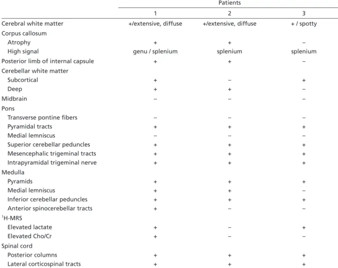
The following structures were affected in all pa-tients: pyramidal tracts at the level of the pons and medulla, pyramids, intraparenchymal trajectory of trigeminal nerves, mesencephalic trigeminal tracts, Table 2. LBSL: MRI findings in three patients.
Patients
1 2 3
Cerebral white matter +/extensive, diffuse +/extensive, diffuse + / spotty Corpus callosum
Atrophy High signal
+ genu / splenium
+ splenium
– splenium Posterior limb of internal capsule + + – Cerebellar white matter
Subcortical Deep
+ +
– +
+ –
Midbrain – – –
Pons
Transverse pontine fibers Pyramidal tracts
Medial lemniscus
Superior cerebellar peduncles Mesencephalic trigeminal tracts Intrapyramidal trigeminal nerve
– + – + + +
– + – + + +
– + – + + + Medulla
Pyramids Medial lemniscus
Inferior cerebellar peduncles Anterior spinocerebellar tracts
+ + + +
+ + + –
+ – + –
1H-MRS
Elevated lactate Elevated Cho/Cr
+ +
– –
+ – Spinal cord
Posterior columns Lateral corticospinal tracts
+ +
+ +
+ +
+, affected ; –, unaffected; Cho, Choline; Cr, Creatine.
inferior and superior cerebellar peduncles (Figs 1D-E and 2C).
Anterior spinocerebellar tracts were mildly in-volved in patient 1. Medial lemniscus was affected at the level of the medulla in patients 1 and 2.
Cerebellum was also affected with diffuse white matter involvement in patient 1, deep white matter involvement in patient 2 (Figs 1E and 2D), and mild subcortical alterations in patient 3.
1
H-MRS showed a doublet at 1.3ppm, compatible with elevated Lac, and higher Cho/Cr within abnor-mal cerebellar white matter in patient 1 (Figure 1F) and within frontoparietal white matter in patient 3. Spinal MRI revealed abnormal signal in the dor-sal columns and lateral corticospinal tracts in all pa-tients (Fig 3).
No contrast enhancement was observed in the post-gadolinium T1-weighted images.
Patient 3 had a follow-up brain and spinal MRI two years apart. There was a mild increase in sub-cortical cerebellar white matter abnormalities as the only change noticed.
DISCUSSION
A novel leukoencephalopathy was described in children by Van der Knaap et al. based on clinical and MRI features7. To our knowledge, only ten additional cases with similar clinical and MRI findings have been demonstrated9,10
. Neurological symptoms frequently start in childhood and progress slowly. Initial com-plaints refer to gait instability and tremor. Muscle weakness, cerebellar ataxia and distal spasticity, most prominent in lower limbs, become more pronounced during disease progression. Cognitive impairment is observed in some patients. Patients typically develop decreased distal position and vibration sense. Clinical features of the present patients were similar to the formerly described cases (Table 1). Laboratory inves-tigation revealed elevated creatine kinase in patients 2 (286 UI/L) and 3 (520 UI/L). To our knowledge this finding has not been previously described and we attributed it to motor axonal neuropathy.
Neurophysiologic findings were indicative of in-volvement of the posterior columns, cortico-spinal tracts, and lower motor neurons (LMN). In two pa-tients ENMG revealed chronic partial denevertion-reinervation in upper and lower limbs, with distal muscles affected more severely than proximal mus-cles, suggesting distal motor neuropathy9
. One pa-tient had diffuse anterior cell dysfunction, probably due to advanced disease stage. These alterations have not been described in LBSL patients.7,9
Border-line reduction of nerve conduction velocities has al-ready been reported7,9. Peripheral sensory system, vi-sual and brainstem auditory pathways were spared in these three patients.
MRI and 1H-MRS findings were unique and cor-related to previous reports (Table 2). Cerebral white matter was involved to a variable extent, with inho-mogeneous, confluent, extensive signal abnormal-ity in patients 1 and 2, and a spotty abnormal signal in patient 3. Correlation of clinical severity and MRI findings have already been verified7,9
. One of our pa-tients (patient 3) had minor cerebral white matter involvement and was the only one with preserved cognitive function. Subcortical U-fibers were spared in all patients.
Posterior corpus callosum had altered signal in-tensity in all cases. Abnormal signal at the genu was detected only in patient one. Diffuse atrophy of the corpus callosum was present in patients 1 and 2.
tient 1 and subcortical signal change was found in patient 3. On the other hand, patient 2 showed mild abnormality affecting merely deep cerebellar white matter, a finding not described on previous series.
Midbrain had no signal intensity abnormality and this is an atypical finding in our patients. Neverthe-less, Serkov et al.10 described normal medial lemnis-cus and pyramidal tracts within the midbrain in two out of five patients described.
Within the pons, neither medial lemniscus nor the transverse pontine fibers were affected. Transverse pontine fibers are not invariably involved in previ-ously reported cases. Altered signal usually becomes apparent in late stages2
. Medial lemniscus signal ab-normality at this level is a common finding, but not always present7. Anatomical structures consistently affected in our cases within the pons included the pyramidal tracts, superior cerebellar peduncles, mes-encephalic trigeminal tracts, and intrapyramidal tri-geminal nerve.
Anterior spinocerebellar tract involvement is a usual finding7, but not invariably present9. The pyra-mids and inferior cerebellar peduncles were always abnormal within the medulla. Medial lemniscus was abnormal in two patients and anterior spinocerebel-lar tract in only one.
Previous reports have emphasized the involve-ment of the sensory and pyramidal tracts over their entire length9,10,12
.Conversely, in our cases we have not detected MRI abnormalities within the midbrain and have not found involvement of the medial lem-niscus at the level of the pons. Actually, one of the previous reports have also found normal signal inten-sity in the pyramidal tracts and medial lemniscus in some cases at the level of the pons and midbrain10.
Spinal cord MRI revealed altered signal in poste-rior columns and lateral corticospinal tracts over the entire length in all cases. These abnormalities are in absolute agreement with earlier reports.
1H-MRS revealed increased Lac in two out of three cases. Elevated Lac is considered a criterion for the definition of this entity, but is not absolutely pres-ent in all cases reported earlier8,12. Elevated Cho has variably been found in previous reported series7,9
and
was detected only in patient 1. It is possibly due to mildly enhanced membrane turnover and myelin loss. 13
The involvement of two siblings in the present cases reinforces the likelihood of an autosomal re-cessive mode of inheritance. Consistent data on the pathogenetic mechanisms of this disease are still lack-ing on current literature. The genetic defect is un-known and no pathology information is available13. In conclusion, we have described three Brazilian patients with overall clinical and imaging features compatible with LBSL. Some features observed by us have not been previously reported, like elevated creatine kinase in the serum and neurophysiologic evidence of distal motor neuropathy. Moreover, we would like to call attention to the discontinuous as-pect of the MRI signal abnormalities in the sensory and pyramidal tracts in our patients.
REFERENCES
1. Van der Knaap MS, Breiter SN, Naidu S, Hart AAM, Valk J. Defining and categorizing leukoencephalopathies of unknown origin: MR im-aging approach. Radiology 1999;213:121-133.
2. Van der Knaap MS, Barth PG, Stroink H, et al. Leukoencephalopathy with swelling and a discrepantly mild clinical course in eight children. Ann Neurol 1995;37:324-334.
3. Hanefeld F, Holzbach U, Kruse B, Willichowski E, Christen H-J, Frahm J. Diffuse white matter disease in three children: an encephalopathy with unique features on magnetic resonance and proton magnetic res-onance spectroscopy. Neuropediatrics 1993;24:244-248.
4. Van der Knaap MS, Barth PG, Gabreëls FJM, et al. A new leukoenceph-alopathy with vanishing white matter. Neurology 1997;48:845-855.Neurology 1997;48:845-855. 5. Cavalcanti CE, Nogueira A. Síndrome de Van der Knaap -
megalence-falia com leucodistrofia: a respeito de dois casos na mesma família. Arq NeuroPsiquiatr 2000;58:157-161.
6. Oliveira HA, Machado ML, Jesus ACF, et al. Leucoencefalopatia mega-lencefálica com substância branca evanescente e cistos subcorticais. Arq Neuropsiquiatr 2004;62:1058-1062.
7. Van der Knaap MS, Van der Voorn P, Barkhof F, et al. A new leukoen-A new leukoen-cephalopathy with brainstem and spinal cord involvement and high lactate. Ann Neurol 2003;53:252-258.
8. Recommendations for the practice of clinical neurophysiology: guide-lines of the International Federation of Clinical Neuroohysiology. Elec-troencephalogr Clin Neurophysiol 1999;52(Suppl):S1-S304.
9. Linnankivi T, Lundbom N, Autti T, et al. Five new cases of a recent-ly described leukoencephalopathy with high brain lactate. Neurology 2004;63:688-692.
10. Serkov SV, Pronin IN, Bykova OV, et al. Five patients with a recently described novel leukoencephalopathy with brainstem and spinal cord involvement and elevated lactate. Neuropediatrics 2004;35:1-5. 11. De Jonghe P, Auer-Grumbach J, Irobi K, et al. Autosomal dominant
ju-venile amyotrophic lateral sclerosis and distal hereditary motor neu-ronopathy with pyramidal tract sings: synonyms for the same disor-der? Brain 2002;125:1320-1325.
12. Chiffmann R, Van der Knaap MS. The latest on leukodystrophies. Curr Opin Neurol 2004; 17:187-192.