EEG coherence allows correlation between differ-ent brain areas to be ascertained by analyzing the mutual relationship between two EEG signals across different frequency bands. As this constitutes a nor-malized value for the power of a given frequency, it is independent from signal amplitude oscillations1,2 Such a property makes coherence a relatively safe method for comparing groups of individuals in as far as differences in EEG powers do not impact estimat-ed coherence values3
. Reduced coherence between two regions is probably indicative of their decreased functional correlations. Ordinary coherence can be calculated between two cerebral electrodes, whether they be placed in the same hemisphere or in homolo-gous areas, by means of a mathematical formula di-viding the cross spectrum of these two channels by the product of self spectrum for each of these chan-nels, as illustrated in the formula below:
The Co value between two electrodes ranges from 0 to 1, where 1 denotes maximum correlation. When correlation is very low or null, the value approach-es 0. Thus, coherence reprapproach-esents a measure of corti-cal interconnectivity. Studies concerning coherence have shown that coherence between closely-placed electrodes is determined mainly by local connections excited by intracortical interneurons (Martinotti and short axoned stellate cells), whereas between distant electrodes such coherence is determined by long ax-onal connections4,5,6
. These two compartments com-pete with one another, influencing the same neuro-nal population in an alternate manner.
The objective of the current investigation was to study the inter-hemispheric coherence between ho-mologous areas in healthy individuals in the 20-50 year age bracket.
METHOD
A total of 190 tests were carried out on individuals with no neurological complaints, where subjects were submitted to digital EEG and then assessed. Subjects had no prior his-tory of neurological problems such as traumatic brain inju-ry, epilepsy, alcohol or drugs use and abuse, or clinical
com-STUDY OF INTERHEMISPHERIC COHERENCE
ON HEALTHY ADULTS
Mario Silva Jorge
1, Ricardo Vieira Botelho
2, Antonio Carlos de Paiva Melo
3ABSTRACT - The interhemispheric coherence of electroencephalogram was studied in a group of healthy individuals in the age range of 20-50 years. The results showed higher coherence for all bands in parietal regions (P3-P4). It was observed that individuals with high values of coherence for a certain frequency band in a pair of electrodes also showed high values of coherence for other bands across other pairs of electrodes. No significant influence on interhemispheric coherence was found for age, gender or hand dominance.
KEY WORDS: coherence, adults, EEG quantitative, normal pattern.
Estudo da coerência interhemisférica em adultos normais
RESUMO - A coerência inter-hemisférica do eletrencefalograma foi estudada em uma população de indiví-duos normais, na faixa etária de 20 a 50 anos. Os resultados mostraram maior valor de coerência para to-das as banto-das nas regiões parietais(P3-P4). Foi observado que em indivíduos com alto valor de coerência para determinada banda em um determinado par de eletrodos, também apresentam alto valor de coerên-cia para outras bandas nos outros pares de eletrodos. Não foi observada influêncoerên-cia da idade, sexo ou do-minância manual no valor da coerência inter-hemisférica.
PALAVRAS-CHAVE: coerência, adulto, EEG quantitativo, padrões de referência.
Setor de Eletroencefalografia do Serviço de Neurologia do Hospital do Servidor Público Estadual, São Paulo SP, Brasil (HSPE): 1
Mé-dico responsável pelo setor de EEG quantitativo; 2Médico Assistente do Serviço de Neurocirurgia do HSPE; 3Diretor do Serviço de
Neurologia do HSPE.
Received 15 September 2006, received in final form 16 January 2007. Accepted 21 February 2007.
Dr. Mário Silva Jorge - Rua José Domingos de Vasconcelos 400 - 12243-840 São José dos Campos SP - Brasil. E-mail: [email protected] Arq Neuropsiquiatr 2007;65(2-B):377-380
[
]
)
(
)
(
2
)
(
2
)
(
f
Gyy
f
Gxx
f
Gxy
f
Co
=
Co denotes coherence function, Gxx and Gyy are spectrum powers for each channel and
378 Arq Neuropsiquiatr 2007;65(2-B)
plaints. All subjects had previously undergone clinical and laboratory tests since they worked for the aeronautics in-dustry which adopts strict medical examination on admis-sion along with periodic check-ups. Laboratory assessment comprised full hemogram, ESR (erythrocyte sedimentation rate), fasting and post-prandial glycaemia, urea, creatinine, hepatic function and total cholesterol exams, as well as tox-icological fractions, spirometric, audiologic and cardiologic assessments. Out of the 190 tests assessed, an initial sam-ple of 82 subjects aged between 22 and 45 years was select-ed. Of these 82 individuals, 72 were included in the study while the remaining 10 were excluded due to undesirable events during signal acquisition or for not having present-ed the long wakeful periods nepresent-edpresent-ed to select the requirpresent-ed epochs during any one state.
Of the 72 subjects studied, 63 were male and 9 female. None of the females were having their menstrual cycles. Age ranged from 21y 3m to 48y 6m, with a mean of 34y 8m.
Twenty-five epochs were selected from each of these 72 exams, where epochs were 2.57 seconds long and in wake state. The chosen epochs then underwent fast Fou-rier transformation, thereby calculating the spectrum for each electrode, and subsequently coherence for electrode pairs.
The electrode placement scheme adopted was the 10-20% system in accordance with the International EEG Fed-eration and the Brazilian Society of Clinical Neurophysiol-ogy (IFSNC-1974)7-10.
The recording period was a minimum of 30 minutes while conforming to all other preconized conditions such as quiet environment, patient at rest, eyes closed.
During the exam, activation maneuvers were performed such as opening and closing of the eyes and hyperventila-tion. Intermittent photo stimulation was not carried out in any of the exams.
The equipment used was an Emsa 20-channel digital EEG with a sampling frequency of 200 samples per second and a digital analogue board with 12 byte processor.
Constant time employed was 0.3 seconds and epoch lengths set at 2.57 seconds, with 70 Hz high-frequency fil-ter and 0.5 Hz for low frequencies.
Signal capture was carried out using concave 3mm-wide silver electrodes, placed on the scalp according to the 10-20 international system using 22 electrodes (Fp1-Fp2, F7-F8, T3-T4, T5-T6, F3-F4, C3-C4, P3-P4, O1-O2, Fz, Cz, Pz, and Oz). Frequency bands were set up according to equipment capa-bilities as follows: delta 1 from 0 to 2 Hz, delta 2 from 2.3 to 3.5 Hz, theta 1 from 3.9 to 5.5 Hz, theta 2 from 5.9 to 7.4 Hz, alpha 1 from 7.8 to 9.8 Hz, alpha 2 from 10.2 to 12.5 Hz, beta 1 from 12.9 to 18 Hz, beta 2 from 18.4 to 23.8 Hz, beta 3 for frequencies greater than or equal to 24.2 Hz.
The reference used was the linked ear reference as-signed to electrodes A1 and A2, where electrode imped-ances were less than 5 kohms and pre-amplification great-er than 100 mOhms.
This study was approved by the Research Ethics Com-mittee of the Hospital do Servidor Publico Estadual-SP, São Paulo. All participants, or guardian relatives, signed
in-formed consent terms prior to enrolment onto the pres-ent study.
Statistical analysis – Coherence values computed using readings from different electrode pairs were statistically compared using the Student t test with a 95% confidence level. Differences amongst coherence values in dextral and sinistral subjects were assessed using the Mann-Whitney U-test, while the Spearman test was employed for correlation between age and coherence for each frequency band. The statistical program utilized was the SPSS II11-12.
Analysis for age, manual dominance and gender was based on F7 F8 coherence values only, in order to avoid ex-cessive redundant data, given that previous analyses had shown high correlation between coherence values.
RESULTS
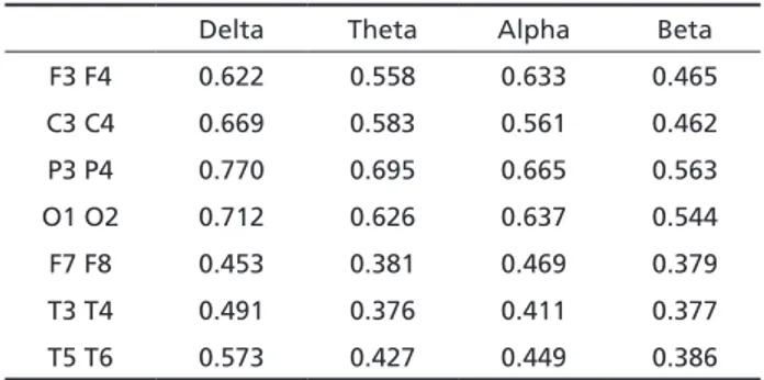
The results of the present study demonstrate a higher coherence value across all frequency bands and sub-bands in parietal regions (electrodes P3P4), showing a statistically significant difference on the Student t test (P ranging from 0.0014 to 0.0056 with a 95% significance level).
Tables 1, 2, and 3 show estimated mean using Hampel’s M-Estimator method for inter-hemispheric coherence of subjects studied.
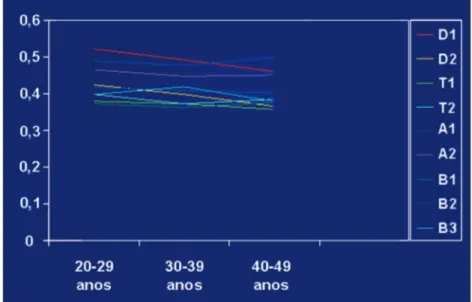
Figures 1 and 2 show that there are no relation in the coherence values between dextrals and sinistrals and its comportment related the age range.
Table 1. Inter-hemispheric coherence value for each frequency band and corresponding electrode pair.
Delta Theta Alpha Beta
F3 F4 0.622 0.558 0.633 0.465
C3 C4 0.669 0.583 0.561 0.462
P3 P4 0.770 0.695 0.665 0.563
O1 O2 0.712 0.626 0.637 0.544
F7 F8 0.453 0.381 0.469 0.379
T3 T4 0.491 0.376 0.411 0.377
T5 T6 0.573 0.427 0.449 0.386
Table 2. Inter-hemispheric coherence value for each electrode pair and frequency sub-band, at rest and with eyes closed.
Delta 1 Delta 2 Theta 1 Theta 2
F3-F4 0.652 0.593 0.544 0.572
C3-C4 0.692 0.646 0.576 0.591
P3-P4 0.792 0.749 0.693 0.697
O1-O2 0.732 0.692 0.637 0.616
F7-F8 0.502 0.405 0.372 0.391
T3-T4 0.549 0.433 0.374 0.379
Arq Neuropsiquiatr 2007;65(2-B) 379
DISCUSSION
In the present study sample, the parietal region (electrodes P3 and P4) tended to present the highest inter-hemispheric Co value, for all frequency bands. However, this finding does not extend to all frequen-cy sub-bands, for which higher values were found
between P3 and P4 electrodes for delta 1 and 2, and theta 1 and 2 sub-bands (Table 2), and alpha 1 and beta 1, 2 and 3 (Table 3), whilst the alpha 2 sub-band presented the highest values in O1 and O2. These data on inter-hemispheric Co is in agreement with results found in the international literature investi-gated13-14.
Thus, as standard and quantitative EEG show variation regarding chronological age, a number of studies have focused on assessing this relationship in coherence. Moreover, despite conflicting results, they show that alterations are different for each frequen-cy band, such that in normal maturation there is an increase in coherence of fast bands, particularly al-pha, with a reduction for slow bands13-16. Concerning the inter-hemispheric coherence value found in the present study for each age bracket of the sample, no significant differences were observed between the 20-29 year, 30-39 year, and 40-49 year age groups. For the 20-50 year age group, EEG spectral characteristics tend to change little, in contrast to both younger (under 10 years) and elderly (over 60 years) groups, according to previous studies in our setting17-18.
Marozi et al.19, in an evolutive study, found in-creased coherence in the frontal region across all bands, except beta, in a population of 46 students followed for 3 years. Aging causes a general re-duction in coherence in all bands, consistent with shrunken corpus callosum, along with reduced neu-ronal population and white matter13-16.
The present study found no differences in inter-hemispheric coherence value in relation to manual dominance in this age bracket, akin to findings by Duffy et al.16
(U Mann-Whitney test with p<0.05). Table 3. Inter-hemispheric coherence value for each electrode
pair and frequency sub-band, at rest and with eyes closed.
Alpha 1 Alpha 2 Beta 1 Beta 2 Beta 3
F3-F4 0.676 0.59 0.474 0.482 0.44
C3-C4 0.607 0.516 0.452 0.468 0.466
P3-P4 0.694 0.637 0.55 0.557 0.583
O1-O2 0.632 0.642 0.518 0.534 0.581
F7-F8 0.482 0.456 0.366 0.402 0.369
T3-T4 0.406 0.416 0.382 0.394 0.356
T5-T6 0.448 0.451 0.365 0.389 0.406
Fig 1. Coherence value for each frequency band at electrode pair F7F8, for dextral and sinistral subjects.
380 Arq Neuropsiquiatr 2007;65(2-B)
The use of a reference for calculating coherence and also for cartographic/mapping display of spectral parameters, have been the subject of investigation for some years, where some studies have compared the use of several types of reference.
The classic study by Rappelsberger20, comparing data obtained through inter-hemispheric and local (adjacent electrodes) coherence analysis, using three types of reference and EEG data simulated by an au-toregressive process of the third order, clearly showed that the use of a linked ears reference (A1+A2) re-sulted in the least distortion to the coherence calcu-lation. Based on this information, the option chosen in the present study was the linked ears reference (A1+A2), although the equipment software was able to use other reference types.
Statistical analysis of the data obtained in this study using non-parametric methods shows that correlation is elevated amongst all frequency bands, meaning that individuals with high coherence in a particular channel and for a given band, also present high coherence across its entire frequency spectrum. Non-parametric correlation of coherence values be-tween different electrode pairs was also calculated. Again, a high correlation between practically all elec-trode pairs was observed, indicating that individuals with a high correlation between electroencephalo-graphic signals of particular inter-hemispheric elec-trode pairs also present high correlation between the other electrode pairs across the whole frequency band analyzed.
A notable finding in the current work was the high inter-hemispheric coherence value found in pa-rietal regions, which may be explained by the an-atomic characteristics of the corpus callosum. The greater thickness of the corpus callosum in these re-gions may be linked to an increased number of inter-hemispheric fiber connections, leading to a higher inter-hemispheric value in these areas, given the sple-nium is anatomically related to the parietal and oc-cipital regions21-24. Studies employing new magnetic resonance techniques such as tractography, may be able to shed further light on these issues.
In conclusion, the current study shows that age, gender and manual dominance in this age bracket (20 to 50 years) do not impact the value obtained for inter-hemispheric coherence, and that all bands have higher coherence values in parietal areas.
REFERENCES
1. Gotman J. The use computers in analysis and display of EEG and evoked potential. In Daly DD, Pedley TA (Eds). Current practice of clinical electroencephalography. 2.Ed. New York: Lippincott-Raven; 1990:51-83.
2. Cibils D. Análise da coerência na doença de Alzheimer e no envelheci-mento normal. In Anghinah R, Luccas FJC (Eds). A neurofisiologia cli-nica no auxílio diagnóstico das demências. ��o Paulo: �iosint�tica, 1999:��o Paulo: �iosint�tica, 1999: 63-68.
3. Towle VL, Carder RK, Khorasani L, Linberger D. Electrocorticograph-ic coherence patterns. J Clin Neurophysiol 1999;16:528-547.
4. Nunez P. Electric fields of the brain: the neurophysics of EEG. New York: Oxford University Press, 1981.
5. Nunez PL, �rinivasan R, Westdorp AF, Wijesinghe R�, Tucker DM, �il-berstein R�. EEG coherence I: statistics, reference electrode, volume conduction, laplacians, cortical imaging, and interpretation at multi-ple scales. Electroencephalogr Clin Neurophysiol 1997;103:499-515. 6. Thatcher RW, Krause PJ, Hrybyk M. Cortico-cortical associations and
EEG coherence: a two-compartmental model. Electroencephalogr Clin Neurophysiol 1986;64:123-143.
7. Lesser RP. Guidelines Committees of American Electroencephalograph-ic �ociety. J Clin Neurophysiol 1994;11:9-11.
8. Nuwer MR, Comi G, Emerson R, et al. IFCN standards for digital re-cording of clinical EEG. Electroencephalograph Clin Neurophysiol 1998;106:259-261.
9. Luccas FJ, �raga NI, Fonseca LC, Froch Tengarten ML. Recomendaç�esRecomendaç�es para o registro e interpretaç�o de mapeamento topográfico do eletren-cefalograma (EEG) e potenciais evocados sensoriais (PE�) parte I: as-pectos gerais. �razilian J Epilepsy Clin Neurophysiol 1996;2:175-182.�razilian J Epilepsy Clin Neurophysiol 1996;2:175-182. 10. Luccas FJ, Anghinah R, �raga NI, et al. Recomendaç�es para o
regis-tro/interpretaç�o do mapeamento topográfico do eletroencefalograma e potenciais evocados. Parte II: correlaç�es clínicas. Ar� Neuropsi�uiatrParte II: correlaç�es clínicas. Ar� Neuropsi�uiatr 1999;57:132-146.
11. Pagano M, Gauvreau K. Coeficientes de correlaç�o de postos de �pear-man. In Pagano M, Gauvreau K (Eds). Principios de �ioestatística. ��o Paulo: Thomson, 2004:357-360.
12. Levin J. Estatística aplicada a ciências humanas. 2.Ed. Traduç�o de �er-gio Francisco Costa. ��o Paulo: Hamburg, 1987:283-288.
13. Duffy FH, Mcanulty G�, Albert M�. Effects of age upon interhemispher-ic EEG coherence in normal adults. Neurobiol Aging 1996;17:587-599. 14. Harmony T, Marosi E, Fernandez T, et al. EEG coherences in patients
with brain lesions. Int J Neurosci 1994;74:203-226.
15. Thatcher RW, Walker RA, Giudice �. Human cerebral hemispheres de-velop at different rates and ages. �cience 1987;236:1110-1113. 16. Duffy FH, Jones KJ, McAnulty G�, Albert M�. �pectral coherence in
normal adults: unrestricted principal components analysis; relation of factors to age, gender, and neuropsychologic data. Clin Electroence-falogr 1995;26:30-46.
17. Anghinah R, Kanda PA, Jorge M�, Lima EE, Pascuzzi L, Melo AC. Es-tudo da coerência do eletrencefalograma para a banda de fre�üência alfa em indivíduos adultos normais e com provável demência do tipo Alzheimer. Ar� Neuropsi�uiatr 2000;58:272-275.
18. Anghinah R, Caramelli P, Takahashi DY, Nitrini R, �ameshima K. Es-tudo da coerência do eletrencefalograma na banda de fre�üência alfa em indivíduos adultos normais. Ar� Neuropsi�uiatr 2005;63:83-86. 19. Marosi E, Harmony T, Reyes A, et al. A follow-up study of EEG
coher-ences in children with pedagogical different evaluations. Int J Psycho-physiol 1997;25:227-235.
20. Rappelsbersger P. The reference problem and mapping of coherence: a simulation study. �rain Topogr 1989;2:63-72.
21. Hwang �J, Ji EK, Lee EK, et al. Gender differences in the corpus callo-sum of neonates. Neuroreport 2004;15:1029-1032.
22. �uganthy J, Raghuram L, Antonisamy �, Vettivel �, Madhavi C, Koshi R. Gender and age-related differences in the morphology of the corpus callosum. Clin Anat 2003;16:396-403.
23. Takeda �, Hirashima Y, Ikeda H, Yamamoto H, �ugino M, Endo �. De-termination of índices of the corpus callosum associated with normal aging in Japanese individuals. Neuroradiology 2003;45:513-518. 24. Laissy JP, Patrux �, Duchateau C, et al. Midsagittal MR measurements