4 5 8
Tatani et al
Transesophageal echocardiography in patients with stroke
Arq Bras Cardiol 2001; 76: 458-61.
OMNI – Centro de Cardiologia Não Invasiva UNIFESP-Escola Paulista de Medicina, John Hopkins University – USA
Mailing address: Solange Bernardes Tatani - Rua Gil Fernandes, 183 - 04148-020 - São Paulo, SP - Brazil
Objective - The purpose of this study is to evaluate
the impact of transeophageal echocardiography on mana-gement of patients at low-risk for cardiogenic embolism to prevent new potential cardiovascular sources of emboli.
Methods - We studied 69 patients with ischemic stroke
at low-risk for cardiogenic embolism. Transeophageal echo-cardiography was performed to access: left atrium enlar-gement; communication or aneurysm of the interatrial septum; patent foramen ovale; spontaneous echo contrast or intracavitary thrombi; the presence of intraaortic atherosclerotic plaques or thrombi; significant valvar morphologic alteration or dysfunction; left ventricle enlargement, hypertrophy, or contractile abnormality. Transesophageal echocardiography altered clinical mana-gement, and we adopted anticoagulant therapy or another procedure apart from the use of acetylsalicylic acid.
Results - Transeophageal echocardiography detected
at least one abnormality in 40 cases (58%). Clinical con-duct was adjusted after the performance of transesopha-geal echocardiography in 11 patients (15.9%); anticoa-gulation was added in 10 cases and surgical correction in one patient.
Conclusion - Transeophageal echocardiography
was a very useful tool in the secondary prevention for stro-ke in patients at low risk for cardiogenic embolism.
Key words: transeophageal echocardiography, stroke, embolism.
Arq Bras Cardiol, volume 76 (nº 6), 458-61, 2001
Solange Bernardes Tatani, Márcia Maiumi Fukujima, João Augusto Costa Lima, Luiz Darcy Cortez Ferreira, Claudia G. Monaco Ghefter, Gilmar Fernandes Prado, Zara Babayan, Lyamara Apostólico de Azevedo
São Paulo, SP – Baltimore, MD
Clinical Impact of Transesophageal Echocardiography in
Patients with Stroke without Clinical Evidence of
Cardiovascular Sources of Emboli
Original Article
The increasingly important role of stroke as a cause of mortality and disability is real. Statistics show that each year approximately 500,000 Americans experience a stroke and 147,800 die of stroke. Moreover, stroke accounts for rou-ghly 2 million cases of permanent disability 1. In Brazil, few
epidemologic studies have been published on cerebrovas-cular diseases, even though they are the most frequent cau-se of death in the country 2,with mortality rates in some
Bra-zilian capitals 4 to 7 times higher than that in other internatio-nal cities 3.
Stroke may occur due to many causes, and a cardio-vascular source of emboli is believed to be responsible for 30% of the cases. Among high-risk patients of embolism-prone cardiac disorders are patients with mitral valve steno-sis, atrial fibrillation (rheumatic and nonrheumatic), dilated myocardial disease, prosthetic valves, and recent myocar-dial infarction 4.
In recent years, the use of transesophageal echocar-diography has greatly contributed to the identification of other cardiac disorders that are being considered new po-tential causes of stroke, such as: patent foramen ovale 5,
atrial septal defect 6, atrial septal aneurysm 7, protruding
aortic atheromatous plaque (>4 - mm thick) 8,9, spontaneous
echo contrast 10, mitral valve strands 11 and intracavitary
thrombi (mainly in the left atrial appex).
The present study aimed at evaluating data obtained from transeophageal echocardiography in patients with is-chemic stroke, who were not included in the therapy adop-ted in the high-risk cardiac group.
Methods
pa-Arq Bras Cardiol 2001; 76: 458-61.
Tatani et al Transesophageal echocardiography in patients with stroke
4 5 9
tients with ischemic stroke who were admitted to the first-aid neurology clinic at the Escola Paulista de Medicina, re-gardless of age and sex. Neurologists performed the diagno-sis and evaluation of patients, following a standard proto-col, including a clinical examination and brain CT. Patients with atrial fibrillation, recent myocardial infarction (last six months), prosthetic cardiac valves, severe impairment of left ventricular function (IF<20%), formal contraindication for TEE, severely ill patients, and those receiving hospital care for more than 15 days were excluded from this study.
After hospital discharge, all patients underwent multipla-ne transeophageal echocardiography with a 5mHz transe-sophageal probe and with Vingmed echocardiographic System V (within 15 days). Images were recorded on videotape for later review by two observers. TEE examinations were performed af-ter administration of topical anesthesia with an aerolized so-lution of lidocaine at 10% and intravenous sedation with mi-dazolan (1.5mg/ml) and meperidine (50mg/ml). Contrast studies were performed by the rapid injection in the peripheral vein of microbubble solution (6ml of isotonic saline 0.9%, 4ml of glucosis 50%, and 1ml of air) at rest, during coughing, and during Valsalva maneuver. A comprehensive transeophageal echocardiography examination was performed with stan-dardized scan planes. The following abnormalities were evaluated: left atrium enlargement, presence of masses, thrombi, or spontaneous contrast either inside the atrium or at the atrial apex or left ventricle. Patients were also evaluated for the following: interatrial septum aneurysm; patent foramen ovale, or any interatrial septal communication; fibrosis, mitral valve strands, calcifications, myxomatous degeneration, significant stenosis, or regurgitation of the mitral or aortic valve; enlargement, hypertrophy, left ventricle segmental or global dysfunction; and also atherosclerotic plaques or thrombi in the thoracic aorta.
Patent foramen ovale was diagnosed if more than three microbubbles were visualized in the left atrium within 5 car-diac cycles, following the opacification in the right atrium (Fig.1). Interatrial septal aneurysm was diagnosed when ex-cessive expansion was observed (>1.5cm of bulging) at least 1.5cm septum basis (do you mean that the bulge had to be measured as 1.5 cm at the base of septum?). Spontaneous echo contrast was characterized by smooth echoes with cir-cular or spiral movement inside cardiac chambers. Mitral valve strands were defined as thin mobile filamentous pro-jections attached to the atrial surface of mitral leflets or sub-valvar apparatus. The left atrium was considered enlarged when it was >40 mm. The left ventricle was considered en-larged when diastolic diameter was >50mm and hypertrophic when diastolic thickness was ≥12mm; atherosclerotic pla-ques of in the ascending aorta, aortic arch, and descending aorta had their maximum thickness measured and were clas-sified into 2 groups: those <4mm and those ≥4mm.
Immediately after the examination, a report with the results obtained was sent to the patient’s personal physician. No events occurred during the echocardiography exam. A neurologist followed up patients within seven days after transeophageal echocardiography had been performed.
Results
Sixty-nine patients with ages ranging from 12 to 90 years (mean age 63.4±14.1 years); 52.2% were women. Of the patients, 44 (63.8%) had hypertension, six (8.7%) had diabe-tes, and two (2.9%) had known coronary disease.
Left atrium enlargement occurred in 20 patients (29%), left ventricle enlargement in 17 (24.6%), myocardial hyper-trophy in 30 (43.5%), and mied or moderated left ventricle systolic impairment in 10 (14.5%). Transeophageal study detected at least one potential cardiovascular source of em-boli in 40 patients (58%). Regarding interatrial septum, 15 (21.7%) patients had patent foramen ovale (fig. 2), 7 (10.1%) had interatrial septum aneurysm, and one had interatrial communication. Of the patients who had interatrial septum aneurysm, four had associated patent foramen ovale. Intra-cardiac thrombus was observed in three patients (4.3%); two thrombi were located in the left ventricle and one on the left atrial appendage. Spontaneous echo contrast was observed in 19 patients (27.5%). Atherosclerotic plaques were detected in 54 patients (78.3%), and in 63.8% of the cases those plaques were smaller than 4mm, and in 14.5% they were 4mm or more thick. Mitral valve strands were not reported in any of the patients. Apart from these findings,
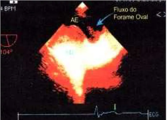
Fig. 2 - Transeophageal echocardiography showing flow through patent foramen ovale.
4 6 0
Tatani et al
Transesophageal echocardiography in patients with stroke
Arq Bras Cardiol 2001; 76: 458-61.
we diagnosed in one patient, descending aorta dissection, mied mitral stenosis (with valvar area estimated at 1.6cm2)
and myxomatous degeneration of the mitral valve. In these cases, no clinical evidence existed to suggest diagnosis of these abnormalities, and another potential source of emboli existed in all cases.
Based on transeophageal study, therapeutics was changed in 11 patients (15.9%). Ten patients received oral anticoagulant (warfarin), and the patient who had aortic dissection was sent to for surgical correction. Anticoagu-lant therapy was indicated in 3 cases of intracavitary throm-bus, 2 cases of isolated patent foramen ovale, 3 cases of patent foramen ovale associated with interatrial septal aneurysm, patent foramen ovale, and one case of global and mied systolic left ventricle dysfunction. Oral anticoagulant was also indicated in one patient with mitral stenosis and patent foramen ovale, who had spontaneous echocardio-graphy contrast inside left atrium. The other patients received acetylsalicylic acid.
Discussion
It is very important to identify the cause of ischemic stroke so that the therapeutics adopted can be defined, because the management of patients with cerebrovascular atherosclerotic stroke is substantially different from that of patients with ischemic stroke due to embolism. It is funda-mental to secondary prevention of stroke because in 12% of these patients has a recurrent stroke.
Platelet antiaggregating drugs, especially acetylsali-cylic acid, are the best option for patients with cerebrovas-cular atherosclerotic stroke in which platelets play an impor-tant role in the development of thrombi on occlusion of arte-ries, the vertebrobasilar system, or cerebral artery. On the other hand, if cerebral artery occlusion occurs from a cardio-genic thrombus made of mainly red cells and fibrin, anticoa-gulants should be used for both treatment and prevention. Anticoagulant therapy prevents the development of new thrombi, thus preventing recurrence of ischemic stroke in patients with embolic diseases. Many studies have shown the benefits of anticoagulant therapy in patients wi-th known embolic sources 14,15 (especially in detecting
intra-cardiac thrombi). No doubts exist concerning the need for in-dication of anticoagulant therapy in these cases.
Although it is important to define the cause of a stroke, the cause is generally presumed and seldom recognized. No consensus exists about the use of imaging strategies to be used in this investigation.
Transeophageal echocardiography has enabled iden-tification of new potential sources of emboli, such as patent foramen ovale, interatrial septum aneurysm, spontaneous echocardiography contrast, and atheromatous aortic pla-ques in patients with ischemic stroke who are at low-risk for cardiac diseases. Transeophageal echocardiography is also more accurate in detecting intracardiac thrombus (especial-ly in the left atrial appendage).
Patent foramen ovale has been related to paradoxical
embolism, in which thrombi (intracavitary or peripheral) pass from the right to the left atrium through a patent fora-men ovale. With transesophageal echocardiography, it is possible to visualize all the extension of the interatrial sep-tum, and also to observe transeptal flow through foramen ovale. However, cases exist in which the orifice is very nar-row with minimum transeptal flow and may not be noticed. To avoid false-negative results in such cases, we make use of contrast injection in the peripheral vein of agitated saline solution, at rest and after maneuvers, which causes an in-crease in pressure in the right side of the heart, thus deter-mining the passage of contrast through patent foramen ova-le. In 1988, Webster et al 16 and Lechat et al 17 reported a
hi-gher prevalence of patent foramen ovale in young patients with cryptogenic stroke (respectively, 40% and 50%) when compared with patients without stroke (10% and 15%, res-pectively). After these studies, other studies using transeo-phageal echocardiography have proven the association of patent foramen ovale and stroke. Patent foramen ovale has also been associated with a higher rate of stroke recurrence. Mas et al 18 found in a retrospective study a 4.4% annual
rate of recurrence of events in patients with cryptogenic stroke and patent foramen ovale. Bogousslavsky et al 19
per-formed a prospective study with stroke patients who were less than 60 years of age, in which they found a 1.9% annual rate of recurrence, in that, patent foramen ovale was one of the predictors of recurrence. However, few studies have been successful in proving venous thrombosis in patients with stroke and patent foramen ovale. Stöllberg et al 20 after
performing of transesophageal echocardiography docu-mented venous thrombosis in 24 of the 42 patients with patent foramen ovale and with suspected paradoxical embo-lism. Nellensen et al 21 detected through transeophageal
echocardiography a thrombus overriding the interatrial septum in patients with deep venous thrombosis. However, other studies, such as the ones of Ranoux et al 22 and Gautier
et al 23, could not document venous thrombosis in patients
with patent foramen ovale and stroke.
Correlation between interatrial septal aneurysm and embolism has been demonstrated 7,24; however, the reason
for embolism is yet unknown. Association with patent fora-men ovale in such patients is frequent 25, thus making them
prone to paradoxical embolism.
The aorta is the most common site of atherosclerosis, surpassing the carotid and vertebral arteries. Risk of emboli increases according to the dimensions of atheromatous pla-ques and is higher in those >4mm thick; it is also higher in the presence of ulcerated plaques and when platelet deposits with thrombi are present, leading to occurrence of mobile debris on the surface 8,9,26-, 28.
Arq Bras Cardiol 2001; 76: 458-61.
Tatani et al Transesophageal echocardiography in patients with stroke
4 6 1
which supports the findings of previous studies in the li-terature. A classic study by Chimowitz et al 29 shows double
embolic events in patients with mitral disease, who had spontaneous contrast on transesophageal echocardiogra-phy compared with those without spontaneous contrast.
Although transesophageal echocardiography has been proven to be a useful tool in the detection of new sour-ces of emboli previously described, the routine use of this procedure in the patient with ischemic stroke is still contro-versial. The use of transeophageal echocardiography in risk stratification for new ischemic events was conducted at Johns Hopkins University. O’Brien and cols have published preliminary results of this study 30, and have proven that it is
possible to define the prognosis of patients with ischemic stroke using transesophageal echocardiography findings, which identify atherosclerotic aortic and spontaneous echo contrast as predictors of new event recurrence.
1. Wolf PA. An overview of the epidemiology of stroke. Stroke 1990; 21(suppl II): II-4-II-6.
2. Sistema de informações hospitalares do SUS (SIH/SUS). Mortalidade e morbida-de hospitalar do SUS. Http://www.datasus.gov.br
3. Duncan BB, Schmidt MI, Polanczyk CA, Mengue SS. Altos coeficientes de mor-talidade em população adulta brasileira: uma comparação internacional. Rev Assoc Med Bras 1992; 38: 138-44.
4. Cardiogenic brain embolism: the second report of the Cerebral Embolism Task Force. Arch Neurol 1989; 46: 727-43.
5. Di Tullio M, Sacco RL, Gopal A, Mohr JP, Homma S. Patent foramen ovale as a risk factor for cryptogenic stroke. Ann Intern Med 1992; 117: 461-5.
6. Harvey JR, Teague SM, Anderson JL, Voyles WF, Thadani U. Clinically silent atrial septal defects with evidence of cerebral embolization. Ann Intern Med 1986; 105: 695-7.
7. Lucas C, Goullard L, Marchan M, et al. Higher prevalence of atrial septal aneurysms in patients with ischemic stroke of unknown cause. Acta Neurol Scand 1994; 89: 210-3.
8. Amarenco P, Cohen A, Tzourio C, et al. Atherosclerotic disease of the aortic arch and the risk of ischemic stroke. N Engl J Med 1994; 331: 1474-9.
9. Amarenco P, Duyckaerts C, Tzourio C, Hérnin D, Bousser M-G, Hauw JJ. The pre-valence of ulcerated plaques in the aortic arch in patients with stroke. N Engl J Med 1992; 326: 221-5.
10. Briley DP, Giraud Gd, Beamer NB, et al. Spontaneous echo contrast and hemo-rheologic abnormalities in cerebrovascular disease. Stroke 1994; : 1564-9. (need volume #)
11. Tice FD, Slivka AP, Walz ET, Orsinelli DA, Pearson AC. Mitral valve strands in patients with focal cerebral ischemia. Stroke 1996; 27: 1183-6.
12. Hart RG. Cardiogenic embolism to the brain. Lancet 1992; 339: 589-94. 13. Leonard AD, Newburg S. Cardioembolic stroke. J Neurosci Nurs 1992; 24: 69-76. 14. Baker RN, Broward JA, Fang HC. Anticoagulant therapy in cerebral infarction:
report on cooperative study. Neurology 1962; 12: 823-35.
15. Report of the veterans administration cooperative study of atherosclerosis: an evaluation pf anticoagulation therapy in the treatment of cerebrovascular disea-se. Neurology 1964; 1: 132-8.
16. Webster MWI, Smith HJ, Sharpe DN, et al. Patent foramen ovale in young stroke patients. Lancet 1988; 2: 11-2.
17. Lechat P, Mas JL, Lascault G, et al. Prevalence of patent foramen ovale in patients with stroke. N Engl J Med 1988; 318: 1148-52.
18. Mas JL, Zuber M. Recurrent cerebrovascular events in patients with patent fora-men ovale or atrial septal aneurysm, or both, and cryptogenic stroke or TIA.
Fren-References
ch Study Group on Patent Foramen Ovale and Atrial Septal Aneurysm. Am Heart J 1995; 140: 1083-8.
19. Bogousslavsky J, Garazi S, Jeanrenaud X, Aebischer N, Van Melle G. Stroke re-currence in patients with patent foramen ovale: The Lausanne Study. Neurology 1996; 46: 1301-5.
20. Stöllberg C, Slany J, Schuster I, Leitner H, Winkler WB, Karnik R. The preva-lence of deep venous thrombosis in patients with suspected paradoxical embo-lism. Ann Intern Med 1993; 119: 461-5.
21. Nellensen U, Daniel WG, Matheis G, Oelert H, Depping K, Lichtlen PR. Impen-ding paradoxical embolism from atrial thrombus: correct diagnosis by transeso-phageal echocardiographic and prevention by surgery. J Am Coll Cardiol 1985; 5: 1002-4.
22. Ranoux D, Cohen A, Cabanes L, Amarenco P, Bousser MG, Mas JL. Patent fora-men ovale: is stroke due to paradoxical embolism? Stroke 1993; 24: 331-4. 23. Gautier JC, Dürr A, Koussa S, Lasclult G, Grosgogeat Y. Paradoxical cerebral
em-bolism with a patent foramen ovale. Cerebrovasc Dis 1991; 1: 193-202. 24. Agmon Y, Khandheria BK, Meissner I, Gentile F, Whisnant JP, Sicks JD.
Frequen-cy of atrial septal aneurysm in patients with cerebral ischemic events. Circulation 1999; 99: 1942-4.
25. Mügge A, Daniel WG, Angermann C, et al. Atrial septal aneurysm in adult pati-ents. A multicenter study using transthoracic and transesophageal echocardio-graphy. Circulation 1995; 91: 2785-92.
26. Karalis DG, Chandrasekaran K, Victor MF, Ross JJ, Mintz GS. Recognition and embolic potential of intraaortic atherosclerotic debris. J Am Coll Cardiol 1991; 17: 73-8.
27. Tunick PA, Rosenzweig BP, Katz ES, Freedberg RS, Perez JL, Kronzon I. High risk for vascular events in patients with protruding aortic atheromas: a prospec-tive study. J Am Coll Cardiol 1994; 23: 1085-90.
28. Stone DA, Hawke MW, LaMonte M, et al. Ulcerated atherosclerotic plaques in the aorta are associated with cryptogenic stroke: a multiplane transesophageal echocardiographic study. Am Heart J 1995; 130: 105-8.
29. Chimowitz MB, DeGeorgia MA, Poole RM. Left atrial spontaneous echo con-trast is highly associated with previous stroke in patients with atrial fibrillation or mitral stenosis. Stroke 1993; 24: 1015-9.
30 O’Brien PJ, Thiemann DR, McNamara RL, et al. Usefulness of transesophageal echocardiography in predicting mortality and morbidity in stroke patients wi-thout clinically known cardiac sources of embolus. Am J Cardiol 1998; 81:. 31. Mc Namara RL, Lima JAC, Whelton PK, Powe NR. Echocardiographic
identifi-cation of cardiovascular sources of emboli to guide clinical management of stroke: a cost-effectiveness analysis. Ann Int Med 1997; 127: 775-87.
Furthermore, according to a study conducted by McNamara et al 31, cost-effectiveness analysis of
transeo-phageal echocardiography in the prevention of secondary stroke has been presented as the best cost-effectiveness me-thod, when compared with other strategies of diagnosis and therapeutics frequently used in such patients.