O
r i g i n a la
rt i c l e3 1 3 Arq Bras Oftalmol. 2017;80(5):313-6 http://dx.doi.org/10.5935/0004-2749.20170076
ABSTRACT
Objective: The aim of this study was to determine the central corneal thickness (CCT) and intraocular pressure (IOP) in full-term newborns and to correlate these values with the following variables: weight, sex, and post-conception age (PCA).
Methods: IOP and CCT were determined in 52 full-term newborns with a mean gestational age of 39.43 ± 1.03 weeks and a mean birth weight of 3,273 ± 558 g. The mean age of the neonates at the time of taking the measurements was 1.15 ± 1.38 days after birth. IOP was determined with a Tono-Pen, and CCT was deter-mined using a handheld ultrasound pachymeter.
Results: The mean overall IOP was 14.0 ± 2.91 mmHg, and the mean IOP in male and female newborns was 13.77 ± 2.88 mmHg and 14.32 ± 3.05 mmHg, respec-tively. The mean overall CCT was 605.87 ± 62.98 µm, and the mean CCT in male and female newborns was 626.70 ± 67.46 µm and 577.45 ± 45.50 µm, respectively; the mean CCT was higher in male newborns than in female newborns. PCA was negatively associated with CCT, IOP, and weight; however, only the correlation with CCT was statistically significant. Comparisons between the sexes showed significant differences for two variables: weight and CCT.
Conclusions: No relationship was found between CCT and IOP; however, we found a positive association between weight and CCT.
Keywords: Glaucoma/genetics; Cornea topography; Intraocular pressure; Tono-metry, ocular; Humans; Infant, newborn
RESUMO
Objetivo: O objetivo deste estudo foi avaliar a espessura central da córnea (ECC) e a pressão intraocular (PIO) em recém-nascidos (RN) a termo, correlacionando seus valores entre as variáveis peso, sexo e idade pós-concepção (PCA).
Métodos: A pressão intraocular e a espessura central da córnea foram
determi-nadas em 52 recém-nascidos a termo com idade gestacional média de 39,43 ± 1,03 semanas, peso médio de nascimento de 3.273 ± 558 g. A média de idade durante as medições após o nascimento foi 1,15 ± 1,38 dias. A pressão intraocular foi determinada com Tono-pen. A espessura central da córnea foi determinada com paquímetro ultrassônico portátil.
Resultados: A pressão intraocular média foi de 14,0 ± 2,91 mmHg em ambos
os sexos, de 13,77 ± 2,88 mmHg nos recém-nascidos do sexo masculino e 14,32 ± 3,05 mmHg nos recém-nascidos do sexo feminino. A espessura central da córnea média foi de 605,87 ± 62,98 μm em ambos os sexos; de 626,70 ± 67,46 μm recém-nascidos do sexo masculino e de 577,45 ± 45,50 μm nos recém-nascidos do sexo feminino. A maior média da espessura da córnea foi no sexo masculino. A variável idade pós-concepção teve uma relação decrescente com espessura central da córnea, pressão intraocular e peso, porém, a correlação só foi significante com a espessura central da córnea. Na comparação entre os sexos houve diferenças significativas para duas variáveis: peso e espessura central da córnea.
Conclusões: Não houve relação entre a espessura central da córnea e a pressão
intraocular neste estudo, entretanto verificou-se que existe uma relação de aumento de peso com a espessura central da córnea.
Descritores: Glaucoma/genético; Topografia da córnea; Pressão intraocular;
Tono-metria ocular; Humanos; Recém-nascido
Intraocular pressure and central corneal thickness in full-term newborns
Medida da pressão intraocular e espessura corneana central em recém-nascidos a termo
Claudia Cardoso Maestri Ferreira1, ivan Maynart tavares1
Submitted for publication: September 23, 2016 Accepted for publication: March 19, 2017
1 Department of Ophthalmology, Escola Paulista de Medicina, Universidade Federal de São Paulo,
São Paulo, SP, Brazil.
Funding: No specific financial support was available for this study.
Disclosure of potential conflicts of interest: None of the authors have any potential conflict of interest to disclose.
Corresponding author: Claudia Cardoso Maestri Ferreira. Rua Gelu Vervloet dos Santos, 280/501 - Vitória, ES - 29090-100 - Brazil - Email: [email protected]
Approved by the following research ethics committee: Universidade Federal de São Paulo (#110.281).
INTRODUCTION
Until the 20th century, little was known about ocular pressure in newborns, mainly because of the lack of suitable equipment for measuring ocular pressure. The physical properties of the neonatal cornea appear to be the main factors associated with diiculty in obtaining reliable measurements of intraocular pressure (IOP) using the majority of available tonometers. Corneal thickness is an inde-pendent risk factor for glaucoma development in adults with ocular hypertension(1). However, few studies have been conducted in Brazil
on IOP values in full-term newborns, and these studies have not re-ported a correlation between IOP and central corneal thickness (CCT)(2).
Glaucoma is an important disease in children, accounting for 2.5%-10% of all cases of childhood blindness(3). Knowledge of the reference
values for IOP in newborns, in association with CCT, weight, sex, and post-conception age (PCA), is important for the early diagnosis of glaucoma, especially in children who have not yet developed the
classic signs of primary congenital glaucoma (e.g., cloudy cornea, bu-phthalmia). The aim of the present study is to ill this knowledge gap.
METHODS
The study protocol was approved by the Ethics Committee of the Federal University of São Paulo. The parents/guardians of the newborns were informed about the tests, and provided their written consent.
In t r a o c u l a rp r e s s u r ea n dc e n t r a lc o r n e a lt h I c k n e s sI nf u l l-t e r mn e w b o r n s
3 1 4 Arq Bras Oftalmol. 2017;80(5):313-6
The newborns were placed on a wheeled stretcher in the supine position. After the administration of topical anesthesia (one drop of 0.5% proparacaine), their eyelids were carefully parted by the operator’s ingers while avoiding placing the weight of the hand on the ocular globe, which can inluence the accuracy of optical measurements. The measurement was only performed when the newborn was quiet and still. The mean IOP was calculated after three measurements with a Tono-Pen AVIA (Reichert, Depew, NY, USA). If the IOP results from the three measurements difered by >3 mmHg, the measurement was repeated. The mean CCT was calculated after eight measurements in each eye with an SP-100 portable pa-chymeter (Tomey, Nagoya, Japan). All measurements were centrally performed, and all tests were performed by the same operator during the same period of the day (between 13 h and 16 h).
Statistical analyses were initiated by the characterization of va-riables through measures of central tendency and variability. The Kolmogorov-Smirnov test was used to verify the normality of the data. Multiple linear regression was used to evaluate the relationship between variables. The premises of normality of the residuals (Kolmo-gorov-Smirnov), absence of serial autocorrelation (Durbin-Watson), absence of heteroscedasticity [(variance inlation factor (VIF)], and the absence of multicollinearity (robust standard error) were veriied. The diferences between the sexes of the tested variable. were deter-mined by the Mann-Whitney test. The level of signiicance adopted was 5%, with a conidence interval of 95.0%. SPSS Statistics software (version 21; IBM Corp., Armonk, NY, USA) was used to perform all statistical analyses.
RESULTS
The overall mean IOP was 14 ± 2.91 mmHg (range 8-20 mmHg) (Table 1), and the mean IOP in male and female newborns was 13.77 ± 2.88 mmHg (range 8-19 mmHg) and 14.32 ± 3.05 mmHg (range 8-20 mmHg), respectively. The overall mean CCT was 605.87 ± 62.98 μm (range 483-794 μm), and the mean CCT in male and female newborns was 626.70 ± 67.46 μm (range 483-794 μm) and 577.45 ± 45.50 μm (range 521-686 μm), respectively. The mean CCT was significantly higher in male newborns (Tables 2 and 3).
Multiple linear regression analysis showed statistical signii cance for weight, female sex, and PCA. There was a positive re lationship between weight and CCT. Female neonates had significantly lower CCT than male neonates. Further, CCT decreased as the PCA increased (Table 4). Male weight was significantly asso ciated with CCT; as the mean weight increased, the CCT also increased (Table 5). Conversely, no significance was found for any of the va-riables in the female neonates; that is, weight, IOP, and PCA were independent of CCT (Table 6).
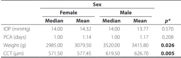
The Mann-Whitney test showed signiicant diferences between the sexes for two variables only, namely weight and CCT, and male sex obtained significantly higher means and median for both va-riables (Table 7).
Table 1. Description of the variables under analysis in newborns
Interval
Minimum Maximum Mean ± SD
Age (days) 0000 7 0001.15 ± 001.38
CCT (µm) 0483 794 0605.87 ± 062.98
IOP (mmHg) 0008 20.0 0014.00 ± 002.91
GA (weeks) 0037 41.6 039.46 ± 001.03
Weight (g) 1960 4435 3273.56 ± 558.20
PCA= post-conception age; CCT= central corneal thickness; IOP= intraocular pressure; GA= gestational age; SD= standard deviation.
Table 2. Mean CCT and sex
Variable n Mean CCT ± SD
Newborns 52 605.87 ± 63.69
Sex
F 22 577.45 ± 45.50
M 30 626.70 ± 67.46
n= number of newborns; CCT= central corneal thickness; SD= standard deviation; F= fe-male; M= male.
Table 3. Relationship between PCA and CCT
PCA
CCT IOP
Mean ± SD Interval Mean ± SD Interval
days n µm µm mmHg mmHg
0 17 632.35 ± 66.69 530-794 14.41 ± 2.67 08-18
1 22 607.36 ± 60.29 521-749 13.64 ± 3.26 08-19
2 09 569.44 ± 48.27 524-661 14.44 ±2.70 12-20
3 01 603.00 - 18
-4 01 483.00 - 13
-6 01 573.00 - 10
-7 01 609.00 - 12
-Total 52 605.86 483-794 14 ± 2.94 08-20
PCA= post-conception age; CCT= central corneal thickness; IOP= intraocular pressure; SD= standard deviation; n= number of newborns.
Table 4. Association between CCT and the variables under analysis (overall)
Independent variable
Model
R² p
95.0% conidence interval for B
VIF (with robust
standard error)
Lower limit
Upper limit
Weight (g) y= 655.83 + 0.05x 0.356 0.005 -00.02 -00.090 1.031
Sex (female) y= 655.83 - 43.40x 0.006 -73.78 -13.013 1.025
IOP (mmHg) y= 655.83 - 1.51x 0.560 0-6.69 -03.670 1.032
PCA (days) y= 655.83 - 11.89x 0.034 -22.83 -0-0.950 1.034
Durbin-Watson= 2.128; Kolmogorov-Smirnov (p) = 0.200.
R²= Adjusting the model; IOP= intraocular pressure; PCA= post-conception age; VIF= variance inlation factor. Dependent variable: CCT= central corneal thickness.
Table 5. Association between CCT and the variables under analysis (male newborns)
Independent variable
Model
R² p
95.0% conidence interval for B
VIF (with robust
standard error)
Lower limit
Upper limit
Weight (g) y= 654.58 + 0.55x 0.303 0.014 -0.12 0.10 1.019
IOP (mmHg) y= 654.58 - 1.42x 0.726 -9.69 6.85 1.104
PCA (days) y= 654.58 -11.86x 0.092 -25.77 2.06 1.115
Durbin-Watson = 1.932; Kolmogorov-Smirnov (p) = 0.200.
Fe r r e i r a CCM, Tava r e s iM
3 1 5
Arq Bras Oftalmol. 2017;80(5):313-6
DISCUSSION
In the present study, we assessed the effect of CCT on IOP, weight, sex, and PCA in full-term newborns. The mean IOP was 14.42 ± 2.91 mmHg, which is lower than that observed in previous similar studies(4-6).
As per the existing literature, IOP values obtained in the supine position tend to be higher than those measured in the sitting po-sition as a result of changes in scleral venous pressure(7). Although
Goldmann applanation tonometry is the gold standard method for measuring IOP, it only provides estimated values(8,9). Accuracy for
this measurement depends on the use of correct technique, and in particular, the use of adequate amounts of luorescein(10). However,
errors resulting from changes in the corneal thickness and rigidity cannot be avoided when using Goldmann applanation tonometry(8,9).
The Tono-Pen is the preferred instrument for pediatric patients be-cause it is less afected by corneal thickness, according to a study by Yildirim et al.(10).
In the present study, the mean CCT in full-term newborns was 605.87 ± 62.98 µm. This value is higher than that reported in several previous studies(4,11-14) but similar to that reported by Rushood et al.(15).
Other studies have shown that CCT is signiicantly lower in children with congenital glaucoma or childhood glaucoma(16,17).
In a study by Uva et al.(4), premature and full-term newborns
were compared; the results showed that IOP increased with an in-crease in CCT, and that CCT dein-creased with an inin-crease in birth weight. The authors concluded that the main factor affecting IOP was CCT. In the present study, there was no correlation between CCT and IOP. Although a correlation between CCT and IOP has not been demonstrated in the literature, there is little information about this correlation in full-term newborns. Studies such as that conducted by Karahan et al.(6) have similarly not shown an effect
of CCT on IOP in premature newborns; however, these previous studies were limited by their small sample sizes.
In the present study, there was a significant negative corre-lation between PCA and CCT (Table 5). This finding corroborates
the results found by Ehlers et al.(19), who observed that corneal
thickness in newborns is highest during the first 24 h and signifi-cantly decreases after 48h. Moreover, they reported a decrease in the mean CCT between the day-of-birth group and the 48h group of full-term newborns (from 632 µm to 569 µm, i.e., a 10% reduction after birth), possibly owing to improved regulation of corneal hydra-tion and evaporahydra-tion and corneal remodeling, with an increase in corneal diameter and a decrease in curvature(12,14). This process of
corneal thinning steadily continues until 3 years of age, when the corneal thickness becomes the same as that of an adult. Thereafter, there is a gradual, but non-significant, decrease in the corneal thickness(18). Remon et al.(14) also observed significant differences in
corneal thickness between 1-day-olds and older children, and Ehlers et al.(19) reported that corneal thickness decreases after birth. CCT
is a sensitive parameter for early diagnosis of congenital glaucoma, and its measurement is important for monitoring suspected cases; CCT measurement is also important for the interpretation of IOP in acute cases, with or without corneal edema, and chronic cases of childhood glaucoma(15).
In the present study, weight was correlated with CCT. There were significant differences between the sexes for the variables of weight and CCT; however, other authors(6) have not reported any
correlation between CCT and sex. We found that the mean CCT and mean weight were higher in male newborns than in female newborns. It is not surprising that weight correlates with CCT, since male newborns are tipically heavier on average than their female counterparts; however, the difference found between the sexes might be explained by differences in GA between these groups.
The few studies published on this topic conirm that there are several variables and many questions to be explored in order to determine the factors that are associated with a predisposition for glaucomatous disease in newborns.
The present study had several limitations, such as the small sample size as a consequence of excluding newborns with syste-mic conditions and abnormalities on biosyste-microscopy, which could have affected the results of the study by introducing selection bias; moreover, the measurements could be performed only when the newborns were quiet or in the absence of any movement that could trigger a Valsalva maneuver. Moreover, the chosen technique of parting the eyelids with the operator’s fingers requires great care to avoid placing the weight of the hand on the ocular globe. Therefore, eyes with intense palpebral edema had to be excluded. Finally, the lack of reliable equipment to quickly measure ocular pressure in newborns and obtain reliable mean values led to the need for repeated measurements, which is problematic because it is difficult to keep newborns quiet and still for long periods of time.
In conclusion, we found a relationship between weight and CCT, that is, as the mean weight increases, the CCT also increases. The mean CCT in Brazilian full-term newborns was 605.87 µm and the mean IOP was 14.42 mmHg, and we found no relationship between these variables in the present study.
REFERENCES
1. Gordon MO, Beiser JA, Kass MA. The ocular hypertension treatment study: baseline factors that predict the onset of primary open-angle glaucoma. Arch Ophthalmol. 2002;120(6):714-20; discussion 829-30.
2. Lindenmeyer RL, Farias L, Mendonça T, Fortes Filho JB, Procianoy RS, Silveira RC. In-traocular pressure in very low birth weight preterm infants and its association with post-conceptual age. Clinics. 2012;67(11):1241-5.
3. Krieglstein GK. Congenital glaucoma-diagnosis and management. Trans Ophthalmol Soc UK. 1986;105 (Pt 5):549-54.
4. Uva MG, Reibaldi M, Longo A, Avitabile T, Gagliano C, Scollo D, et al. Intraocular pres-sure and central corneal thickness in premature and full-term newborns. J AAAPOS. 2011;15(4):367-9.
5. Muslubas IB, Oral AY, Cabi C, Caliskan S. Assessment of the central corneal thickness and intraocular pressure in premature and full-term newborns. Indian J Ophthalmol. 2014;62(5):561-4. Comment in: Indian J Ophthalmol. 2015;63(3):288; Indian J Oph-thalmol. 2015;63(2):172-3.
Table 6. Association between CCT and the variables under analysis (female newborns)
Independent variable
Model
R² p
95.0% conidence interval for B
VIF (with robust
standard error)
Lower limit
Upper limit
Weight (g) y= 599.93 + 4.25x 0.167 0.814 -33.26 41.77 1.086
IOP (mmHg) y= 599.93 - 1.63x 0.652 -9.08 5.82 1.087
PCA (days) y= 599.93 - 10.78x 0.467 -41.24 19.69 1.175
Durbin-Watson = 2.214; Kolmogorov-Smirnov (p) = 0.200.
R²= Adjusting the model; IOP= intraocular pressure; PCA= post-conception age; VIF= variance inlation factor; Dependent variable: CCT= central corneal thickness.
Table 7. Comparison of the variables between the sexess
Sex
p*
Female Male
Median Mean Median Mean
IOP (mmHg) 0014.00 0014.32 0014.00 0013.77 0.570
PCA (days) 0001.00 0001.14 0001.00 0001.17 0.208
Weight (g) 2985.00 3079.50 3520.00 3415.80 0.026
CCT (µm) 0571.50 0577.45 0619.50 0626.70 0.005
In t r a o c u l a rp r e s s u r ea n dc e n t r a lc o r n e a lt h I c k n e s sI nf u l l-t e r mn e w b o r n s
3 1 6 Arq Bras Oftalmol. 2017;80(5):313-6
6. Karahan E, Zengin MO, Tuncer I, Zengin N. Correlation of intraocular pressure with central corneal thickness in premature and full-term newborns. Eur J Ophthalmol. 2015;25(1):14-7.
7. Weber AK, Price J. Pressure diferential of intraocular pressure measured between supine and sitting position. Ann Ophthalmol. 1981;13(3):323-6.
8. Herndon LW, Choudhri AS, Cox T, Damji KF, Shields MB, Allingham RR. Central corneal thickness in normal, glaucomatous, and ocular hypertensive eyes. Arch Ophthalmol. 1997;115(9):1137-4.
9. Stodtmeister R. Applanation tonometry and correction according to corneal thick-ness. Acta Ophthalmol Scand. 1998;76(3):319-24.
10. Yıldırım N, Sahin A, Basmak H, Bal C. Efect of central corneal thickness and radius of the corneal curvature on intraocular pressure measured with the Tono -Pen and noncontact tonometer in healthy schoolchildren. J Pediatr Ophthalmol Strabismus. 2007;44(4):216-22.
11. Insler MS, Cooper HD, May SE, Donzis PB. Analysis of corneal thickness and corneal curvature in infants. CLAO J. 1987;13(3):182-4.
12. Autzen T, Bjornstrom L. Central corneal thickness in full-term newborns. Acta Oph-thalmol (Copenh). 1989;67(6):719-20.
13. Portellinha W, Belfort R Jr. Central and peripheral corneal thickness in newborns. Acta Ophthalmol (Copenh). 1991;69(2):247-50.
14. Remon L, Cristobal JA, Castillo J, Palomar T, Palomar A, Pérez J. Central and peripheral corneal thickness in full-term newborns by ultrasonic pachymetry. Invest Ophthal-mol Vis Sci. 1992;33(11):3080-83.
15. Rushood AA, Zahrani MH, Khamis A. Central corneal thickness in full-term Saudi newborns. Acta Ophthalmol. 2012;90(5):e355-8.
16. Henriques MJ, Vessani RM, Reis FA, de Almeida GV, Betinjane AJ, Susanna R Jr. Corneal thickness in congenital glaucoma. J Glaucoma. 2004.13(3):185-8.
17. Wygnanski-Jafe T, Barequet IS. Central corneal thickness in congenital glaucoma. Cornea. 2006;25(8):923-5.
18. Sobottka Ventura AC, Walti R, Bohnke M. Corneal thickness and endotelial density before and after cataract surgery. Br J Ophthalmol. 2001;85(1):18-20.
19. Ehlers N, Sorensen T, Bransen T, Poulsen EH. Central corneal thickness in newborns and children. Acta Ophthalmol (Copenh). 1976;54(3):285-90.