J. Evid. Based Med. Healthc., pISSN- 2349-2562, eISSN- 2349-2570/ Vol. 3/Issue 66/Aug. 18, 2016 Page 3567
CORRELATION BETWEEN BLOOD PRESSURE AND INTRAOCULAR PRESSURE IN CENTRAL
KERALA POPULATION
Saritha V. Nair1, Bindhu Vasudevan2
1Assistant Professor, Department of Physiology, Government T. D. Medical College, Alappuzha.
2Associate Professor, Department of Community Medicine, Government T. D. Medical College, Alappuzha.
ABSTRACT
BACKGROUND
Intraocular pressure (IOP) is the key modifiable risk factor in glaucoma, a major public health problem, if undiagnosed can ultimately lead to irreversible blindness. The role of blood pressure (BP) in genesis of increased IOP has attracted attention besides being a modifiable problem.
AIMS AND OBJECTIVES
To find outthe correlation between blood pressure and intraocular pressure in a population of central Kerala and to assess the age and gender related changes in IOP.
METHODS
A cross sectional study was conducted in apparently healthy 487 patients aged between 20-80 years who attended Ophthalmology Outpatient Department at Kottayam Medical College. Blood Pressure was measured using sphygmomanometer in the right arm in sitting posture after 5 minutes of rest and IOP was measured using Schiotz tonometer.
RESULTS
Positive correlation between BP (Systolic and diastolic BP) and IOP was obtained. The correlation was statistically highly significant in males of all age groups except 41-60 age group and females of all age groups except 20-40 years. (p value< 0.05).Rise in IOP with age was statistically significant in males. There was no statistically significant difference in mean IOP values of males and females. Systolic and diastolic BP increased with age in both genders and found to be statistically significant.
CONCLUSION
The study showed a positive correlation of SBP, DBP and IOP in both males and females. Hence, a routine evaluation of BP and IOP in outpatient departments becomes essential in this pretext and a population based screening for elevated IOP and BP could reduce the risk of glaucoma and hypertension in future by controlling elevated IOP and pre-hypertensives.
ABBREVIATION LIST
IOP - Intraocular Pressure, SBP - Systolic Blood Pressure; DBP - Diastolic Blood Pressure.
KEYWORDS
Correlation, Blood Pressure, Intraocular Pressure, Glaucoma, Kerala.
HOW TO CITE THIS ARTICLE: Nair SV, Vasudevan B.Correlation between blood pressure and intraocular pressure in central Kerala population. J. Evid. Based Med. Healthc. 2016; 3(66), 3567-3571. DOI: 10.18410/jebmh/2016/765
INTRODUCTION: Glaucoma is a major public health problem.[1,2,3] causing visual impairment and even blindness
which becomes irreversible.[2] if diagnosed late. It is the
largest cause of bilateral blindness, second only to the cataract. The World Health Organization recommended to its member countries to combat this public health problem through a program approach. To plan the strategies, it is of utmost importance that the prevalence, distribution and risk factors of glaucoma have to be identified.
Moreover, there are only a few population-based studies on glaucoma in India. Hypertension is equally damaging and associated with atherosclerosis which can lead to ischaemic heart disease,stroke or such dangerous
morbidities eventually. The correlation between blood
pressure and IOP in normal population and Open angle glaucoma were evaluated in 2 major studies, the Blue mountain eye study.[4,5,6,7] and Rotterdam study.[8,9,10]
respectively. A lot of studies were done in different ethnic populations demonstrating variable correlations between systolic and diastolic blood pressure with intraocular pressure. The physiologic correlation between BP and IOP may be complex, influenced by ethnic differences in genetic makeup and environmental factors.[11,12] The intra-ocular
pressure (IOP) is maintained by an equilibrium between the aqueous production from the ciliary body and its drainage via the trabecular complex.
Financial or Other, Competing Interest: None. Submission 01-08-2016, Peer Review 06-08-2016, Acceptance 11-08-2016, Published 17-08-2016. Corresponding Author:
Dr. Saritha V. Nair,
Assistant Professor, Department of Physiology, Government T. D. Medical College,
Alappuzha, Kerala.
J. Evid. Based Med. Healthc., pISSN- 2349-2562, eISSN- 2349-2570/ Vol. 3/Issue 66/Aug. 18, 2016 Page 3568 The mean IOP varies between 10 and 21 mmHg (mean
16±2.5).[13] The mean IOP in a Japanese.[14] survey showed
13.3 mm of Hg for normal people more than 40 years, while Korean.[15] study showed mean IOP in younger people more
than 20 years to be 15.5 mmHg. So it would be necessary to implement studies on IOP distribution of different populations to determine its normal range. Many factors have been reported to influence IOP. Age, sex, iris colour, diabetes and blood pressure are the most frequent factors, but the results have been inconsistent. Thus, we undertook the present study to examinein a cross sectional setting, associations among systolic BP, diastolic BP, and IOP in different age groups without having the influences of glaucoma or systemic hypertension in a normal population in central Kerala. Thus, the important risk factors in the development of glaucoma can easily be elucidated and appropriate treatment started before irreversible damage occurs.
MATERIALS AND METHODS: The present study was conducted at Kottayam, Kerala, India, after getting the approval of the institutional ethical committee. A cross sectional study was conducted in 487 apparently healthy patients who attended Ophthalmology OPD in Kottayam Medical College. The patients were grouped on the basis of age and sex. Normotensive and newly diagnosed hypertensive (Systolic BP up to 160 mmHg and diastolic BP up to 100 mmHg) subjects who were not on any medication and who were asymptomatic were recruited for the study. After the examination of the IOP, these patients were referred to the Medicine OPD for the management of their BP. Persons with known hypertension who were on treatment were not included in the study. Patients who had undergone intraocular surgery or known history of glaucoma or blind were excluded from the study. Persons with diabetes and any other medical or surgical illness on medication were not recruited for the study.
The written informed consent of the subjects was obtained. After taking a brief history and after a clinical examination, the blood pressure recording was done in with the subjects in the sitting posture after a 5 min. rest, with a mercury sphygmomanometer, in the right upper limb by both palpatory and auscultatory methods. The IOP was recorded using a Schiotz indentation tonometer. The instrument was calibrated before each use by placing it on a polished metal sphere and checking it to be sure that the scale reading was ‘Zero’.
If the reading was not zero, it was readjusted to zero. The patient was laid in the supine position and he/she was asked to look straight upwards on an overhead target or on a mark on the ceiling with a fixed gaze. The cornea was anaesthetised with 2-3 drops of 4% topical lignocaine. The tonometer tip and the footplate were wiped carefully with an
alcohol swab and they were allowed to air dry. The subject’s
eye lids were retracted gently with the left hand without placing tension on the globe. The footplate of the tonometer was placed directly over the cornea by holding its handle with the right hand.
The handle of the tonometer was lowered to a position midway between the top and the footplate of the cylinder. Thus, the instrument would act independently by its own weight. The reading on the scale was recorded as soon as the needle became steady. The scale of the Schiotz Indentation tonometer is calibrated in such a fashion that each scale unit represents a 0.05 mm protrusion of the plunger. The recording of the IOP was started with a 5.5-gram weight; however, if the scale reading was less than three, an additional weight was added to the plunger to make it 7.5 grams or 10 grams as indicated. The IOP measurement was repeated until three consecutive readings agreed within the 0.5 scale units. The average scale reading and the plunger weight were then converted into the IOP in mmHg by using a conversion chart, the Friedenwald Nomogram.
After each use, the tonometer plunger and the footplate were rinsed with water, followed by alcohol and then they were wiped dry by using a lint–free material. After the procedure, a prophylactic antibiotic, Ciprofloxacin eye drops was instilled in both the eyes to prevent infections.
Statistics: Data was coded and entered into Microsoft Excel. Data was analysed using SPSS statistical package. The results were presented as mean±standard deviation (SD).
The Student’s t test was used for comparing means of the
two groups. Correlation was performed to assess the relationship between the different quantitative variable. A
‘p’-value of ≤0.05 was considered for statistical significance.
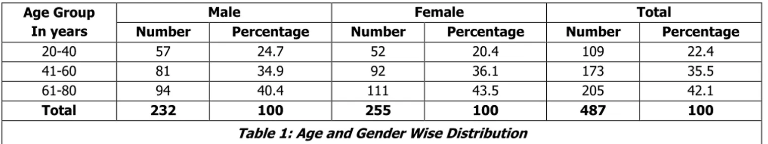
RESULTS: The study was conducted among 487 subjects which include 232 males and 255 females. The age of the subjects ranged between 20 to 80 years. The distributions of the subjects in the different age groups are shown in Table – 1 with percentages.
Age Group In years
Male Female Total
Number Percentage Number Percentage Number Percentage
20-40 57 24.7 52 20.4 109 22.4
41-60 81 34.9 92 36.1 173 35.5
61-80 94 40.4 111 43.5 205 42.1
Total 232 100 255 100 487 100
Table 1: Age and Gender Wise Distribution
J. Evid. Based Med. Healthc., pISSN- 2349-2562, eISSN- 2349-2570/ Vol. 3/Issue 66/Aug. 18, 2016 Page 3569
Gender Age Mean IOP Standard Deviation 95% CI for mean upper lower
Limit
ANOVA P value F value
Males
20-40 14.6 2.7 13.9 15.3
0.001 10.12
41-60 16.6 2.6 16.1 17.2
61-80 15.7 2.7 15.2 16.3
Total 15.8 2.8 15.4 16.1
Females
20-40 15.8 3.0 14.9 16.6
0.825 0.2
41-60 15.9 2.9 15.3 16.5
61-80 15.7 2.8 15.1 16.2
Total 15.8 2.9 15.4 16.1
Table 2: Variations of IOP with Gender & Age
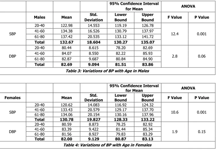
The mean IOP was highest in 40-60 yrs. in both genders. In males and females, mean IOP was higher in higher age group as compared to 20-40 years. The difference in mean IOP in different age group was statistically significant in males but not in females. The mean IOP in males were 15.8±2.8 mmHg. The mean IOP in females were 15.8±2.9 mmHg. There was no statistically significant difference in IOP between males and females. The variations of BP with age in both gender is shown in Table 3 and Table 4.
95% Confidence Interval
for Mean ANOVA
Males Mean Std.
Deviation
Lower Bound
Upper
Bound F Value P Value
SBP
20-40 122.98 14.553 119.19 126.78
12.4 0.001
41-60 134.38 16.526 130.79 137.97
61-80 137.42 20.535 133.12 141.72
Total 132.67 18.604 130.27 135.07
DBP
20-40 80.44 8.615 78.20 82.69
2.8 0.06
41-60 84.07 8.550 82.22 85.93
61-80 82.87 9.687 80.84 84.90
Total 82.69 9.094 81.51 83.86
Table 3: Variations of BP with Age in Males
95% Confidence Interval
for Mean ANOVA
Females Mean Std.
Deviation
Lower Bound
Upper
Bound F Value P Value
SBP
20-40 120.62 14.083 116.92 124.32
10.6 0.001
41-60 133.43 20.579 129.17 137.70
61-80 134.06 20.154 130.16 137.96
Total 130.78 19.827 128.33 133.22
DBP
20-40 80.59 8.873 78.25 82.92
1.9 0.15
41-60 83.39 9.422 81.44 85.34
61-80 81.56 8.927 79.83 83.29
Total 82.00 9.129 80.87 83.13
Table 4: Variations of BP with Age in Females
The results in Table 3 show statistically significant increase in SBP with age in both genders. DBP was highest in 40-40 years in both genders. The difference in diastolic BP with age was not statistically significant in both genders. The SBP and DBP was higher among males as compared to females, but the difference was not statistically significant.
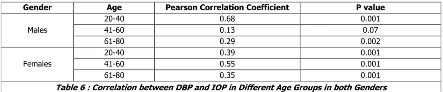
Gender Age Pearson Correlation Coefficient P value
Males
20-40 0.49 0.001
41-60 0.16 0.15
61-80 0.27 0.01
Females
20-40 0.22 0.1
41-60 0.54 0.001
61-80 0.45 0.001
J. Evid. Based Med. Healthc., pISSN- 2349-2562, eISSN- 2349-2570/ Vol. 3/Issue 66/Aug. 18, 2016 Page 3570 A correlation analysis was performed to assess the relationship between the age, systolic BP, diastolic BP and IOP. Table 4 & 5 shows a positive correlation between systolic BP and IOP and also between DBP & IOP in all age groups in both genders. The correlation was not significant regarding the SBP & IOP in males in 41-60 yrs. age group and 20-40 yrs. age group in females.
Gender Age Pearson Correlation Coefficient P value
Males
20-40 0.68 0.001
41-60 0.13 0.07
61-80 0.29 0.002
Females
20-40 0.39 0.001
41-60 0.55 0.001
61-80 0.35 0.001
Table 6 : Correlation between DBP and IOP in Different Age Groups in both Genders
Linear regression analysis was done to predict IOP from the variables of the present study. The significant model predicted by the statistical programme with highest adjusted
R2 value was. IOP = 3.567+0.029 SBP+0.102 DBP.
The adjusted R2 value of the model was 0.21. So 21% of the
variability of IOP could be explained by the Systolic and diastolic BP of the study subjects.
DISCUSSION: Although controversial, systemic hypertension has been postulated to be one of the risk factors in the genesis of primary open angle glaucoma. There are a number of studies relating blood pressure and intraocular pressure in different population subgroups like the Japanese, Korean, Chinese and Arabs.[3,10,11,13-16] The
difference may be because of ethnic differences between the populations and the differences in the anterior chamber depth and aqueous outflow mechanisms. The IOP is widely regarded as the most important modifiable risk factor which is associated with the development of glaucomatous optic neuropathy.[16,17] Therefore, the factors that influence the
IOP and its measurement are of great relevance in understanding the pathogenesis of the disease and in reducing the burden of blindness. The findings from our study indicated that systolic BP and diastolic BP were positively independently correlated to the IOP and that the correlations were statistically significant.
Some studies have found that a change in the IOP was directly and significantly associated with a change in the BP.[6,7,11,14,18,19] A positive association between the systolic
BP and a raised IOP has constantly been shown in both cross sectional and longitudinal studies.[4,5,8,18,19] Some studies
have shown that the diastolic BP was positively associated with a raised IOP. The IOP may have been increased in patients with an increased BP due to an increased retinal blood volume after a rise in the central retinal vein pressure because of an increased pressure in the adjacent central retinal artery.[20] An increased blood volume in the ciliary
body and a decreased facility of the aqueous outflow, owing to an increase in the resistance in the episcleral and the anterior ciliary veins.[20] An increased ultrafiltration of the
aqueous fluid in the ciliary body, owing to the increased perfusion pressure in the ciliary arteries. Obstruction to the
aqueous drainage at the anterior chamber angle due to the increasing episcleral venous pressure.[20]
The IOP shows a tendency to increase with age which may be due to slight sclerosis of the trabecular meshwork at the angle reducing the aqueous outflow. The findings from this study indicate that the IOP increased with age in both men and women and that it was statistically significant. The systolic BP and the diastolic BP were positively and significantly correlated with the IOP.
CONCLUSION: It can be concluded that persons with hypertension and advancing age need to be monitored for high IOP. Similarly, periodic BP monitoring may be indicated in persons with an increased IOP. Hence, a population based screening for an elevated IOP and its control could reduce the number of people who are at the greatest risk of glaucoma.
ACKNOWLEDGEMENTS: I am immensely indebted to Dr Gopal S Pillai for his valuable directions in carrying out the study.
REFERENCES
1. Thomas R, Sekhar GC, Kumar RS. Glaucoma
management in developing countries: medical, laser and surgical options for glaucoma management in countries with limited resources. Curr Opin Ophthal 2004;15(2):127-131.
2. Wong TY, Loon SC, Saw SM. The epidemiology of age
related diseases in Asia. Br J Ophthal
2006;90(4):506-511.
3. Palimkar A, Khandekar R, Venkatraman V. Prevalence
and distribution of glaucoma in central India (Glaucoma survey 2001). Indian J of Ophthalmol 2008;56(1):57-62.
4. Lee AJ, Wang JJ, Kifley A, et al. Open angle glaucoma and cardiovascular mortality: the Blue Mountain eye study. Ophthalmology 2006;113(7):1069-1076. 5. Chandrasekharan S, Cumming RG, Rochtchina E, et
al. Associations between elevated IOP and glaucoma, use of glaucoma medications and 5 year incident
cataract: the Blue Mountain eye study.
J. Evid. Based Med. Healthc., pISSN- 2349-2562, eISSN- 2349-2570/ Vol. 3/Issue 66/Aug. 18, 2016 Page 3571 6. Mitchell P, Lee AJ, Wang JJ, et al. Intraocular pressure
over the clinical range of blood pressure: Blue Mountain study findings. Am J Ophthalmol 2005;140(1):131-132.
7. Mitchell P, Lee AJ, Wang JJ, Rochtchina E. Open angle glaucoma and systemic hypertension: the Blue Mountains eye study. J Glaucoma 2004;13(4):319-326.
8. Ikram MK, de Voogd S, Wolfs RC, et al. Retinal vessel diameters and incident open angle glaucoma and optic disc changes: the Rotterdam study. Invest Ophthalmol Vis Sci 2005;46(4):1182-1187.
9. Wolfs RC, Borger PH, Ramrattan RS, et al. Changing
views on open angle glaucoma: definitions and prevalences- the Rotterdam study. Invest Ophthalmol Vis Sci 2000;41(11):3309-3321.
10. De Voogd S, Ikram MK, Wolfs RC, et al. Incidence of open angle glaucoma in a general elderly population:
the Rotterdam eye study. Ophthalmology
2005;112(9):1487-1493.
11. Wong TT, Wong TY, Foster PJ, et al. The relationship of intraocular pressure with age, systolic blood pressure and central corneal thickness in an Asian
population. Invest Ophthalmol Vis Sci
2009;50(9):4097-4102.
12. Shimmoyo M, Ross AJ, Moy A, et al. Intraocular pressure, Goldmann applanation tension, corneal thickness and corneal curvature in Caucasians, Asians, Hispanics and African Americans. Am J Ophthalmol 2003;136(4):603-613.
13. Khurana AK, Khurana I. Anatomy and physiology of
the eye. 2nd edn. New Delhi: CBS Publishers and
Distributors 2006:44-81.
14. Kawase K, Tomidokoro A, Araie M, et al. Ocular and
systemic factors related to IOP in Japanese adults: Tajimi study. Br J Ophthalmol 2008;92(9):1175-1179. 15. Kim SK, Cho BJ, Hong S, et al. Pulsatile ocular blood flow in healthy Koreans. Korean J Ophthalmol 2008;22(1):6-9.
16. Leske MC, Connell AMS, Wu SY, et al. Incidence of open–angle glaucoma: the Barbados eye studies. Arch Ophthalmol 2001;119(1):89-95.
17. Le A, Mukesh BN, McCarty CA, et al. Risk factors which are associated with the incidence of open-angle glaucoma: the visual impairment project. Invest Ophthalmol Vis Sci 2003;44(9):3783-3789.
18. Kisan R, Kisan SR, Anitha OR, et al. Correlation between the IOP and BP in different age groups. JCDR 2012;6(4 suppl 2):581-585.
19. Klein BE, Klein R, Linton KL. Intraocular pressure in an American community: the Beaver Dam eye study. Invest Ophthalmol Vis Sci 1992;33(7):2224-2228. 20. Bulpitt CJ, Hodes C, Everitt MG. The intraocular