1
Arquivos Brasileiros de Cardiologia - Volume 85, Nº 2, Agosto 2005
Case Report
Diffuse Atherosclerotic Disease Unmasked by Invasive
Physiologic Assessment of Coronary Flow
Fernando Mendes Sant’Anna, Expedito E. Ribeiro da Silva, Leonardo Alves Batista,
Fábio Machado Ventura, Carlos Alberto Mussel Barrozo, Nico H.J. Pijls
Santa Helena Hospital do Coração - Cabo Frio, RJ - Instituto do Coração do Hospital das Clínicas
(FMUSP) - São Paulo, SP e Catharina Hospital - Eindhoven, The Netherlands
Mailing address: Fernando Mendes Sant’Anna - Rua Safira 20, Portinho, Cabo Frio - 28915-400 - Cabo Frio, RJ - Brazil E-mail: [email protected]
Sent for publishing on 11/07/2004 Accepted on 03/04/2005
It is well known that coronary atherosclerosis is often a diffuse process poorly visible at angiography. This paper describes a patient with persisting stable angina after acute myocardial in-farction (AMI) 3 months earlier and a severe lesion in the left anterior descending artery (LAD) at coronary angiography. Frac-tional flow reserve (FFR), measured by coronary pressure mea-surements, was 0.37 during hyperemia, unequivocally demons-trating the presence of ischemia. A stent was placed in the LAD and despite excellent angiographic result, post FFR was only 0.75, the lower limit for ischemia. When the pressure sensor was slowly pulled back from distal to proximal LAD there was a graded, continuous increase in coronary pressure, which clearly indicates diffuse atherosclerosis, not focal stenosis. Across the stent no hyperemic gradient was present. The patient was treated medically and remained event free thereafter.
Coronary circulation is generally considered a two compartment model, which consists of epicardial vessels, also referred as “con-ductance vessels” and microcirculation, arteries <400 mm or “resistive vessels”1. When there is no stenosis, myocardial flow is
primarily controlled by resistive vessels.
Pathological and intravascular ultrasound studies have shown that when a stenosis is visible at angiography, the remainder of the coronary tree is often diffusely involved by atherosclerosis, although this may not be identified by coronary angiography2-5.
De Bruyne et al. showed that diffusely atherosclerotic epicardial coronary arteries in contrast to truly normal coronary arteries often cause a continuous pressure decline along their length, reduce fractional flow reserve, contribute to myocardial ischemia and abnor-mal perfusion during exercise and pharmacological vasodilatation, and are identifiable by intracoronary pressure measurements6.
Fractional flow reserve (FFR) is defined as the ratio of maximal hyperemic blood flow in the presence of a stenosis divided by normal hyperemic blood flow without stenosis and is calculated as the ratio of distal coronary pressure (Pd) divided by aortic
pres-sure (Pa) at maximum hyperemia (FFR=Pd/Pa)7. The larger the
resistance to blood flow, the larger the decline in pressure and, thus, the smaller FFR. Therefore, FFR is an index of resistance to flow along the epicardial vessel and is not affected by changes in blood pressure, heart rate and other pathologic conditions. Even if microcirculatory disease is present, FFR still gives the (abnormal) resistance to flow along the epicardial artery, given that state of microcirculatory disease. FFR and its properties have been well validated over recent years8-10. Importantly, FFR below 0.75-0.80
discriminate lesions which are associated with inducible ischemia with a diagnostic accuracy of almost 100%9,10.
The present report describes a patient with stable angina who had a severe stenosis in the left anterior descending (LAD) coronary artery. Measured FFR was 0.37 and thus indicative of important ischemia. A major, focal gradient was present across the stenosis itself. After treating this lesion by stent implantation, FFR improved significantly but still remained inside the area for inducible ische-mia. However, coronary pressure tracings obtained by the pullback curve under maximal hyperemia showed no gradient across the stent individually deployed well, but a continuous increase from distal to proximal LAD typical of diffuse atherosclerotic disease. This report might demonstrate how FFR can unmask diffuse athe-rosclerotic disease after treatment of a focal lesion.
Case Report
A 58-year-old male, suffering from AMI 3 months earlier, pre-sented at outpatient clinic with typical recurrent chest pain at moderate exercise. Known risk factors were hypercholesterolemia and arterial hypertension. Physical examination was normal. Res-ting ECG showed Q-waves with absent R waves from V1 to V4. Echocardiography showed a mild anterior hypokinesia of the left ventricle with a slightly depressed left ventricular function.
At cardiac catheterization, there was a very tight stenosis in the LAD, approximately 90% by visual assessment (Figure 1). As the patient was symptomatic and left ventricular function was almost normal, coronary angioplasty followed by stent implantation was chosen as the best treatment option.
Sys-2
Arquivos Brasileiros de Cardiologia - Volume 85, Nº 2, Agosto 2005
Diffuse Atherosclerotic Disease Unmasked by Invasive Physiologic Assessment of Coronary Flow
tems, Upsala, Sweden) was introduced into the LAD and, after intravenous administration of adenosine 140 mg/kg per minute, the recordings were made as presented in Figure 2. At maximal hyperemia, FFR of the LAD equaled 0.37 and the pressure pullback curve showed a clear spot inside the LAD at the place of the stenosis where a sudden drop of the pressure was recorded by the pressure wire (Figure 2).
The lesion was then predilated with a 2.5 mm balloon and two stents (3.0 and 2.5 mm diameters) were implanted in order to cover the entire diseased segment. After excellent angiographic result (Figure 3), FFR was measured again and its value was 0.75, around the threshold for inducible ischemia. When the pressure wire was slowly pulled back from distal to proximal LAD, there was no residual gradient across the stent itself but a graded, continuous increase in coronary pressure (Figure 4), which clearly indicates diffuse atherosclerosis, not focal stenosis, and thus not amenable for further stenting. The patient was kept in medical treatment and remained event free thereafter.
Discussion
Normal coronary arteries are characterized by the absence of any decline of pressure along its course, not even at maximal hyperemia9. In diffuse disease, hyperemic decline is often observed
and can even be responsible for inducible ischemia6.
In this patient, a very tight (anatomic and physiologic) stenosis was present in the LAD with large focal hyperemic pressure drop and little further decline (Figure 2).
After stenting the focal spot, blood flow increased by more than 100% (0.37 → 0.75) and due to this increase in flow the
diffuse disease (not observable before) was unmasked.
It is also important to mention that distal embolization due to stent implantation could not have been responsible for the low FFR; it would make the FFR higher than expected yielding a false result.
This case teaches two important lessons:
1. Pressure measurements proved that stents were placed well (there were no gradients across them). Without that observation, residual ischemic complaints could be attributed to insufficient stent deployment and might have provoked further action in the stents. 2. The diffuse disease was unmasked by successful stenting of a focal stenosis, as proven by the hyperemic pullback curve.
These findings have important implications for evaluation of coronary stenting. It has already been demonstrated by intravascular ultrasound and also by pressure measurements, that the presence of a focal stenosis is almost always associated with diffuse athe-rosclerosis of coronary vasculature3-6. Even after successful
sten-ting of a focal stenosis, a residual gradient may remain with an abnormal FFR if pressure is measured in the distal part of the artery. Therefore, to evaluate whether the stent has fully re-esta-blished the conductance of a previously stenotic stented segment, FFR should be calculated from the ratio of the pressure just distal to just proximal to the stented segment during a pullback maneuver under maximal hyperemia. The pressure gradient between the two edges of the stent indicates the status of the stented segment alone, whereas the pullback pressure recording along the length of the artery indicates the conductance of the entire epicardial artery, including the stented segment6, and also indicates if
is-chemia will be inducible at exercise.
Although it is well known, by recently published studies11, that
post stent FFR has important prognostic implications, we have to be aware that, specially in patients with multivessel disease and certain types of associated diseases (like diabetes), sometimes it is impossible to reach optimal physiologic results and this is due to diffuse atherosclerosis not to inadequate stent deployment.
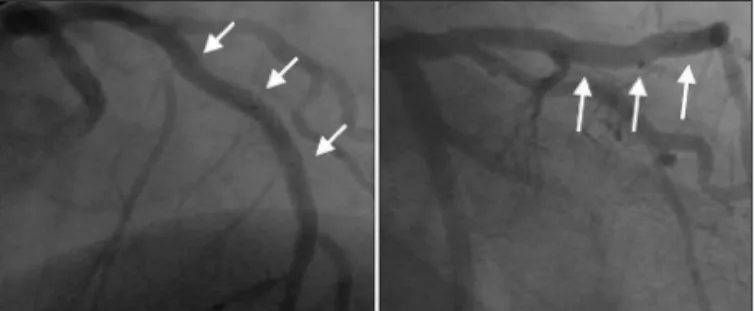
Fig. 3 - LAD after stent implantation. Excellent angiographic result with less than 10% residual lesion.
Fig. 4 - Pressure tracings in the LAD after the intervention. The pressure pullback curve shows a continuous, graded increase in the coronary pressure from distal to proximal LAD, a typical pattern of diffuse atherosclerosis. There are no more focal pressure drops inside the artery.
Fig. 1 - Coronary angiogram showing a 90% stenosis in the middle left anterior descending artery, after the first septal branch.
3
Arquivos Brasileiros de Cardiologia - Volume 85, Nº 2, Agosto 2005
Diffuse Atherosclerotic Disease Unmasked by Invasive Physiologic Assessment of Coronary Flow
1. Maseri A, Crea F, Kaski JC, Crake T. Mechanisms of angina pectoris in syndrome X.
J Am Coll Cardiol 1991;17(2):499-506.
2. Glagov S, Weisenberg E, Zarins CK, Stankunavicius R, Kolettis GJ. Compensatory enlargement of human atherosclerotic coronary arteries. N Engl J Med
1987;316(22):1371-1375.
3. McPherson DD, Hiratzka LF, Lamberth WC, Brandt B, Hunt M, Kieso RA, Marcus ML, Kerber RE. Delineation of the extent of atherosclerosis by high-frequency epi-cardial echocardiography. N Engl J Med 1987;316(6):304-309.
4. Nissen SE, Gurley JC, Grines CL, Booth DC, McClure R, Berk M, Fischer C, DeMaria AN. Intravascular ultrasound assessment of lumen size and wall morphology in normal subjects and patients with coronary artery disease. Circulation 1991; 84(3):1087-1099.
5. Mintz GS, Painter JA, Pichard AD, Kent KM, Satler LF, Popma JJ, Chuang YC, Bu-cher TA, Sokolowicz LE, Leon MB. Atherosclerosis in angiographically “normal” co-ronary artery reference segments: an intravascular ultrasound study with clinical correlations. J Am Coll Cardiol 1995;25(7):1479-1485.
6. De Bruyne B, Hersbach F, Pijls NHJ, Bartunek J, Bech JW, Heyndrickx GR, Gould KL, Wijns W. Abnormal epicardial coronary resistance in patients with diffuse atherosclero-sis but “normal” coronary angiography. Circulation 2001;104(20): 2401-2406.
References
7. Pijls NH, van Son JA, Kirkeeide RL, De Bruyne B, Gould KL. Experimental basis of determining maximum coronary, myocardial and collateral blood flow by pressure measurements for assessing functional stenosis severity before and after percuta-neous transluminal coronary angioplasty. Circulation 1993;87(4):1354-1367. 8. De Bruyne B, Baudhuin T, Melin JA. Coronary flow reserve calculated from
pressu-re measupressu-rements in humans; validation with positon emission tomography. Circu-lation 1994;89:1013-1022.
9. Pijls NHJ, Van Gelder B, Van der Voort P, Peels K, Bracke F, Bonnier HJRM, El Gammal, MIH. Fractional flow reserve: a useful index to evaluate the influence of an epicardial coronary stenosis on myocardial blood flow. Circulation
1995;92(11):3183-3193.
10. Pijls NHJ, De Bruyne B, Peels K, Van Der Voort PH, Bonnier HJ, Bartunek J, Koolen JJ. Measurement of fractional flow reserve to assess the functional severity of co-ronary-artery stenoses. N Engl J Med 1996; 334:1703-1708.
11. Pijls NHJ, Klauss V, Siebert U, Powers E, Takazawa K, Fearon W, Escaned J, Tsurumi Y, Akasaka T, Samady H, De Bruyne B for the Fractional Flow Reserve (FFR) Post-Stent Registry Investigators. Coronary pressure measurement after stenting predicts adverse effects at follow-up. A Multicenter Registry. Circulation