1 8 1 8 1 8 1 8 1 8
Instituto Dante Pazzanese de Cardiologia
Mailing address: Jarbas Jakson Dinkhuysen - Av. Dr. Dante Pazzanese, 500 - 10º andar Cep 04012-180 - São Paulo, SP, Brazil - E-mail: [email protected] Received 8/11/02
Accepted
English version by Stela Maris C. e Gandour
Arq Bras Cardiol, volume 82 (nº 1), 18-26, 2004
Jarbas Jakson Dinkhuysen, Tarcisio Luiz Valle de Almeida, Ibraim Masciarelli Francisco Pinto, Luiz Carlos Bento de Souza
São Paulo, SP - Brazil
Surgical Treatment of Coarctation of the Aorta Using
Trapezoidal Aortoplasty
Advances in the diagnosis and surgical treatment of coarctation of the aorta changed the natural history of the disease, causing a significant improvement in patients’ life expectancy and quality of life. Several surgical techniques have been proposed and used; however, recurrences and other complications have been reported.
Currently, dilation of the coarctation of the aorta by an-gioplasty is performed and provides good results. It is the first therapeutic modality to be considered for children, ex-cept for neonates and infants, and for postsurgical recoarc-tations. Surgery has become more and more restricted to the correction of complex coarctations, completely allevia-ting the obstruction with low morbidity and mortality, allo-wing aortic growth proportional to the patient’s growth. Ne-vertheless, the exact moment of surgical indication is still controversial to an asymptomatic, normotensive, apparen-tly healthy child. On the other hand, it is a consensus that symptomatic patients should undergo surgery promptly.
Although all techniques already described have appli-cations, the 3 following methods have emerged as excellent for routine repair of coarctation of the aorta: 1) aortoplasty with a subclavian flap 1 preserving or not blood flow to the
ipsilateral (left) upper limb; 2) end-to-end anastomosis with resection of the coarctation of the aorta 2,3; and 3) extended
aortoplasty with widening of the stenotic area with synthe-tic tissue 4. All methods have applications and limitations.
Aortoplasty with a subclavian flap or subclavian’s flap angioplasty includes the use of autologous natural tissue to widen the stenotic area, avoids tension in the suture line, does not require large regional dissection, and leaves no cir-cumferential scar. However, this technique not only inter-rupts blood flow to the left upper limb with a potential growth delay 5, but also maintains the tissue of ductus
arte-riosus enabling the appearance of an aneurysm in the long run. On the other hand, subclavian’s flap angioplasty with blood flow preservation to the ipsilateral upper limb 6 has the
characteristics of aortoplasty with a subclavian flap, but does not require a section of the subclavian artery to serve as a flap. The origin of the subclavian artery in the aorta is re-located, widening the stenotic area.
Objective - Trapezoidal aortoplasty is a technical
va-riant of end-to-end anastomosis, which, based on elements of geometry, aims at increasing the diameter of the aorta at the level of the suture, therefore reducing the occurrence of residual or recurrent pressure gradients in the short and long run.
Methods - After resecting the coarcted area and
duc-tal tissue, 3 trapezoids are confected in each aortic stump, which, when confronted, create a suture line with a sinu-soidal aspect (zigzag). Thirty-three patients underwent surgery with this technique, 22 (66.7%) males, with ages ranging from 3 months to 36 years (mean of 9.84 ± 9.69).
Results - No immediate or late deaths occurred.
Follow-up ranged from 1.1 to 7.6 years (mean of 3.6 ± 3.4). Most patients became asymptomatic with normal blood pressure levels, enabling the discontinuation of antihyper-tensive therapy (P<0.0001). A significant reduction in the pressure gradients was observed on Doppler echocardio-graphy and during cardiac catheterization (P<0.001). The analysis of the images of aortography showed good anato-mical continuity in the region of the anastomosis, and the morphometric study of the aorta revealed the beneficial ef-fects of the technique indicated by the increase in the cali-ber of the aorta in the distal segment of the arch, isthmus, and descending portion.
Conclusion - Trapezoidal aortoplasty showed
satis-factory clinical results that allow its application in all cases indicated for end-to-end anastomosis.
In addition to removing the coarcted area and the ductal tissue 7, which are potentially pathologic, end-to-end
anastomosis has the advantage of encompassing the proxi-mal tubular hypoplasias. Its weak points include tension in the suture line, occasional technical difficulties with more extensive dissections, and the presence of potentially re-tractile circumferential scars.
The advantages of extended aortoplasty with wide-ning with synthetic tissue are similar to those of aortoplasty with a subclavian flap, except for the smaller need for regio-nal surgical dissection and preservation of the blood flow to the left upper limb. The disadvantages of that technique are as follows: maintenance of the coarcted area and the ductal tissue; the need for synthetic material for widening of the stenotic region; and the incidence of aneurysm close to the aortic wall opposite the flap 8.
The pros and cons of these techniques should be mea-sured in terms of results indicated by mortality; residual or recurrent pressure gradients; complications related to the method itself, such as aneurysms, damage to nerve trunks, chylothorax, dysfunction of the left upper limb, paraplegia, and bleeding.
Until the present time, distinctions between these me-thods have not been possible, despite the great experience acquired over the years. The major criterion of distinction is the incidence of residual or recurrent coarctation, which has been the motive of many modifications and new proposals, in an attempt to suppress or minimize these events. One of these technical proposals is trapezoidal aortoplasty 9, which
aims at optimizing the end-to-end anastomosis and at avoiding the appearance of residual or recurrent gradients that result from surgical technical problems or from the inca-pacity of the aorta to grow at the level of the anastomosis proportionally to the patient’s development.
This study aimed at retrospectively assessing the im-mediate and late clinical results, the occurrence of residual pressure gradients and recoarctations, and the growth of the anatomical portions of the aorta determined by trapezoi-dal aortoplasty, a new technique for the surgical treatment of coarctation of the aorta.
Methods
The study comprised 33 patients with coarctation of the aorta, 22 (66.7%) males, with ages ranging from 3 months to 36 years (mean of 9.8 ± 9.7). The patients were referred to or were seeking medical care in the Medical Section of Con-genital Cardiology of the Instituto Dante Pazzanese de Cardiologia, in São Paulo. They were operated on from 04/ 15/1993 to 02/18/2000 by the same surgeon.
Seventeen (51.5%) patients had the isolated anatomi-cal form of the disease, and 16 (48.5%) had one or more asso-ciated congenital cardiac defects, fibroelastosis (3 - 18.7%) and persistent ductus arteriosus (3 - 18.7%) being the most frequent ones.
The preoperative clinical data of the 33 patients refer to the presence of symptoms resulting from the disease; 27 (81.8%) patients were symptomatic, and the remaining 6
(18.2%) were oligosymptomatic. Twenty (60.6%) patients had hypertension, 5 (15.2%) had mild hypertension, and 8 (24.2%) were normotensive. Twenty-three (69.7%) patients were using hypotensive medication, only 1 (3%) patient used no medication, and no information about use of medi-cation existed in 9 (27.3%) cases. The electrocardiogram showed sinus rhythm in all (33 - 100%) patients, 15 (45.6%) of whom had left ventricular hypertrophy, 4 (12%) had nor-mal tracings, and more than half (18 - 54.5%) had other elec-trocardiographic changes. The preoperative Doppler echo-cardiogram performed in all patients assessed the pressure gradient at the level of the coarctation of the aorta in 22 (66.7%) patients. Thirty-one (93.9%) patients underwent cardiac catheterization with obtainment of angiographic images in all of them and assessment of the respective gra-dients in only 16 (48.4%) patients. Aortography, in the left anterior oblique view, enabled the morphometric analysis of the proximal and distal portions of the aortic arch, isthmus, and descending aorta in 31 patients, according to the me-thodology of Moulaert et al 10.
The inclusion criteria for trapezoidal aortoplasty were based on the following anatomies of the coarctation of the aorta: 1) in the diaphragm or usually preductal shelf, which could obstruct the entire aortic lumen or leave an eccentric orifice; 2) in ring, hourglass, or waist, with a localized intro-flexion of the aortic wall with thickening of the intima or me-dia; 3) an anatomy of the defect different from those pre-viously specified, but to which the application of the end-to-end anastomosis technique may be cogitated. The exclu-sion criteria were as follows: 1) segmentary, tubular, or hypo-plastic coarctation of the aorta, ie, extensive narrowing af-fecting the entire aortic wall, with a reduction in the lumen in a segment, usually the aortic isthmus (isthmic hypoplasia); and 2) defects in atypical locations in the transverse arch, and in the thoracic or abdominal portion of the aorta.
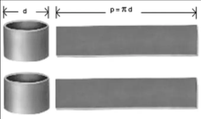
The technique proposed was based on geometric considerations conceived in a simple model, in which a tube of circular section with a d diameter was transversally cut at its axis to create 2 cylinders with orifices in its extremities. After longitudinally opening these cylinders and exposing their lateral surfaces, 2 rectangles with a p base equal to the perimeter of the tube were obtained, as shown in figure 1.
2 0 2 0 2 0 2 0 2 0
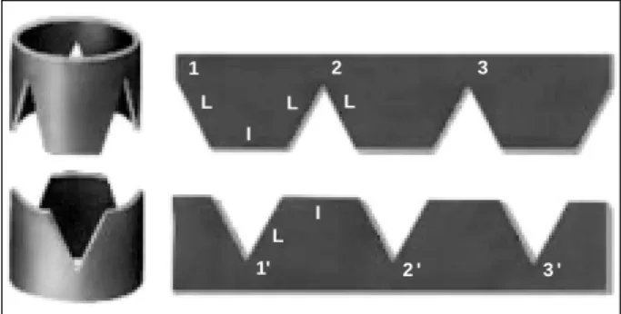
The extremities of the first cylinder and of the rectangle were cut; 3 pieces in the form of a triangle were removed and intercalated every 120°. The triangles were equilateral with sides of length L, forming 3 trapezoids with a smaller base of length l and larger base of length π.d/3 (fig. 2).
Observing the cylinder opened in a rectangle, the angles 0°, 120°, 240°, and 360° represent the positions of the vertices of the equilateral triangles with L sides. This proce-dure was repeated, a second cylinder was cut from the same tube, and 3 triangles were cut in the extremity and from the rectangle, this time, however, from its upper face, intercala-ted in the same manner and also forming 3 trapezoids. This second cylinder was a specular image of the first. The 2 cy-linders were juxtaposed resembling the coupling of the teeth of 2 gearwheels, ie, the teeth of 1 penetrating the spaces between the teeth of the other (fig. 3). In this figure, the vertices of the triangles of the first cylinder are indica-ted by the numbers 1, 2, and 3, and the vertices of the trian-gles of the second cylinder by the numbers 1’, 2’, and 3’.
If the material is elastic, the perimeter of each rectangle may be extended by traction, so that the trapezoids of 1 cy-linder may perfectly fit within the triangles of the other cylin-der (fig. 4).
During this procedure, each vertex of the triangles is extended until a length equal to the smaller base of the trapezoid (L) and, consequently, the perimeters (p) of the rectangles are extended to P, causing a significant increase in its perimeter (P > p). As 3 vertices of zero length are replaced by 3 segments of L length, the new perimeter will be P= π d + 3L. Transferring the fit to the cylindrical form, a
greater perimeter (P) results, and its diameter (d) will also be greater, passing to D, which may be calculated by the fol-lowing formula: P= π D= π d + 3L, and, therefore, D=d +3L.
π
The final diameter will be proportional to the L side of the triangles, which is a measure directly associated with the depth of the indentations. It is worth noting that the indenta-tions should not be very shallow, because, if so, they would hinder a reasonable coupling. On the other hand, being deeper would not be advantageous, because a good cou-pling would also be difficult, due to the differences between the sides of the triangles and the bases of the trapezoids.
Figure 4 shows triangles with L sides and trapezoids with an l smaller base and a 2l larger base. Adopting these measures, the value of L as a function of the diameter of the original tube could be calculated. The depth of the indenta-tions is the height of the triangles; therefore, the height of the triangle should be approximately half of the diameter of the tube.
πd = 2L, ie, L = πd 3 6
h = √3 L and replacing L: h = π√3.d or h = 0.45 d
2 12
Transporting these principles for application in biolo-gical tissues, whose capacity of spatial adaptation to the most diverse forms is full, one may conclude that the final result of the coupling to the cylinders has a sinusoidal as-pect, such as in figures 5 and 6, where a significant increase in the perimeter and diameter (P > p and D > d) is observed. In the operating room, the initial approach to the pa-tient consisted of monitoring the cardiac rhythm and cathe-terizing the right radial artery, providing continuous control of mean blood pressure. After anesthetic induction, the subclavian vein was catheterized and vesical probing was performed. The patient was put in the right lateral decubitus position, and the surgical access was performed through left posterolateral thoracotomy in the 4th intercostal space with strict control of hemostasis. Then, the region of the dis-tal transverse arch, the isthmus, and the descending portion of the aorta until the coarcted area were dissected in the me-diastinal pleura and the following structures were isolated:
1
L
I L
2
L
3
1' L
I
2 ' 3 '
Fig. 2 - Two cylinders with the corresponding rectangles, from where 3 triangular pieces were cut, forming 3 trapezoids in specular images.
1 2 3
1' 2 ' 3 '
Fig. 3 - Approximation of the 2 cylinders and rectangles to fit similarly to the teeth of a gearwheel.
1'
2
2 ' 3
3 '
Fig. 4 - Elastic coupling of the 2 rectangles.
the ligamentum arteriosum (or patent ductus arteriosus), the coarcted region, the left subclavian artery, the distal portion of the aortic arch, including the left carotid artery, and a large part of the thoracic aorta (fig. 7).
After systemic heparinization (1 mg/kg of weight), the proximal and distal stumps were pinched with tweezers, the ligamentum arteriosum was sectioned (or the ductus arterio-sus was sutured), and the coarcted region, including the ductal tissue, was resected, which resulted in the formation of free stumps (fig. 8). Then, 3 trapezoids were cut by resec-ting 3 identical and equidistant wedges in specular image in each stump (fig. 9). Anastomosis was then initiated with continuous or separated stitches, approximating the vertex of 1 trapezoid to the intertrapezoidal depression of the other
stump, like a gearwheel (fig. 10). Figure 11 illustrates the fi-nal anastomosis, a sinusoidal line of suture (zigzag) being observed.
Despite the smaller caliber of the aorta in children, this technique could be performed according to the methodolo-gy described. In addition to the usual anesthesia concerns, mean blood pressure was always maintained around 60/70 mmHg, and, many times, the use of peripheral vasodilators (sodium nitroprusside) was necessary. Once the suture was concluded, and according to the activated coagulation time, protamine was administered to neutralize the heparin. The following steps were then performed: reassessment of he-mostasis, synthesis of the parietal pleura, drainage of the cavity, and closure of the thorax by planes. The patient was then sent to the postoperative recovery unit, under the regi-men of constant surveillance for a period of 48 hours. Once the clinical parameters were stabilized, the pleural drain was removed and the patient transferred to the ward, from where, if good postoperative conditions were maintained, the patient was discharged on approximately the 7th posto-perative day (PO).
Clinical assessments and the residual gradients and stenoses were precociously analyzed during the in-hospital phase, and later, the patients were followed up, the last am-bulatory visit information being considered.
The hospitalization phase was assessed based on the following clinical and surgical parameters: events occurring
Fig. 7 - Illustrative drawing of the region of the coarctation isolated with patent duc-tus arteriosus.
Fig. 8 - Illustrative drawing of the aortic stumps pinched with tweezers and resection of the coarcted region, including the ductal tissue.
Fig. 9 - Illustrative drawing of the creation of the trapezoids (3) in each stump.
Fig. 10 - Illustrative drawing of the beginning of the suture with approximation of the trapezoids in each aortic stump.
2 2 2 2 2 2 2 2 2 2
in the operating room, in the intensive care unit, and in the ward until hospital discharge, and mortality. The late phase of evolution was assessed based on the following clinical and laboratory parameters: presence of symptoms; presen-ce of blood pressure levels above the normal levels for the patients’ age; presence of residual pressure gradients (mmHg) on the Doppler echocardiogram obtained on the last ambulatory visit; presence of residual pressure gra-dients determined through intra-aortic blood pressure mea-surement (mmHg) in the region of the anastomosis on the occasion of the hemodynamic re-study; presence of residu-al stenoses (%) based on the images of the site of the anas-tomosis and its relation to the segments of the thoracic aorta obtained through magnetic resonance imaging during early or late evolution.
Morphometric analysis was performed by comparing the images of the ascending aorta with those of the seg-ments of the transverse and descending aorta on the left an-terior oblique view obtained on angiography and magnetic resonance. Percentage values were attributed to the diffe-rent segments of the aorta as follows: 100% to the caliber of the normal ascending portion of the aorta; a minimum of 60% to the proximal segment of the aortic arch; 50% to the distal segment; 40% to the isthmus; and 70% to the descen-ding aorta 10. Lower percentage values in each anatomical
portion were considered hypoplasias.
Because this was a retrospective analysis aiming at demonstrating only the results obtained with the method, comparisons with a control group were not performed.
The statistical analysis was performed in regard to blood pressure levels and need for medication by using the McNemar test 11. In the descriptive analysis, the data
refer-ring to blood pressure gradients and the morphological and anatomical parameters were summarized as means, medians, standard deviation (SD), and minimum and maximum values (annex). To assess the distribution of the variables and to identify the presence of discrepant values, diagrams of the box-plot type were built 12. In the comparative analysis
bet-ween the results before and after surgery, the values were expressed as means ± SD. The paired Student t test was used for statistical comparison 13, and the hypothesis that
the data follow a normal distribution was assessed with the Shapiro Wilk test 14. In the cases where normality could not
be assumed, the comparison was also performed by the nonparametric Wilcoxon signed rank test 13. In all studies,
only the patients assessed in both situations, before and after surgery, were considered. The results whose descrip-tive levels (P values) were lower than 0.05 were considered statistically significant. SPSS for Windows, version 8.0, was used for calculation.
Results
Surgery was uneventful in all 33 patients. No distor-tion or bleeding was observed in the dissecdistor-tions and suture lines of the anastomoses. In 1 patient who had poor collate-ral circulation, after the coarcted area was pinched with
tweezers, the mean blood pressure measured in the descen-ding thoracic aorta was approximately 20 mmHg, while in the transverse aorta it was 70 mmHg. In these circumstances, a surgical bypass was installed between the aortic arch and the thoracic aorta to ensure blood flow and distal blood pressure and to avoid possible subsequent medullary le-sions. The immediate evolution of this patient was ordinary with no signs of neurological impairment.
Considering the trend towards hypertension in the first moments after surgery, hypotensive medications (IV sodium nitroprusside) were used in more than 90% of the patients.
Two patients had complications in this phase. The first developed abdominal distension that regressed after the 2nd PO day, and the 2nd complained of paresthesia in the lower limbs that disappeared on the 5th PO day. Except for these 2 patients who were discharged from the hospital on the 17th and 33rd PO days, the other patients were dis-charged between the 7th and the 11th PO days, all in good clinical condition. No in-hospital deaths occurred.
The patients were followed up in the outpatient care clinics of the Medical Section of Congenital Heart Diseases of the Instituto Dante Pazzanese, which allowed obtainment of middle- and long-term evolutionary information. Follow-up ranged from 1 year and 1 month to 7 years and 5 months (mean of 3.6 years; SD of 2.2 years).
Doppler echocardiography was performed in 30 pa-tients, magnetic resonance imaging in 27, and hemodynamic re-study by catheterization in 15, with obtainment of data and assessment of the presence of residual gradients and stenoses, in addition to the morphological and anatomic as-pect of the aortic arch and segments, including the region of the anastomosis and descending aorta.
Regarding pressure gradients on Doppler echocar-diography and on cardiac catheterization, preoperative Doppler echocardiography was performed in 30 (91%) patients, and in 22 (67%) the pressure gradient was asses-sed at the level of the defect, the mean value of 66.1 mmHg with SD of 17.6 being obtained. After surgery, these data were assessed in most patients (30 - 91%), its mean value being 20.1 mmHg with SD of 8.2 mmHg. These values were not assessed in 11 patients in the preoperative phase and in 3 (9%) patients in the postoperative phase.
For comparing the mean gradients before and after surgery, 21 patients with measurements in both phases were considered. No violation of the normal distribution was ob-served in either phase. The paired t test indicated a signifi-cant reduction (P < 0.001) of 40.8 mmHg (fig. 12).
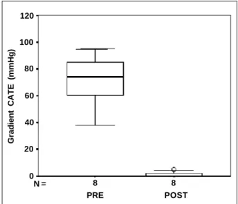
In the preoperative period, cardiac catheterization was performed in 31 (94%) patients, and in only 16 (48.6%) were the pressure gradients assessed (mean of 66.1 mmHg; SD of 20.9). After surgery, catheterization was performed in 15 (45.5%) patients, the mean value of the pressure gradient and SD being, respectively, 7.1 and 10.8 mmHg. These va-lues were not measured in 17 (51.5%) and 18 (54.5%) pa-tients, respectively, before and after the procedure.
Eight patients who had undergone the examination in the preoperative phase did not undergo it after surgery; on the other hand, 7 patients with postoperative assessments had no preoperative measurements. Therefore, for compa-rative analysis, only 8 patients were left with measurements in both periods. The analysis indicated that the patients had a significant reduction in the pressure gradient, changing from 71.5 ± 18.8 to 1.1 ± 2.1 mmHg.
Because many patients had no pressure gradient after surgery (value equal to 0), the distribution of values was asymmetric, and, consequently the Shapiro Wilk test rejec-ted the hypothesis of normality in this phase. For this rea-son, the values were compared using the nonparametric Wilcoxon test, which also indicated a significant reduction in the pressure gradient (P = 0.01) (fig. 13).
As the patients did not undergo magnetic resonance imaging in the preoperative phase, comparison between the results before and after the intervention could not be perfor-med. Therefore, 27 patients were studied after surgery, 21 (63.6%) of whom showed no stenoses at the site of the anas-tomosis, and 6 (18.2%) showed a percentage reduction in the caliber ranging from 9% to 46% (mean of 9.5±3.7).
Morphometric analysis was performed using angio-graphic images of the contrasted aorta in the left anterior oblique view and magnetic resonance imaging, considering the methodology recommended by Moulaert et al. 10.
Aortography was performed in 31 patients in the preo-perative phase. After surgery, only 15 patients underwent aortography, 27 underwent magnetic resonance, and the mean values are shown in table I.
The values obtained in aortography before and after surgery were compared in 14 patients. In the box-plot dia-gram (fig. 14), some data were identified as discrepant becau-se they had values below and above the others.
Analyzing the results with the paired t test, a signifi-cant increase in the caliber of the isthmus and of the descen-ding aorta was observed, a mean of 20% and 16%, respecti-vely (tab. II).
In the long-term evolution, no patient died.
In regard to the analysis of residual stenoses and growth of the anastomosis, 2 cases of trapezoidal
aorto-Gradient ECO (mmHg)
100
80
60
40
20
0
N = 21 21
Fig. 12 - Gradient measured on Doppler echocardiography before and after surgery with values expressed as mean ± SD, median and graph of the box-plot type.
Gradient CATE (mmHg)
120
100
80
60
40
20
0
N = 8 8
PRE POST
Fig. 13 - Gradient measured on catheterization in the preoperative and postoperative phases with values expressed as mean ± SD, median, and graph of the box-plot type.
Table I - Percentage values of the segments of the aortic arch, isthmus, and descending aorta obtained on angiography before and after
surgery and on postoperative magnetic resonance imaging
Segment of the Aortography Aortography POP Magnetic Aorta % before surgery after surgery resonance imaging
(N = 30) (N = 15) (N = 27)
Proximal 67.1±15.1 68.9±08.1 71.9±11.1 Distal 52.2±14.3 62.8±09.8 57.5±13.3 Isthmus 40.8±16.7 61.3±15.6 59.4±14.5 Descending aorta 74±22.5 88.1±12.4 83.6±16.1
2 4 2 4 2 4 2 4 2 4
plasty stood out, because they had postsurgical residual pressure gradients on Doppler echocardiography and on cardiac catheterization, and stenosis on magnetic resonan-ce imaging. One of these patients had been diagnosed with coarctation of the aorta + fibroelastosis, and was 3 months old at the time of surgery. Three years and 1 month after sur-gery, the patient was asymptomatic and without medication, his blood pressure was 130/70 mmHg, and his pressure gra-dient was 28 mmHg on Doppler echocardiography and 25 mmHg on cardiac catheterization. On angiography, a certain degree of residual isthmic hypoplasia was observed. The second case was a female patient, who had been diag-nosed with coarctation of the aorta + dextrocardia and ope-rated upon at the age of 15 years. Right after hospital dis-charge, she had pressure gradients of 23 mmHg and of 35 mmHg, respectively, on Doppler echocardiography and on cardiac catheterization, and a 46% stenosis on magnetic resonance imaging. The preoperative aortography also showed isthmic hypoplasia, which persisted after surgical correction, although the caliber of the anastomotic region was satisfactory. Nevertheless, she was asymptomatic and normotensive on her last follow-up visit. Instead of under-going trapezoidal aortoplasty, these 2 cases would be better treated with a surgical technique that considered isthmic hypoplasia.
The evolutionary data on a female patient diagnosed with isolated coarctation of the aorta and aged 11 years at the time of surgery are worth noting. On magnetic resonan-ce imaging performed on the 11th PO day, a 37% stenosis was observed; however, in the 7th month, on re-study with catheterization and magnetic resonance imaging, a signifi-cant increase in the caliber of the isthmus and of the anasto-mosis was observed, with a pressure gradient of 11 mmHg and stenosis of 10%. These findings suggest the occurren-ce of growth during that period. On the last clinical revision, the patient was asymptomatic, using no medication, and no pressure difference was observed between the upper and lower limbs.
Discussion
For the patient with coarctation of the aorta, the effect of surgery is dynamic 15 and encompasses complete
sup-pression of the stenosis, and residual transient or definitive or even recurrent coarctation may occur. The surgical tech-nique to be used depends on each case and influences the clinical evolution of the patient. The surgery may have com-plications, both immediately after surgery and in the long run, and, despite the good results of the extended aortoplas-ty, the appearance of dilations in the wall opposite the ex-tension has been shown, probably due to the exaggerated resection of the diaphragm, and only 1 case of dilation in the grafting itself was described 16,17.
One of the consequences of the lack of blood irrigation caused by ligature and section of the left subclavian artery in aortoplasty with a subclavian flap is the ischemic lesion of the left upper limb, which results from the surgical me-thod used. In noninvasive analysis with Doppler echocar-diography and temperature assessment in the upper limbs of patients undergoing ligature and transection of the left subclavian artery 5, an immediate drop in blood pressure
with a rapid return to approximately 70% of that in the oppo-site arm was observed, as was low temperature in the first week with subsequent normalization, suggesting recruit-ment in the collateral circulation. The impaired arm showed good tolerance to exercise and a mild reduction in muscle mass, suggesting that ligature of the subclavian artery may be performed without greater consequences.
Persistence of arterial hypertension after correction has been attributed to the late indication for surgery in coarcta-tion of the aorta 18,19. That is why the patient, once diagnosed
with coarctation of the aorta, should be referred for surgical or percutaneous treatment. Ebaid and Afiune 20 have drawn
attention to situations that pass unnoticed in clinical history and on patient’s physical examination, resulting in the lack of a diagnosis and of adequate management.
From the clinical point of view, patients undergoing trapezoidal aortoplasty had a good response to surgical treatment, because according to data on the last clinical re-vision, the symptoms disappeared in most patients, with blood pressure normalization and reduction in the need for antihypertensive medication. However, in some cases,
inde-Fig. 14 - Segments of the aorta obtained on aortography before and after surgery. Va-lues expressed as percentages considering the descending aorta as 100%, mean ± SD, median and graph of the box-plot type.
PRE
% da aorta
120
100
80
60
40
20
0
N = 14 14 14 14 14 14 14 14
PROXIMAL DISTAL ISTHMUS DESCENDING
POST
Table II - Comparisons of the different segments of the aorta on aortography before and after surgery
Segment of the Before After Paired t test Mean difference
Aorta % (P) 95% CI
Proximal 69.3 ± 15.3 67.6 ± 06.8 0.72 -1.7 [-11.8; 8.4] Distal 53.8 ± 13.2 61.2 ± 07.8 0.12 7.4
[-2.1; 16.8] Isthmus 40.7 ± 17 60.6 ± 15.9 0.001 19.9
[10.4; 29.3] Descending 72.2 ± 17 88.6 ± 12.7 0.02 16.4
[3.6; 29.1]
pendent of age, maintenance of therapy was required, with eventual discontinuation over time. These data suggest the nonoccurrence of residual or recurrent coarctation. Despite the good results, the performance of subsidiary examina-tions for anatomical and functional assessment of trapezoi-dal aortoplasty was useful, and a statistically significant drop in the gradients was observed on Doppler echocardio-graphy, on cardiac catheterization, and on magnetic reso-nance imaging. Greater gradient values were observed on echocardiography as compared with those on catheteriza-tion, but a good statistical correlation between these me-thods was not possible.
These results reinforce the idea that Doppler echocar-diography, given its facilities, is a good method for long-term follow-up of patients undergoing surgical correction of coarctation of the aorta. However, a detailed clinical exami-nation is required, with assessment of arm/leg pressure gra-dients, pressure and angiographic determination on cathe-terization, accompanied or not by magnetic resonance, to safely establish the diagnosis of recoarctation. Our patients who had pressure gradients > 20 mmHg on echocardiogra-phy, except for 2 cases, showed no pressure gradients on catheterization nor a reduction in the caliber on magnetic re-sonance imaging.
Several studies have drawn attention to recoarctation, trying to correlate it to factors, such as the technique used, patient’s age at the time of surgery, and the presence of car-diac defects associated.
Metzdorff et al 21, considering the low index of
recur-rence and absence of long-term potential adverse effects with the end-to-end anastomosis technique, recommend it for infants below the age of 8 weeks, and present a literature review for those below the age of 2 months, in whom the end-to-end anastomosis and aortoplasty with a subclavian flap techniques were used. The number of children treated with aortoplasty with a subclavian flap was small, with a ma-ximum 2-year follow-up and 13% recurrence, the results with end-to-end anastomosis being similar, with no statistical dif-ference. Mortality for both techniques is around 20%. These authors have concluded that, when the end-to-end anastomosis cannot be performed, the aortoplasty with a subclavian flap may be useful in severely ill newborn in-fants and in special circumstances.
Körfer et al 22 believed that recoarctation was due to a
flaw in surgical reconstruction, because, frequently, a small portion of the stenosis is removed because the surgeons usually fear that the proximal and distal stumps may not be approximated. Incomplete resection leads to thickening and a reduction in the elasticity of the margins, which does not allow the growth of the anastomosis. Other authors 23, 24
agree and refer to the study by Elzenga and Gittenberger-de Groot 25, who histologically showed the ductal tissue
exten-ding to half of the circumference of the aorta at the level of the coarctation. These authors believe that that tissue is a pathological component of the aortic wall, and, if not remo-ved, it may cause recoarctation due to retraction and fibro-sis. Therefore, they concluded that, when the end-to-end anastomosis is used, recurrence is due to lack of growth in
the circular anastomosis, contrary to that which occurs when using aortoplasty with a subclavian flap and extended aortoplasty, in which the fibrosis and internal proliferation of the coarcted tissue, if untouched, can cause restenosis. Trapezoidal aortoplasty 9 is a surgical method for
cor-recting coarctation of the aorta. It is a technical modification of the end-to-end anastomosis that, in addition to removing the coarcted area eliminating the presence of the ductal tis-sue and the inner spur, it causes an increase in the inner aortic diameter at the site of anastomosis. It is not a method of indiscriminate application for either adults or children, and its methodology is identical to that of end-to-end anas-tomosis. It cannot be used in the cases of tubular hypopla-sia with or without involvement of the aortic arch. It is worth emphasizing that, when performing trapezoidal aortoplasty in a vessel whose original diameter is identical to the diame-ter of the stumps, according to the already described princi-ples, the final result is an increase in the caliber of the region of suture. On the other hand, when the diameters of the stumps are smaller than the original diameter of the vessel, anatomical recomposition of the caliber of the vessel is ob-served after the anastomosis is concluded. From the mor-phological point of view, it provides an increase in the aortic lumen, aiming at avoiding the appearance of residual pres-sure gradients due to restenoses in the long run. The sinu-soidal and nonlinear disposition of the suture may be a pro-tective factor from fibrous retraction, because a radial distri-bution of the lines of force exists and acts in this type of unusual suture, in addition to allowing the increase in the diameter of the vessel as the patient grows.
Factors, such as associated cardiac anomalies and age below 2 weeks, significantly contribute to increased mortality in the short and long run. In a series with 333 chil-dren, 54% under the age of 1 year, Tawes et al 26 showed
that surgical mortality was small (2.8%) for the patients older than 6 months, and that almost half of those with heart failu-re could be saved with early surgery. Other authors 27
con-cluded that the low surgical mortality rate was due to aggressive therapy with cardiac catheterization and emer-gency surgery, and to the fact of having avoided hypother-mia, in addition to adequate relief of the obstruction.
However, no consensus exists regarding the idea that as-sociated cardiac anomalies may influence surgical mortality, which is low when complete relief of the obstruction is obtai-ned, associated with appropriate postoperative cares. The use of aortoplasty with a subclavian flap has been considered by some authors as a factor that decreases surgical risk 28-30.
Despite the existence of several patients aged < 2 years in the trapezoidal aortoplasty series, no early or late mortali-ties were observed after surgical repair of the anomaly.
According to Moulaert et al 10 and based on the
mor-phometric analysis of the aorta, the proximal segment of the transverse arch is hypoplastic if its diameter is lower than 60% of the diameter of the ascending aorta, than 50% of the diameter of the distal segment, and than 40% of the diameter of the isthmus.
Siewers et al 31, morphometrically analyzing a group of
2 6 2 6 2 6 2 6 2 6
1. Waldhausen JA, Nahrwold DL. Repair of coarctation of the aorta with a subcla-vian flap. J Thorac Cardiovasc Surg 1966; 51:532-3
2. Craaford G, Nylin G. Congenital coarctation of aorta and its surgical treatment. J Thorac Surg 1945;14:347-61.
3. Gross RE, Hufnagel CA. Coarctation of the aorta: experimental studies regarding its surgical correction. N Engl J Med 1945;233:287-93.
4. Vosschulte K. Surgical correction of coarctation of the aorta by an “isthmusplas-tic” operation. Thorax 1961;16:338-45.
5. Lodge FA, Lamberti JJ, Goodman AH, et al. Vascular consequences of subclavian artery transection for the treatment of congenital heart disease. J Thorac Cardio-vasc 1983;86:18-23.
6. Mendonça JT, Carvalho MR, Costa RK, Franco E. Coarctation of the aorta: a new surgical technique. J Thorac Cardiovasc Surg 1985;90:445-7.
7. Jonas R A. Coarctation: do we need to resect ductal tissue? Ann Thorac Surg 1991;52:604.
8. Mcgoldrick JP, Brown IW, Ross DN. Coarctation of the aorta: late aneurysm for-mation with dacron onlay patch grafting. Ann Thorac Surg 1988;45:89. 9. Dinkhuysen JJ, Souza LCB, Chaccur P, et al. Aortoplastia trapezoidal: proposta
técnica para correção cirúrgica de coarctação de aorta. Rev Bras Cir Cardiovasc 1994;9:205-12.
10. Moulaert AJ, Bruins CC, Oppenheiner-Dekker A. Anomalies of the aortic arch and ventricular septal defects. Circulation 1976;53:1011-5.
11. Singer MI, Rowen M, Dorsey TJ. Transluminal aortic balloon angioplasty for coarctation of the aorta in the newborn. Am Heart J 1982;103:131-2. 12. Bussab WO, Morettin PA. Estatística Básica. 4ª Ed, São Paulo: Atual, 1987:34-9. 13. Soares JF, Siqueira AL. Introdução À Estatística Médica. Belo Horizonte,
Depar-tamento de Estatística - Ufmg, 1999.
14. Conover WJ. Practical Nonparametric Statistics. 2nd Ed, New York: John Wiley & Sons, 1980
15. Waldman JD, Lamberti JJ, Goodman AH, et al. Coarctation in the first year of life. patterns of postoperative effect. J Thorac Cardiovasc Surg; 1983;86:9-17. 16. Goldrick JPM, Brown IW, Ross DN. Coarctation aorta: late aneurysm formation
with dacron onlay patch grafting. Ann Thorac Surg 1988;45:89-90. 17. Marcial MB, Verginelli G, Sirera JC, Ebaid M, Zerbini EJ. Surgical treatment of
coarctation of the aorta in the first year of life. immediate and late results in 35 pa-tients. Thorac Cardiovasc Surg 1982;30:75-8.
18. Maia MMCS, Aiello VD, Barbero-Marcial M, Ebaid M. Coartação de aorta corrigi-da na infância. aspectos clínicos evolutivos. Arq Bras Cardiol 2000; 74:167-73.
References
19. Nanton MA, Olley PM. Residual hypertension after coartectomy in children. Am J Cardiol 1976;37:769-72.
20. Ebaid M, Afiune JY. Coarctação de aorta. do diagnóstico simples às complicações imprevisíveis. Arq Bras Cardiol 1998;71:647-8.
21. Metzdorff MT, Cobanoglu A, Grunkemeier GL, Sunderland CO, Starr A. Influen-ce of age at operation on late results with subclavian flap aortoplasty. J Thorac Cardiovasc Surg 1985; 89:235-41.
22. Körfer R, Meyer H, Kleikamp G, Bircks W. Early and late results after ressection and end-to end anastomosis of coarctation of the thoracic aorta in early infancy. J Thorac Cardiovasc Surg 1985;89:616-22.
23. Fenchel G, Steil E, Seybold-Epting W, Seboldt H, Apitz J, Hoffmeister HE. Re-pair of symptomatic aortic coarctation in the first three months of life. early and la-te results afla-ter resection and end-to-end anastomosis and subclavians of flap an-gioplasty. J Cardiovasc Surg 1988;29:257-63.
24. Harlan JL, Doty DB, Brandt III B, Ehrenhaft J. Coarctation of the aorta in infants. J Thorac Cardiovasc Surg 1984;88:1012-9.
25. Elzenga N, Gittenberger de Grott AC. Localised coarctation of the aorta: an age dependent spectrum. Br. Heart J 1983;49:317-23.
26. Tawes RL, Aberdeen E, Waterston DJ, Carter B. Coarctation of the aorta in infants and children: a review of 333 operative cases including 170 infants. Circulation 1969; 39(suppl.I):173-84
27. Kamau P, Miles V, Toews W, et al. Surgical repair of coarctation of the aorta in in-fants less than six months of age: including the question of pulmonary artery ban-ding. J Thorac Cardiovasc Surg 1981;81:171-9.
28. Bergdahl LAL, Blackstone EH, Kirklin JW, Pacifico AD, Bargeron LM. Deter-minants of early success in repair of aortic coarctation in infants. J Thorac Cardio-vasc Surg 1982;83:736-42.
29. Campbell DB, Waldhausen JA, Pierce WS, Fripp R, Whitman V. Should elective repair of coarctation of the aorta be done in infancy? J Thorac Cardiovasc Surg 1984;88:929-38.
30. Kopf GS, Hellenbrand W, Kleinman C, Lister G, Talner N, Laks H. Repair of aortic coarctation in the first three months of life: immediate and long-term results. Ann Thorac Surg 1986;41:425-30.
31. Siewers RD, Ettedgui J, Pahl E, Tallman T, Del Nido PJ. Coarctation and hypopla-sia of the aortic arch: will the arch grow? Ann Thorac Surg 1991;52:608-14. 32. Lacour-Gayet F, Bruniaux J, Serraf A, et al. Hypoplastic transverse arch and
coarctation in neonates. surgical reconstruction of the aortic arch: a study of six-ty-six patients. J Thorac Cardiovasc Surg 1990;100:808-16.
aorta, reported that the so-called “widened” correction 32
should be used for those whose diameter of the transverse arch/ascending aorta ratio (aortic arch ratio) was lower than 0.25 and those who had undergone correction of the defects by the techniques of aortoplasty with a subclavian flap or end-to-end anastomosis of the transverse arch.
On morphometric analysis, the effect of trapezoidal aortoplasty in the growth of the aortic arch and isthmus was clearly positive. The caliber of the proximal segment of the