59
Original Article
Doppler Echocardiographic Variables and the Type
of Surgery to be Performed in Rheumatic Mitral
Valve Regurgitation
Jorge Eduardo Assef, Leopoldo Soares Piegas, Sérgio Cunha Pontes Junior, Rodrigo Bellio
de Mattos Barretto, Mercedes Maldonado, Mohamed Hassam Saleh, David Le Bihan,
Vera Marcia Lopes Gimenes, Zilda Machado Meneghelo, Paulo Paredes Paulista
São Paulo, SP - Brazil
In our country, rheumatic disease is still one of the most pre-valent causes of mitral valve regurgitation. Unlike mitral valve regurgitation caused by myxomatous degeneration or ischemic disease, that caused by rheumatic disease may have restriction and thickening of the leaflets, and marked abnormalities in the subvalvular region 1, which are also commonly associated with
valvular stenosis. Rheumatic mitral valve regurgitation usually affects young individuals and should be assessed in a differentiated way in its clinical and surgical approaches. Some studies have reported the superiority of mitral valvuloplasty (conservative surgery) over valvular replacement (prosthesis implantation), when surgical treatment is required. The former has a smaller incidence of infective endocarditis and complications secondary to anticoagu-lation, and has also been associated with a better preservation of left ventricular systolic function after surgery 2-6.
Currently, Doppler echocardiography plays an important role in the early determination of the type of surgery that may be performed for correcting mitral valve regurgitation 7-9. Morphological
and functional aspects obtained on Doppler transthoracic and tran-sesophageal echocardiography usually allow estimating at 85% the possibility of performing mitral valvuloplasty and its success in patients with myxomatous degeneration, particularly when the posterior leaflet is the most impaired one 10-12. However, the studies
that try to validate Doppler echocardiography as a powerful ins-trument for predicting the type of surgery to be performed in patients with rheumatic disease lack an expressive number of patients. In fact, the unique characteristics of rheumatic disease decrease the probability of performing mitral valvuloplasty, with rates esti-mated at 50% 13. So far, the variables obtained on Doppler
echo-cardiography for predicting the surgery to be performed in mitral valve regurgitation of different etiologies are not applicable to pa-tients with the disease of rheumatic etiology.
Previous knowledge of the surgery to be performed, determined by the identification of Doppler echocardiographic variables, may help the cardiologist even in deciding the ideal moment for surgical treatment. The subgroups that will benefit most from that infor-mation are the patients with severe chronic regurgitation with normal left ventricular function and the following characteristics: a) asymptomatic patients referred for surgery with a high chance of valvuloplasty; b) young patients with a low socioeconomic con-dition, who have a low adherence to the anticoagulant therapy, if that treatment is mandatory due to metallic prosthetic
implanta-Instituto Dante Pazzanese de Cardiologia
Mailing address: Jorge Eduardo Assef - Rua Cravinhos 114/101 Cep 01408-020 - São Paulo, SP, Brazil - E-mail: jorgeassef@uol.com.br Received for publication: 06/22/2004
Accepted for publication: 09/03/2004 English version by Stela Maris Costalonga
Objective
To identify the Doppler echocardiographic variables associated with the type of surgery performed in rheumatic mitral valve regurgitation and to determine the relation between those va-riables and the medium-term results of valvuloplasty.
Methods
Doppler echocardiographic variables were assessed in 68 pa-tients with severe rheumatic mitral valve regurgitation on the day preceding surgery. The patients were divided, according to the surgery performed, into 2 groups: 1) valvuloplasty group; and 2) valve replacement group. The valvuloplasty group also underwent Doppler echocardiography before hospital discharge and 6 months after that. The Doppler echocardiographic variables of the preope-rative period were assessed considering the type of surgery per-formed and the degree of regurgitation detected 6 months later.
Results
The groups did not differ in regard to their demographic charac-teristics and ventricular function. The valve replacement group had a smaller mitral area (P=0.001). In univariate analysis, the variables related to valve replacement were as follows: restricted mobility of the anterior (P=0.01) and posterior (P=0.01) leaflets; calcification of the anterior leaflet (P=0.01); and fusion of the chordae tendineae (P=0.018). Restricted mobility of the anterior leaflet and area remained as independent determining factors of the prosthetic implantation after multivariate analysis. Of the 7 patients with mitral regurgitation greater than mild detected 6 months after valvuloplasty, 6 had, before surgery, restricted mobility of the anterior leaflet and 4 had fusion of the chordae tendineae.
Conclusion
The chance of valve replacement is 3.8 times greater when restricted mobility of the mitral valve anterior leaflet exists and 2.2 times greater for each 0.5 cm2 reduction in the mitral valve
area. Restricted mobility of the anterior leaflet and fusion of the chordae tendineae are associated with regurgitation greater than mild, observed 6 months after valvuloplasty.
Keywords
60
tion; and c) young females with a relative contraindication to the use of anticoagulants during pregnancy. In the latter 2 subgroups, previous knowledge about the impossibility of valvuloplasty may sometimes result in a delay in surgical treatment, because implantation of biological prostheses in young patients may often be accompanied by rapid degeneration of the prosthetic tissue. Another group that could benefit from the previous knowledge about the surgery to be performed is formed by patients with severe chronic mitral regurgitation and New York Heart Association functional class III or IV, who have very reduced ejection fraction (< 0.30) 14. In that situation, mitral valvuloplasty would be the
most indicated surgery (if not the only surgery), because it preser-ves left ventricular function better than other surgeries do.
This study aimed at identifying the Doppler echocardiographic variables associated with the type of surgery to be performed (valvuloplasty or valve replacement) in patients with mitral valve regurgitation of rheumatic origin, and also at identifying the Doppler echocardiographic determining factors of success of mitral valvu-loplasty 6 months after conservative surgery.
Methods
This study was approved by the Committee on Ethics and Research at the Instituto Dante Pazzanese de Cardiologia, and all patients signed the written consent. From 1999 to 2002, 87 patients were admitted to our institution for surgical treatment of a probable mitral valve regurgitation of rheumatic origin, and the indications for surgery were based on the guidelines of the American College of Cardiology/American Heart Association (ACC/AHA) 14.
The inclusion criteria were as follows: severe chronic mitral re-gurgitation of rheumatic origin, diagnosed based on clinical or Doppler echocardiographic criteria, isolated or associated with mild mitral valve stenosis, with an area ≥ 2.0 cm2. The exclusion
criteria were as follows: acute mitral valve regurgitation; active rheumatic fever; acute infective endocarditis; moderate or severe aortic valve regurgitation; and contraindication to transesophageal echocardiography.
All patients underwent transthoracic and multiplanar transeso-phageal Doppler echocardiography 15-18 one day before surgery, with
commercially available ultrasonographic equipment. The exami-nations were performed according to the guidelines of the Ameri-can Society of Echocardiography 15. All studies were recorded on
videotapes for later analysis, when necessary. The rheumatic etio-logy was established when, in association with the clinical history, the following characteristics were found on Doppler echocar-diography: thickening and occasional calcification of the mitral valve components; occasional reduction in the mobility of the leaflets, or commissural fusion, or both 19. Assessment of the severity of mitral
valve regurgitation was obtained through the transthoracic and transesophageal techniques, by using the following: calculation of the effective regurgitant orifice area (ERO) 20; the width of the vena
contracta21; the flow pattern of the pulmonary vein 22; the ratio
between the areas of the regurgitant jet and the left atrium 23; and
the length of the regurgitant jet into the left atrium 24. Mitral valve
regurgitation was considered severe when at least 2 of the following findings were present: ERO > 0.4 cm2; width of the vena contracta
> 0.5cm; reverse systolic flow in the pulmonary vein(s); ratio between the areas of the regurgitant jet and the left atrium > 40%; and length of the regurgitant jet into the left atrium > 4.5 cm. Mitral valve regurgitation was considered mild in the presence
of the following: ERO < 0.2 cm2; width of the vena contracta
< 0.2cm; absence of reverse systolic flow in any pulmonary vein; ratio between the areas of the regurgitant jet and left atrium < 20%; and length of the regurgitant jet < 1.5 cm. Mitral valve regurgitation was considered moderate when greater than mild, but smaller than severe regurgitation.
The Doppler echocardiographic variables obtained through the transesophageal technique and used for mitral valve assessment were as follows: mobility of the leaflets; presence of calcification in the leaflets; thickening of the leaflets; area and presence of mitral ring calcification; and characteristics of the chordae tendineae. The latter were as follows: thickening (in the absence of fusion and calcification); fusion (in the absence of calcification); calcification; and stretching 25. The area of the mitral valve ring was calculated
by using the formula: pi x r1 x r2, where r1 and r2 were the radii obtained at 0o and 90o, respectively. Transthoracic Doppler
echo-cardiography was used to calculate the mitral valve area, obtained by measuring the atrioventricular pressure half time 26, and to
de-termine the left ventricular ejection fraction, using the Simpson method 27. The mitral valve area was estimated by using the mean
of 5 consecutive measurements.
Five surgeons took part in the study. They decided the type of surgery to be performed based on the degree of distortion of the mitral valve observed during surgery. All patients underwent intraoperative transesophageal Doppler echocardiography. Accor-ding to the surgery performed, the patients were divided into the 2 following groups: valvuloplasty group and valve replacement group. The patients treated with valvuloplasty underwent 2 other Doppler echocardiographic studies: on the day of hospital dis-charge (exam II) and 6 months after surgery (exam III). These examinations aimed at detecting and quantifying the severity of the possible mitral valve regurgitation, and the results were com-pared with those of the Doppler echocardiographic studies per-formed before surgery.
The continuous variables were expressed as mean ± standard deviation or median, when appropriate, and the categorical varia-bles were described as percentages. The SPSS 8.0 computer program was used for statistical calculation. For comparing the valvuloplasty and valve replacement groups, the Student t test and ANOVA were used for the continuous variables, and the chi-square test or the Fisher exact test was used for the categorical variables, when appropriate. Logistic regression was used to establish the association between the Doppler echocardiographic variables and the type of surgery performed. The value of P < 0.05 was adopted as the significance level.
Results
Of the 87 patients, 19 were excluded from the study due to the following findings on Doppler echocardiography: 11 due to a mitral valve area smaller than 2.0 cm2; 6 due to lack of rheumatic
characteristics in the mitral valve; and 2 due to mitral valve re-gurgitation degree less than severe. The 68 patients included in this study had a mean age of 31.5 ± 13.2 years, and 38 (55.9%) were women. The comparison of the clinical characteristics of the 2 groups is shown in table I.
61
The surgical findings confirmed the rheumatic etiology and the severe mitral valve regurgitation in all patients. Twenty-nine patients underwent conservative surgery (valvuloplasty) and 39 underwent valve replacement. The most used techniques in the 29 patients undergoing valvuloplasty were as follows: annuloplasty with implantation of a prosthetic ring in 21 (72.4%) patients and with bovine pericardium strips in 7 (24.1%) patients; quadrangular resection of the posterior leaflet in 7 (24.1%) patients; shortening of the chordae tendineae in 14 (48.2%); commissurotomy in 11 (37.9%); resection of secondary chordae in 6 (20.7%); and im-mobilization of the posterior leaflet in one (3.5%). None of these patients died. Of the 39 patients undergoing valve replacement, 27 (69.2%) received a biological prosthesis implantation, and 12 (30.8%) received a metallic prosthesis implantation. In this latter group, 3 patients died, all of them during the in-hospital phase as follows: 2 due to multisystem organ failure, and one due to stroke. Table II shows the Doppler echocardiographic findings. The patients undergoing prosthetic implantation had a smaller mitral valve area, and, more often, restriction on mobility of the leaflets, calcification of the anterior leaflet, and fusion of the chordae tendineae. Measurements of the thickness of the leaflets, the thickening and stretching of the chordae tendineae, as well as the characteristics of the mitral valve ring were not associated with the type of surgery performed.
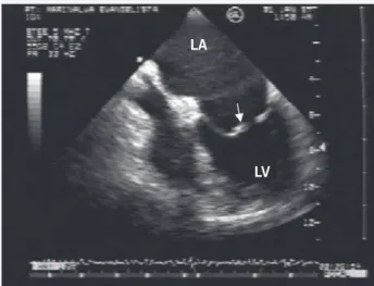
Logistic regression identified the smaller valvular areas and the restricted mobility of the anterior leaflet (fig. 1) as the only independent determinants for mitral valve replacement (tab. III). All patients treated with mitral valvuloplasty underwent the second Doppler echocardiographic examination before hospital discharge (7.97±3.16 days after surgery), and 26 patients underwent another examination 157.43±47.34 days after surgery. Doppler echocardiography showed that none of the 29 patients studied before hospital discharge had a mitral valve regurgitation degree greater than mild. Seven (24.13%) had no regurgitation, 10 (34.48%) had minimum regurgitation, and 12 (41.37%) had mild regurgitation.
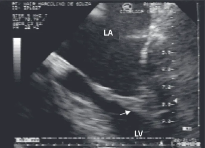
However, 7 (26.9%) of the 26 patients undergoing the last examination showed regurgitation greater than mild, all of which were moderate. Of those, 6 (85.71%) had restricted mobility of the anterior leaflet (P < 0.001), and 4 (57.14%) had fusion of the chordae tendineae (P = 0.002) on the Doppler echocardiography performed before surgery. These data represent 100% of the patients with restricted mobility of the anterior leaflet and fusion of chordae tendineae (fig. 2) referred for reparative surgery (tab. IV).
The interobserver concordance of the categorical Doppler echocardiographic variables was established using the Kappa sta-tistic, whose results showed the values of 0.74, 0.86, and 0.69 for thickening, calcification, and stretching of the chordae tendi-neae, respectively. The remaining variables showed Kappa values equal to one.
Discussion
The advantages of valvuloplasty over valvular prosthesis im-plantation include smaller mortality, a low risk of thromboembolism,
Table I – Clinical characteristics
Mitral valvuloplasty Valvular replacement p
n=29 n=39
Female sex 19 (65.5%) 19 (48.7%) 0.17 Age (years) 29.0±2.7 33.4±1.9 0.18 BS (m2) 1.54±0.06 1.59±0.02 0.45
Sinus rhythm 24 (82.8%) 28 (71.8%) 0.29 NYHA Functional 18 (62.1%) 18 (46.2%) 0.19 class III/IV
BS- body surface; n- number; NYHA- New York Heart Association.
Table II – Characteristics of the mitral valve
Mitral valvuloplasty Valvular replacement p
n=29 n=39
Valvular area (cm2) 3.32±0.13 2.81± 0.08 0.001
Restriction on mobility of the leaflets
Absent (n=21) 13 (61.9%) 8 (38.1%) 0.275 Anterior* (n=26) 6 (23.1%) 20 (76.9%) 0.01 Posterior* (n=46) 15 (32.6%) 31 (67.4%) 0.01 Thickness of the leaflets (mm)
Anterior 4.34±0.16 4.50± 0.15 0.462 Posterior 4.49±0.16 4.56± 0.13 0.733 Calcification of the leaflets
Absent (n=48) 21 (43.7%) 27 (56.3%) 0.386 Both (n=10) 2 (20.0%) 8 (80.0%) 0.058 Isolated posterior (n=9) 6 (66.6%) 3 (33.3%) 0.317 IIsolated anterior (n=1) 0 1 ( 100%) -Ring area (cm2) 15.55±0.85 15.32±0.70 0.830
Calcification of the 8 (20.0%) 12 (80.0%) 0.776 mitral ring (n=20)
Alterations in chordae tendineae
Thickening (n=38) 20 (52.6%) 18 (47.4%) 0.75 Fusion (n=18) 4 (22.2%) 14 (77.8%) 0.018 Calcification (n=6) 0 6 (100%) -Stretching (n=17) 10 (58.8%) 7 (41.2%) 0.617
*isolated or associated with the other leaflet.
Table III – Logistic regression
Variables Coefficient Standard p value Odds Ratio 95% confidence deviation interval
Age 0.025 0.02 0.257 1.03 [0.98; 1.07] MVA -0.787* 0.26* 0.002* 2.20* [1.32; 3.65]* RMAL 1.325 1.65 0.03 3.76 [1.11; 12.7]
MVA- mitral valve area; RMAL- restriction on mobility of the anterior leaflet; * for each 0.5 cm2.
Fig. 1 – Transesophageal echocardiogram showing restricted mobility of the mitral valve leaflets; impairment of the anterior leaflet (arrow) is more evident. LA – left atrial; LV – left ventricle.
LA
62
no need for anticoagulation, and better preservation of the left ventricular function after surgery. These advantages make mitral valve reconstruction not only an alternative for valve replacement, but also the ideal choice of treatment in the cases where it is feasible. In addition, the recent questions related to the possibility and durability of the repairs were satisfactorily answered, and excellent results were demonstrated in patients with mitral valve regurgitation due to degenerative and ischemic processes. On the other hand, in lesions with a rheumatic etiology, merely acceptable results are a reflex of the high index of significant residual regur-gitation with a subsequent elevated incidence of reoperations.
The results of the present study have shown that Doppler echocardiography plays an important role in predicting the type of surgery to be performed in patients with mitral valve regurgitation of rheumatic etiology. Our number of patients is the greatest re-ported in the publications comparing the Doppler echocardiographic findings obtained before surgery with the type of surgery effectively performed for correcting mitral valve regurgitation of rheumatic origin. Therefore, the usefulness of Doppler echocardiography is confirmed for the management of patients with that type of disease. The population studied was representative of patients with rheu-matic disease, the majority of whom were young women, and the results were comparable to those of the study by Sand et al 28.
Chauvaud et al 29 also assessed rheumatic patients with severe
mitral valve regurgitation undergoing conservative surgery. The population of their study had characteristics similar to those of the population of the present study, as follows: mean age, 26 years and 29 years, respectively; prevalence of sinus rhythm, 63% and 82%, respectively; and prevalence of restricted mobility of
the leaflets, 60% and 55%, respectively. In addition, the percentage of individuals undergoing the techniques of annuloplasty was almost identical, approximately 95% of the patients in both studies.
Valvuloplasty could be performed in only 43% of our patients, a finding similar to that reported by Chavez et al 30, who also
assessed patients with mitral valve regurgitation exclusively of rheumatic etiology.
The analysis of the Doppler echocardiographic features of mitral valves showed important variables, which certainly corre-lated with the type of surgery performed. The mitral valve area was smaller in individuals undergoing valve replacement. The lo-gistic regression showed that 0.5-cm2 decreases in valvular area
were associated with a 2.2-fold greater probability of the patients being treated with prosthetic implantation. Usually this is related to the fact that the smaller the mitral valve area, the more intense the anatomic impairment of the different regions of the valvular apparatus. This means that the greater the degree of thickening and calcification of the leaflets and chordae tendineae, as well as the fusion of the latter structure and the region of the commissures, the smaller the opening degree of the leaflets.
The direct and inversely proportional relation between valvular opening and the final result of anatomic impairment, ie, restricted mobility of the leaflets, is as important as the association of ana-tomic abnormalities and valvular opening. Therefore, the greater the restricted mobility of the leaflets, the smaller the opening area of the mitral valve. Thus, a significant and direct association between the presence of restricted mobility of the leaflets and valvular replacement should be expected. The presence of restricted mobility of the anterior leaflet reduced the chance of mitral valvu-loplasty by 3.8 times. This result reflects the difficulty of the surgeons in repairing the valves in patients whose major pathoge-netic factor for mitral valve regurgitation is the anterior leaflet alteration. This difficulty may be partially explained by the dimen-sions and shape of that leaflet, which do not favor the performance of resections, both triangular and quadrangular. Another motive of this difficulty is the fact that part of the ring related to the ante-rior leaflet is located between the fibrous trigones of the heart (left and right). This area is difficult to access, and, therefore, difficult to manage surgically. It is also an extremely important region, because it is related to valve support. In the present study, all patients with restricted mobility of the anterior leaflet who underwent valvuloplasty had mitral valve regurgitation greater than mild 6 months after surgery, despite the satisfactory results in the operating room and on hospital discharge. According to Carpentier et al 13, the fusion of chordae tendineae is the most important
cause determining the restricted mobility of the leaflets. The findings in this study are in accordance with those reported by Turner et al 31, who consider mitral reparative surgery formally
contraindicated in individuals with restricted mobility of the anterior leaflet. In our study, which assessed the morphological and functio-nal aspects of patients who required reoperation, none of the 51 patients with severe mitral regurgitation exclusively of rheumatic etiology and undergoing initial reparative surgery had that type of functional alteration. However, 49% of the patients had restricted mobility of the posterior leaflet at that moment (before the first surgery). Lessana et al 32 have suggested extreme caution when
performing mitral valvuloplasty in patients with restricted mobility of the leaflets, but they did not distinguish between the impaired leaflets.
Table IV – Degree of mitral valve regurgitation 6 months after valvuloplasty
Variables Absent or Greater than p mild n=19 mild n=7
Restriction on mobility of the anterior leaflet
Absent 19 1
Present* 0 6 <0.001
Fusion of the chordae tendineae
Absent 19 3
Present** 0 4 0.002
* all patients with this alteration who underwent valvuloplasty. ** all patients with this alteration who underwent valvuloplasty. Fig. 2 - Magnified image (“zoom”) of a transverse view of transesophageal echocardiography, showing fusion of the chordae tendineae (arrow). LA – left atrial; LV – left ventricle.
LA
63
Of the 26 patients in our study with restricted mobility of the anterior leaflet, only one did not show on Doppler echocardiography the concomitant restricted mobility of the posterior leaflet. On the other hand, 21 patients were identified as having isolated restricted mobility of the posterior leaflet of the mitral valve, data that suggest that rheumatic impairment in the mitral valve may begin in the posterior region of the valve.
Our study is the first report in the literature about the quanti-tative analysis of thickening in the valvular leaflets in patients with severe chronic mitral valve regurgitation. Both leaflets had similar and only mild thickening (approximately 4.5 mm), being characterized as degree I when using the valvular score proposed by Wilkins et al 25, which is largely used in the Doppler
echocar-diographic studies performed in patients with mitral stenosis. Such a mild leaflet thickening observed in the patients did not seem to be the cause of the restricted leaflet mobility, which would explain the lack of a significant relation to the surgery of valvular prosthesis implantation. However, calcification of the anterior leaflet did relate to valvular replacement, although the statistical analysis was not performed due to the reduced number of patients with that alteration. On the other hand, the presence of calcification in the posterior leaflet did not interfere with the surgeon’s decision in regard to the type of surgery to be performed.
The area of the mitral valve ring in the patients was 2.5-fold greater than that observed in healthy individuals 33, but did not
influence the type of surgery performed. The increase in the dia-meters of the mitral valve ring is usually consequent to the marked left ventricular dilation observed in patients with dilated cardio-myopathy or volume overload of that cavity, such as aortic or mitral valve regurgitation. Thus, once installed, ring dilation may not only cause mitral valve regurgitation, but may even exacerbate the degree of the regurgitation already present, which can dilate even more the left ventricle and the valvular ring itself, typical aspects of a vicious circle. Turner et al 31have also reported a
high prevalence of dilation of the valvular ring in patients with mitral valve regurgitation of rheumatic etiology undergoing repa-rative surgery. The prevalence, determined by surgeons during surgery, was 92.1%. In a multicenter study performed in the Ne-therlands 8,9, the prevalence of that alteration was 62%, and the
patients with severe mitral valve regurgitation of different etiologies were diagnosed with dilation of the ring by use of Doppler echo-cardiography performed just before surgical correction. Using the surgeon’s assessment as the “reference pattern,” 81% sensitivity was obtained for the mentioned examination.
Abnormalities in the chordae tendineae were frequently observed and diagnosed in more than half of the patients in the present study. One of the motives of this high prevalence was the inclusion only of patients with mitral valve regurgitant lesion of the rheumatic etiology. Another motive was the subjectivity of Doppler echocar-diography for diagnosing those abnormalities. Subjectivity may account for diagnostic divergences among echocardiographers, mainly in cases of less severe impairments. It is worth noting that, during the Doppler echocardiographic assessment of the subvalvular region, one of the authors of this study (or at least strong evidence existed) already knew the etiology of the disease, a fact that may have induced false-positive diagnoses. Thickening of the chordae tendineae is considered by Carpentier 13 the major
cause of restricted mobility of the leaflets. In our study, thickening
of the chordae tendineae showed no type of association with the surgical treatment used. This lack of association may have been partially due to the fact that only the presence or absence of that abnormality was considered. Although thickening limited to the juxtavalvular region was not considered (which corresponded to degree I of the Wilkins et al 25 score), several other degrees of
impairment that could have different repercussions on the mobility of the valvular leaflets were only considered as present. Thus, patients with more discrete thickening may have undergone re-parative surgery, while those with severe thickening (and probably with more marked restricted mobility of the leaflets) may have undergone prosthetic implantation.
More advanced forms of thickening may result in fusion of the chordae tendineae. The prevalence of that abnormality in a study analyzing patients with mitral valve regurgitation of different etiologies was 13% 9, ie, two times smaller than that observed in
the present study (27.9%). Extensive fusions that cause restricted mobility of the leaflets may be considered one of the indications for prosthesis implantation. In this study, univariate analysis showed the existence of a relation between the presence of fusion of the chordae and prosthesis implantation. It is worth emphasizing that 14 of 18 patients with that type of anatomic abnormality under-went valve replacement. However, an unfavorable postoperative evolution was observed in the 4 patients undergoing valvuloplasty and who had fusion of the chordae tendineae before surgery. All patients with that type of alteration undergoing conservative surgery had mitral valve regurgitation greater than mild on the Doppler echocardiographic study performed 6 months after surgery. Ac-cording to Duran et al, 34 the major causes of the unsatisfactory
results after valvuloplasty are as follows: a) use of inadequate technique in regard to the degree of valvular distortion; 2) valvular reconstruction performed in an inadequate manner; 3) the techniques of valvuloplasty are unstable and the results may dete-riorate over time; and 4) progression of the underlying disease.
Carpentier et al 13 reported that 40% of the patients undergoing
surgical correction of severe mitral valve regurgitation of any etiology had stretching of the chordae tendineae, and that was the major cause of mitral valve regurgitation observed in patients undergoing conservative surgery. In the present study, that alteration was not associated with the type of surgery performed.
64
may identify patients with severe mitral valve regurgitation of rheumatic etiology who will receive valvular prosthesis implantation. The advantages of that technique over the transesophageal one are as follows: not invasive; lower cost; greater availability; easier to perform; no need for previous preparations; better tolerated by patients; and performed by a greater number of echocardiographers.
Of the variables subjectively assessed in this study, the following stand out: restricted mobility of the leaflets, calcification of the components of the mitral valve, and thickening and stretching of the chordae tendineae. Such subjectivity may have caused different assessments, both performed by a single observer at different times and between different echocardiographers. However, it is worth emphasizing that even the assessment of anatomic and functional abnormalities of the mitral valve performed by surgeons and pa-thologists is performed in a subjective manner. Although quantitative methods of analysis are desired, acceptable objective ways of
measuring certain variables are still lacking, such as the mobility of the posterior leaflet of the mitral valve and stretching of the chordae tendineae. Another limitation of this study was the fact that several surgeons with different skills for performing reparative surgery took part in this research. It is worth emphasizing that the relation between the echocardiographic variables and the type of surgery performed also depends on the individual skill of each surgeon, and this may have influenced our results. The per-centage of valvuloplasties performed in this study should not be extrapolated to other institutions, the possibility of performing reparative surgery being inherent to each institution. Finally, the absence of 3 patients in the medium-term Doppler echocardio-graphic assessment may have affected the percentage of patients with mitral valve regurgitation observed 6 months after surgery. However, the loss of only 10% of the patients of this case series may not have significantly altered the result.
1. Marcus RH, Sareli P, Pocock WA, Barlow, J.B. The spectrum of severe mitral valve disease in a developing country: correlations among presentation, surgical patho-logic findings and hemodynamic sequelae. Ann ntern Med. 1994;120:177-83. 2. Duran CG, Pomar JL, Revuelta JM. Conservative operation for mitral insufficiency:
critical analysis supported by postoperative hemodynamic studies of 72 patients. J Thorac Cardiovasc Surg 1980;79:326-37.
3. Yacoub M, Halim M, Radley-Smith R, Mckay R. Nijveld A, Towers, M. Surgical treatment of mitral regurgitation caused by floppy valves: repair versus replace-ment. Circulation. 1981;64:210-6.
4. David TE, Uden DE, Strauss HD. The importance of mitral apparatus in left ventri-cular function after correction of mitral regurgitation. Circulation. 1983;8:76-82. 5. Goldman ME, Mora F, Guarino T, Fuster V, Mindich BP. Mitral valvuloplasty is superior to valve replacement for preservation of left ventricular function: an intraoperative two-dimensional echocardiographic study. J Am Coll Cardiol 1987; 10:568-75.
6. Enriquez-Sarano M, Schaff HV, Orszulak TA, Tajik AJ, Bailey KR, Frye RL. Valve repair improves the outcome of surgery for mitral regurgitation: a multivariate analysis. Circulation. 1995;91:1022-8.
7. Enriquez-Sarano M, Freeman WK, Tribouilloy CM. Functional anatomy of mitral regurgitation: accuracy and outcome implications of transesophageal echocardio-graphy. J Am Coll Cardiol.1999;34:1129-36.
8. Hellemans IM, Pieper EG, Ravelli AC. Comparison of transthoracic and transeso-phageal echocardiography with surgical findings in mitral regurgitation. The ESMIR research group. Am J Cardiol. 1996;77:728-33.
9. Hellemans IM, Pieper EG, Ravelli AC. Prediction of surgical strategy in mitral valve regurgitation based on echocardiography. Interuniversity Cardiology Institute of the Netherlands. The ESMIR research group. Am J Cardiol. 1997; 79: 334-8. 10. Craver JM, Cohen C, Weintraub WS. Case-matched comparison of mitral valve
replacement and repair. Ann Thorac Surg. 1990;49:964-9.
11. Fernandez J, Joyce DH, Hirschfeld K, Chen. Factors affecting mitral valve reopera-tion in 317 survivors after mitral valve reconstrucreopera-tion. Ann Thorac Surg. 1992; 54:440-7.
12. Perier P, Stumpf J, Gotz C. Isolated prolapse of the posterior leaflet of the mitral valve: results of reconstructive surgery. Arch Mal Coeur Vaiss. 19980; 91: 831-6. 13. Carpentier A. Cardiac valve surgery - the “french correction”. J Thorac Cardiovasc
Surg. 1983;86:323-37.
14. Guidelines For The Management Of Patients With Valve Heart Disease: Executive summary. Circulation 1998;98:1949-84.
15. Henry WL, Demaria A, Gramiak R. Report of the American Society of Echocardio-graphy Committee on nomenclature and standards in two-dimensional echocar-diography. Circulation. 1980;62:212-5.
16. Wilson N, Goldberg SJ, Dickinson DF, Scott O. Normal intracardiac and great ar-tery blood velocity measurements by pulsed doppler echocardiography. Br Heart J. 1985;53:451-8.
17. Omoto R, Kasai C. Physics and instrumentation of doppler color flow mapping. Echocardiography 1987;4:467-83.
References
18. Seward JB, Khandheria BK, Oh JK. Transesophageal echocardiography: techni-que, anatomic correlations, implementations and clinical applications. Mayo Clin Proc. 1988;63:649-80.
19. Wannwann LS, Feigenbaum H, Weyman AE, Dillon JC. Cross-sectional echocardio-graphic detection of rheumatic mitral regurgitation. Am J Cardiol. 1978; 41:1258-63. 20. Enriquez-Sarano M, Seward JB, Bailey KR, Tajik AJ. Effective regurgitant orifice area: a noninvasive doppler development of an old hemodynamic concept. J Am Coll Cardiol. 1994;23:443-51.
21. Hall AS, Brickner E, Willett DL, Irani WN, Afridi I, Grayburn PA. Assessment of mi-tral regurgitation severity by doppler color flow mapping of the vena contracta. Cir-culation. 1997;95:636-42.
22. Klein AL, Obarsky TP, Stewart WJ. Transesophageal doppler echocardiography of pulmonary venous flow: a new marker of mitral regurgitation severity. J Am Coll Cardiol. 1991;18:518-26.
23. Helmcke F, Nanda NC, Hsiung MC. Color doppler assessment of mitral regurgita-tion with orthogonal planes. Circularegurgita-tion. 1987;75:175-83.
24. Miyatake K, Izumi S, Okamoto M. Semiquantitative grading of severity of mitral re-gurgitation by real-time two-dimensional doppler flow imaging technique. J Am Coll Cardiol. 1986;7:82-8.
25. Wilkins GT, Weyman AE, Abascal VN, Block PC, Palacios IF. Percutaneous balloon dilatation of the mitral valve: an analysis of echocardiographic variables related to outcome and mechanism of dilatation. Br Heart J. 1988;60:299-308. 26. Libanoff AJ, Rodbard S. Atrioventricular pressure half-time: measurement of
mi-tral valve area. Circulation. 1968;38:144-50.
27. American Society Of Echocardiography Committee On Standards. Recommenda-tions for quantification of the left ventricle by two-dimensional echocardiography. J Am Soc Echocardiogr. 1989;2:358-67.
28. Sand ME, Naftel DC, Blackstone EH, Kirklin JW, Karp RB. A comparison of repair and replacement for mitral valve incompetence. J Thorac Cardiovasc Surg.1987; 94:208-9.
29. Chauvaud S, Fuzellier JF, Berrebi A, Deloche A, Fabiani JN, Carpentier A. Long-term (29 years) results of reconstructive surgery in rheumatic mitral insufficiency. Circulation. 2001;104:12-5.
30. Chavez AM, Cosgrove DM, Lytle BW. Applicability of mitral valvuloplasty techni-ques in a north american population. Am J Cardiol. 1988;62:253-6.
31. Turner E, Wisenbaugh T, Sinovich V, Cronje S, Sareli P. Morphologic patterns in patients undergoing reoperation after repair of rheumatic mitral regurgitation. Am J Cardiol. 1993:71;455-7.
32. Lessana A, Carbone C, Romano M. Mitral valve repair: results and the decision-making process in reconstruction. J Thorac Cardiovasc Surg. 1990; 99: 622-30. 33. Ormiston JA, Shah PM, Chuwa T, Wong M. Size and motion of the mitral valve
an-nulus in man. Circulation. 1982;65:713-9.