Assessing bone thickness in the infrazygomatic crest
area aiming the orthodontic miniplates positioning:
a tomographic study
Aline Rode Santos1, Marcelo Castellucci1, Iêda Margarida Crusoé-Rebello2, Márcio Costa Sobral1
Introduction: Due to the increasing use of miniplates for anchorage purposes in orthodontics, it is very important to know more about infrazigomatic crest anatomy (thickness), in adult patients. Objectives: Evaluate the infrazygomatic crest region thickness, in adult (male and female) patients. Methods: Cone-beam computerized tomography (CBCT) images from 40 patients were used to assess cross-sectional measurements of the infrazygomatic crest region. Measurement 1 considered thickness 2 mm above the distobuccal root of the permanent maxillary first molar, while measurement 2 was taken 2 mm above the first measurement. Results: The mean thickness of the infrazygomatic crest in males was 3.55 mm for measurement 1 and 2.84 mm for measure-ment 2, while in females these were 2.37 mm and 2.24 mm, respectively. Conclusion: The authors concluded that the overall mean thickness of the infrazygomatic crest was 2.49 mm with respect to measurement 1, and 2.29 mm for measurement 2, with no statistically significant differences between gender.
Keywords:Measurements. Tomography. Orthodontics.
1 Universidade Federal da Bahia, Departamento de Ortodontia
(Salvador/BA, Brasil).
2 Universidade Federal da Bahia, Departamento de Radiologia Bucal
(Salvador/BA, Brasil).
» The authors report no commercial, proprietary or financial interest in the products or companies described in this article.
» Patients displayed in this article previously approved the use of their facial and in-traoral photographs.
DOI: https://doi.org/10.1590/2177-6709.22.4.070-076.oar
How to cite: Santos AR, Castellucci M, Crusoé-Rebello IM, Sobral MC. As-sessing bone thickness in the infrazygomatic crest area aiming the orthodontic miniplates positioning: a tomographic study. Dental Press J Orthod. 2017 July-Aug;22(4):70-6. DOI: https://doi.org/10.1590/2177-6709.22.4.070-076.oar
Submitted: October 27, 2016 - Revised and accepted: March 31, 2017
Contact address: Aline Rode Santos E-mail: [email protected]
Introdução: devido ao aumento do uso de miniplacas para ancoragem em Ortodontia, torna-se de fundamental importância compreender melhor a anatomia da crista infrazigomática (espessura) em pacientes adultos. Objetivos: avaliar a espessura da crista infrazigomática em pacientes adultos (sexos feminino e masculino). Métodos: foram utilizadas tomografias computadorizadas de feixe cônico (TCFC) de 40 pacientes, para avaliar medidas da região da crista infrazigomática, no sentido transversal. A Medida 1 verificou a espessura mm acima da raiz distovestibular do primeiro molar permanente superior, enquanto a Medida 2 foi realizada 2 mm acima da primeira medida. Resultados: a espessura média da crista infrazigomática encontrada no sexo masculino foi de 3,55 mm para a Medida 1 e de 2,84 mm para a Medida 2, e no sexo feminino foi de 2,37 mm e de 2,24 mm, respectivamente. Conclusão: os autores puderam constatar que a espessura média da crista infrazigomática foi de 2,49 mm para a Medida 1, e de 2,29 mm para a Medida 2, sem diferença estatisticamente significativa entre os sexos.
INTRODUCTION
One of the most frequently problems in orthodon-tics is to achieve the anchorage necessary to obtain a
desired tooth movement.1-4 Conventional approaches
employ the anchorage potential of existing teeth when a large number of these can resist the movement of a small number. This usually requires the use of auxiliary devic-es, such as intermaxillary elastics and/or headgear, but a negative aspect is that these devices depend on patient’s
cooperation.1,2,3,5-8 The need to eliminate undesirable
ef-fects and, at the same time, maximize anchorage, has led to the development of skeletal anchorage systems utiliz-ing osseointegrated implants, implants and mini-plates. These devices do not allow for the movement of
the anchorage unit during orthodontic mechanics9 and
they can be used 24 hours a day, ofering an alternative
method that better controls side efects.8
Titanium miniplates are temporary anchorage devices that not only provide better stability than mini-implants, but also are more resistant to stronger
forces.7,10 They are placed at a greater distance from
the root apexes, allowing distal movement around the arch as there is no interference between the fixed
device and dental roots.7,8,11 These miniplates can be
used for multiple purposes, such as direct or indirect
anchorage for different types of tooth movement,12 in
addition to providing skeletal anchorage for
maxil-lary protraction.13 However, they require surgical
procedures for placement and removal, which must be performed by a qualified surgeon due to increased
complexity at this anatomical site.7,10
According to De Clerck et al,12 due to the
loca-tion and solid bone structure, the inferior border of the maxillary zygomatic buttress, also known as the infra-zygomatic crest, located between the irst and second molars, is the chosen site for the placement of miniplates with the purpose of using the skeletal anchorage system, placing the miniplates at a safe distance from the roots of the maxillary molars.
Anatomically, the infrazygomatic crest has two cor-tical plates, a vestibular one and the lateral wall of the maxillary sinus. This anatomical advantage allows bi-cortical ixation and it contributes to improved primary
stability of the screw.14 However, the infrazygomatic
crest area is 2-5 mm thick, while miniscrews are ap-proximately 5-7 mm long, which may cause perforation
of the maxillary sinus during its placement.8
Due to the frequent use of skeletal anchorage, it is extremely important to conduct studies to assess the thickness of the infrazygomatic crest to better under-stand its anatomical dimensions, providing safer surgical procedures and minimizing possible failures.
The aim of this study was to verify the thickness of the infrazygomatic crest and compare it between male and female adult subjects, by using coronal slices from cone-beam computerized tomography (CBCT) imaging.
MATERIAL AND METHODS
The present study employed CBCT images of 40 patients from a post-graduate course in orthodontics. Of the included patients, 18 were male (45%), 22 fe-male (55%), aged 22-56 years (mean age of 31 years), and all full-filled the following criteria: need for max-illary bone anchorage during orthodontic treatment, presence of the permanent maxillary first molars; over 21 years of age; no presence of bone lesions in the maxillary region.
In order to test the sample power, it was performed a power analysis using R-software (www.r-project. org, version 3.3.2). It was found a power of 80%, based on the significance level of alpha of 0.01 and the effect size of 0.85.
This project was approved by a Institutional Ethic’s Committee, protocol number 905.596. All included patients were required to sign a informed consent form, allowing their exams to be used for research purposes.
The study assessed the thickness of the infrazy-gomatic crest by obtaining cross-sectional measure-ments using coronal slices from cone-beam computer-ized tomography (CBCT) images. CBCT images were
obtained using an i-CAT® device (Imaging Sciences
International, Hatield, PA, USA) with an acquisi-tion protocol of 120 Kvp, 47 mA, 0.4 mm-thick slices, 0.4-mm voxel size, 20 x 25 cm ield of view (FOV) and an acquisition time of 40 seconds. CT scans were per-formed with patients seated so that the Frankfort hori-zontal plane was parallel to the ground, in a maximum intercuspation position.
The Digital Imaging and Communications in Medicine (DICOM) ile format was used to compose three-dimensional reconstructions of each patient’s
fa-cial structure using Dolphin Imaging® sotware, version
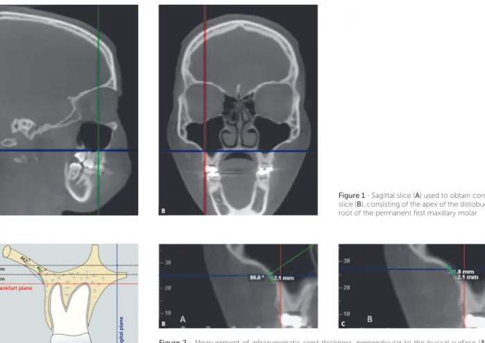
Figure 1 - Sagittal slice (A) used to obtain coronal slice (B), consisting of the apex of the distobuccal root of the permanent first maxillary molar.
Figure 2 - Measurement of infrazygomatic crest thickness, perpendicular to the buccal surface (A). Il-lustrative picture at 2 mm (B) and at 4 mm (C) above the apex of the distobuccal root of the permanent first maxillary molar.
the head orientation in each digital image was standard-ized according to the sagittal, coronal and axial planes. In the frontal view, the patient’s median line was aligned in accordance with the orientation line of the sotware and the right and let frontozygomatic sutures were marked. In the lateral view, the right orbital and right porion points were located and positioned to coincide
with the Frankfort horizontal plane.15
Next, the apex of the distobuccal root of each per-manent irst maxillary molar was located using a sagittal CT slice to obtain a coronal view on each side (Fig 1).
Two measurements were obtained along the infrazy-gomatic crest with the aid of the Digitize-measurement tool in the Dolphin Imaging program. The irst measure-ment (measuremeasure-ment 1) was performed at 2 mm above the distobuccal root apex of the permanent irst maxillary
molar along the buccal wall of the infrazygomatic crest, speciically where the horizontal axis of the program co-incided with the oral surface of the infrazygomatic crest. The next measurement (measurement 2) was performed 2 mm above the irst one, maintaining the same proce-dure used in measurement 1 (Fig 2).
Statistical analysis
Prior to taking measurements, in order to calibrate the examiner, 10 scans were selected. All digital mea-surements were performed by a single previously cali-brated operator under identical conditions at two dif-ferent times, with a two-week interval. To verify agree-ment among the measureagree-ments, Lin’s concordance test was used, obtaining a result of 0.98, considered an al-most perfect concordance.
A B
A
B C
M2
M1
Frankfurt plane
Midsagital plane
2 mm
A database was created in Excel 2003 and analyzed in the R sotware version 3.1.0 (R Foundation for sta-tistical computing, Wien, Österreich). A descriptive analysis (absolute/relative frequency, mean, standard deviation and median) was performed to identify the general and speciic characteristics of the study sample. The normality assumptions were veriied using Kol-mogorov-Smirnov and Shapiro-Wilk tests and the Lev-ene’s test of variance homogeneity. With respect to the data presenting normal distribution, parametric testing was employed, while in the non-normal distributions, non-parametric testing was used.
With the objective of comparing the median thickness of the measured crest with the hypoth-esized measurement of a standard 5-mm long minis-crew, one-sample Wilcoxon test was used. Student’s
t-test was used to verify significant differences among
the different heights. Mann-Whitney test was used to verify if there were any significant differences in measurements according to gender. Wilcoxon test for paired samples was used to compare the measure-ments between the two sides.
The signiicance level for this study was 5%. The re-sults are presented in comparative tables.
RESULTS
The present study found that the overall mean thick-ness of the infrazygomatic crest was 2.49 mm with respect to measurement 1, and 2.29 mm for measure-ment 2. Table 1 shows that no statistically signiicant diferences were observed when comparing genders.
Table 2 delineates the statistically signiicant
difer-ences seen between measurements 1 and 2 (p = 0.019).
Table 3 indicates that statistically signiicant difer-ences were found between the right and let sides with
respect to measurement 2 (p = 0.002).
DISCUSSION
The Schneiderian membrane, which is attached to the bordering bone of the maxillary sinus, is charac-terized by a periosteum overlaid with a thin layer of pseudociliated stratified respiratory epithelium, con-stituting an important barrier for the protection and defense of the sinus cavity. Its integrity is essential to
normal sinus function.16
According to Reiser et al,17 when implants extend
less than 2 mm into the maxillary sinus, the sinus mem-brane becomes elevated, which favors healing as it allows for the formation of a blood clot that provides a scafold
VARIABLES
SEX
p-value
MALE FEMALE
Mean Standard
deviation
Median Mean Standard
deviation
Median
Measurement 1 2.62 1.41 2.2 2.37 1.0 2.2 0.747
Measurement 2 2.34 1.13 1.85 2.24 0.73 2.1 0.692
Table 1 - Comparison of thickness of the infrazygomatic crest between sexes (Mann-Whitney test).
Table 2 - Comparison between measurements 1 and 2 (Student’s t-test).
Measurement 1 Measurement 2
p-value
Mean Standard
deviation
Median Mean Standard
deviation
Median
2.49 1.21 2.2 2.28 0.94 2.0 0.019
Table 3 - Comparison between the right and left sides (Wilcoxon test).
VARIABLES
Right side Left side
p-value
Mean Standard
deviation
Median Mean Standard
deviation
Median
Measurement 1 2.24 0.9 2.0 2.74 1.42 2.4 0.111
for bone formation in this region. When the implant extends further into the maxillary sinus, i.e. greater than 2 mm, the Schneiderian membrane becomes perforat-ed, which may result in the discharge of bone fragments inside the maxillary sinus, thus compromising healing ability, increasing the occurrence of sinusitis.
In a clinical and experimental study of the efects caused by the penetration of osseointegrated dental implants into the nasal cavity and the maxillary sinus,
Brånemark et al18 noted that titanium implants did
not cause side efects and were well-anchored in bone. The authors believe that no side efects were seen be-cause these implants became osseointegrated into the bone structure, which promoted direct contact be-tween the implant, bone and sot tissue. This direct connection between the implant and the hard and sot tissues creates a barrier against the migration of mi-croorganisms, and inhibits the inlammatory process around the implant. In essence, osseointegration pro-tects the implant. On the other hand, according Adell
et al,19 when osseointegration does not occur, a ibrous
tissue covers the implant, normally leading to the in-duction of an inlammatory process followed by bone resorption and implant loss.
In order for implants to be placed into a suitable bone surface, the anatomy of the infrazygomatic crest must be known by means of speciic exams, such as CBCT.
The present study demonstrates that the infrazy-gomatic crest is signiicantly thinner than the length of the miniscrews commonly used in this region, as the mean thickness of the crest was found to be 2.49 mm for measurement 1, and 2.29 mm for measurement 2.
The present indings corroborate those of Liou et al,14
who found a thickness of 2.9 ± 0.9 mm in the lateral wall
of the maxillary sinus (where miniplates were placed)
and Baumgaertel and Hans,20 who found a mean
thick-ness of 3.87 mm at 2 mm from the apex of the distobuc-cal root of the irst molar and 2.98 mm at 4 mm from the apex of the distobuccal root.
Liou et al14 and Lee et al21 found no statistically
sig-niicant diferences between the measurements on the right and let sides. However, the present results show a statistically signiicant diference between the right and let sides with respect to measurement 2, among female patients — although this data is not clinically relevant, since the diferences found were very small in compari-son to the 5-mm miniscrew size.
Lee et al21 found that the infrazygomatic crest was
clinically thicker in male patients than in female pa-tients. In the present study, no statistically signii-cant diferences were detected between sexes. In fact, marked individual variations in these measurements were observed irrespective of sex, which is in agreement
with the indings of Farnsworth et al.22
The present study found that the average thickness of the infrazygomatic crest was smaller when measured further from the root apex, corroborating the results
re-ported by Baungaertel and Hans,20 who found greater
risk of maxillary sinus perforation when miniscrews were placed in a more cranial orientation.
Baungaertel and Hans20 stated that great individual
variation exists in the thickness of the infrazygomatic crest. Indeed, the present study also found measure-ments ranging from 0.9 to 7.4 mm, which is probably due to difering root lengths, maxillary sinus pneuma-tization, buccolingual inclination of the maxillary irst molar, and the height of the alveolar processes among the individuals studied, all of which are determinants to the available bone depth for miniscrew placement.
However, according to Kravistz and Kusnoto,23 if the
maxillary sinus membrane is perforated during mini-screw placement, immediate removal must not occur due to its small diameter. Orthodontic therapy must continue and the patient should be followed to avoid the possible development of sinusitis and mucocele.
Kim et al24 conducted a computed tomographic
study to observe the placement of 31 miniplates, placed between the roots of the posterior teeth of 18 patients. To conduct the study, 74 screws, 4-mm long and 1.5 mm in diameter, were used. Their results showed that, of 74 miniscrews, 39 perforated the maxillary
si-nus, with mean exposure of 1.31 ± 0.72 mm. Among
these, only 3 miniscrews protruded more than 2 mm into the maxillary sinus (2.37, 2.95, and 3.41 millime-ters). No miniscrews presented mobility or caused any further complications, such as sinusitis, swelling or peri-implant inlammation. However, to conduct the study,
Kim et al.24 only selected patients who had presented
De Cleck et al,12 an internationally renowned author for his studies of miniplates for orthodontic anchorage, have indicated that this region is the best option for the placement of miniplates.
Miyawaki et al25 observed in their studies that the
sta-bility of the miniscrew is not related to its length, but rath-er to its diametrath-er, as 1-mm thick miniscrews demonstrated
less stability than 1.5 and 2.3-mm miniscrews. Kim et al2
also agree that the interface between the miniscrew and cortical bone is an important factor afecting the stability
of the miniscrew. Myawaki et al25 further suggest that the
thickness of the cortical bone should be veriied by CT scan prior to the placement of these anchoring devices, which could indicate the use of miniscrews with a diam-eter greater than 2.3 mm in the case of thin cortical bone, thereby providing greater stability by increasing the con-tact between the cortical bone and the miniscrew.
CONCLUSION
The authors concluded that the overall mean thickness of the infrazygomatic crest was 2.49 mm with respect to measurement 1, and 2.29 mm for measurement 2, with no statistically signiicant diferences between sexes.
CLINICAL CONSIDERATIONS
1. Turley PK, Kean C, Schur J, Stefanac J, Gray J, Hennes J, et al. Orthodontic force application to titanium endosseous implants. Angle Orthod. 1988 Apr;58(2):151-62.
2. Kim HJ, Yun HS, Park HD, Kim DH, Park YC. Soft-tissue and cortical bone thickness at orthodontic implant sites. Am J Orthod Dentofacial Orthop. 2006 Aug;130(2):177-82.
3. Kuroda S, Sugawara Y, Deguchi T, Kyung HM, Takano-Yamamoto T. Clinical
use of miniscrew implants as orthodontic anchorage: Success rates and postoperative discomfort. Am J Orthod Dentofacial Orthop. 2007 Jan;131(1):9-15.
4. Cornelis MA, Scheler NR, Mahy P, Siciliano S, De Clerck HJ, Tulloch JF. Modiied miniplates for temporary skeletal anchorage in orthodontics: placement and removal surgeries. J Oral Maxillofac Surg. 2008 July;66(7):1439-45.
5. Haas AJ. Headgear therapy: the most eicient way to distalize molars. Semin Orthod. 2000;6(2):79-90.
6. Araújo TM. Ancoragem esquelética com mini-implantes. In: Lima Filho RMA,
Bolognese AM. Ortodontia: arte e ciência. Maringá: Dental Press; 2007. p. 394-448.
7. Chen YJ, Chang HH, Huang CY, Hung HC, Lai EH, Yao CC. A retrospective
analysis of the failure rate of three diferent orthodontic skeletal anchorage systems. Clin Oral Implants Res. 2007 Dec;18(6):768-75.
8. De Clerck HJ, Cornelis MA. Biomechanics of skeletal anchorage. Part 2:
Class II nonextraction treatment. J Clin Orthod. 2006 May;40(5):290-8; quiz 307.
9. Southard TE, Buckley MJ, Spivey JD, Krizan KE, Casko JS. Intrusion anchorage potential of teeth versus rigid endosseous implants: a clinical and radiographic evaluation. Am J Orthod Dentofacial Orthop. 1995 Feb;107(2):115-20.
10. Ruellas ACO. Biomecânica aplicada à clínica. Maringá: Dental Press; 2013. p. 232-72.
11. Ramos LA, Zange ES, Terada HH, Hoshina FT. Anchorage miniplates on anterior open-bite treatment. Dental Press J Orthod. 2008;13(5):134-43. 12. De Clerck H, Geerinckx V, Siciliano S. The zygoma anchorage system. J Clin
Orthod. 2002 Aug;36(8):455-9.
13. De Clerck H, Cevidanes L, Baccetti T. Dentofacial efects of bone-anchored maxillary protraction: a controlled study on consecutively treated Class III patients. Am J Orthod Dentofacial Orthop. 2010 Nov;138(5):577-81. 14. Liou EJ, Chen PH, Wang YC, Lin JC. A computed tomographic image study
on the thickness of the infrazygomatic crest of the maxilla and its clinical implications for miniscrew insertion. Am J Orthod Dentofacial Orthop. 2007 Mar;131(3):352-6.
REFERENCES
15. Azevêdo MS, Machado AW, Barbosa IS, Esteves LS, Rocha VC,
Bittencourt MAV. Evaluation of upper airways after bimaxillary orthognathic surgery in patients with skeletal Class III pattern using cone-beam computed tomography. Dental Press J Orthod. 2016 Jan-Feb;21(1):34-41.
16. Ardekian L, Oved-Peleg E, Mactei EE, Peled M. The clinical signiicance of sinus membrane perforation during augmentation of the maxillary sinus. J Oral Maxillofac Surg. 2006 Feb;64(2):277-82.
17. Reiser GM, Rabinovitz Z, Bruno J, Damoulis PD, Griin TJ. Evaluation of maxillary sinus membrane response following elevation with the crestal osteotome technique in human cadavers. Int J Oral Maxillofac Implants. 2001 Nov-Dec;16(6):833-40.
18. Brånemark PI, Adell R, Albrektsson T, Lekholm U, Lindström J, Rockler B. An experimental and clinical study of osseointegrated implants penetrating the nasal cavity and maxillary sinus. J Oral Maxillofac Surg. 1984 Aug;42(8):497-505.
19. Adell R, Lekholm U, Rockler B, Brånemark PI. A I5-year study of
osseointegrated implants in the treatment of the edentulous jaw. Int J Oral Surg. 1981 Dec;10(6):387-416.
20. Baumgaertel S, Hans MG. Assessment of infrazygomatic bone depth for mini-screw insertion. Clin Oral Implants Res. 2009 June;20(6):638-42. 21. Lee HS, Choi HM, Choi DS, Jang I, Cha BK. Bone Bone thickness of the
infrazygomatic crest area in skeletal Class III growing patients: A computed tomographic study. Imaging Sci Dent. 2013;43(4):261-6.
22. Farnsworth D, Rossouw PE, Ceen RF, Buschang PH. Cortical bone thickness at common miniscrew implant placement sites. Am J Orthod Dentofacial Orthop. 2011 Apr;139(4):495-503.
23. Kravitz ND, Kusnoto B. Risks and complications of orthodontic miniscrews. Am J Orthod Dentofacial Orthop. 2007 Apr;131(4 Suppl):S43-51. 24. Kim GT, Kim SH, Choi YS, Park YJ, Chung KR, Suk KE, et al. Cone-beam
computed tomography evaluation of orthodontic miniplate anchoring screws in the posterior maxilla. Am J Orthod Dentofacial Orthop. 2009 Nov;136(5):628.e1-10; discussion 628-9.