A c c e t t a A c c e t t a A c c e t t a A c c e t t a A c c e t t a
Pancreatic pseudocyst with splenic involvement. Case report 457
Rev. Col. Bras. Cir. 2010; 37(6): 457-459
Case Report Case ReportCase Report Case Report Case Report
Pancreatic pseudocyst with splenic involvement. Case report
Pancreatic pseudocyst with splenic involvement. Case report
Pancreatic pseudocyst with splenic involvement. Case report
Pancreatic pseudocyst with splenic involvement. Case report
Pancreatic pseudocyst with splenic involvement. Case report
Pseudocisto pancreático com envolvimento esplênico. Relato de caso
Pseudocisto pancreático com envolvimento esplênico. Relato de caso
Pseudocisto pancreático com envolvimento esplênico. Relato de caso
Pseudocisto pancreático com envolvimento esplênico. Relato de caso
Pseudocisto pancreático com envolvimento esplênico. Relato de caso
PIETRO ACCETTA, TCBC-RJ1; ITALO ACCETTA, TCBC-RJ2; RENATO ACCETTA3; KEILA BORBA CAMPOS4;
MANOEL FERNANDO O. RODRIGUES, TCBC-RJ5
Work performed at the Department of General Surgery I, Hospital Universitário Antonio Pedro – UFF – Niterói – Rio de Janeiro – Brazil. 1. Professor, General Surgery, Faculdade de Medicina – Universidade Federal Fluminense (UFF) – Niterói – Rio de Janeiro – Brazil; 2. Professor, Surgery, Faculdade de Medicina - UFF – Niterói – Rio de Janeiro – Brazil; 3. Intensivist, Hospital Universitário Antonio Pedro – UFF – Niterói – Rio de Janeiro – Brazil; 4. Resident, Endocrinology, Hospital Geral de Bonsucesso (HGB) – Rio de Janeiro – Rio de Janeiro – Brazil; 5. Associate Professor, General Surgery, Faculdade de Medicina- UFF – Niterói – Rio de Janeiro – Brazil.
INTRODUCTION
INTRODUCTION
INTRODUCTION
INTRODUCTION
INTRODUCTION
P
ancreatic pseudocysts are a common complication of pancreatitis, but most of them, when small and uncomplicated, do not require treatment. However, some may cause many problems depending on location and the effect caused by the lytic enzyme activity of its contents. Their disruption to the peritoneum, hemorrhage, intracystic infection, compressions and fistulas to neighboring organs are the most common complications1. Other unusual cases found in the literature report leaks to distant sites such as the neck, mediastinum, chest cavity and scrotum2.The spleen, although very close to the pancreas, is rarely involved in this process and until 2001 there were fewer than 50 cases reported in the English scientific literature3. Heavy bleeding from the spleen is the most common and most serious outcome and may require urgent surgical treatment.
This case and its features, although having been handled by an elective operation, dully characterize this type of complication and the difficulties encountered in its treatment.
CASE REPPORT
CASE REPPORT
CASE REPPORT
CASE REPPORT
CASE REPPORT
A Caucasian man, aged 42, native of Rio de Janeiro (RJ), was admitted to Antonio Pedro University Hospital (HUAP) diagnosed with left lung atelectasis and pneumonia. He complained of pain in the left subscapular region radiating to the shoulder on the same side for 45 days; the pain was unrelated to respiration, but got worse at bedtime. He had been treated at another hospital with a diagnosis of pneumonia with left pleural effusion. He was treated with antibiotics and thoracentesis and was discharged after 15 days. Eight days later, high fever, malaise, weight loss and pallor ensued. There was no cough, neither sputum. He reported as antecedents: laparotomy for acute
458
Rev. Col. Bras. Cir. 2010; 37(6): 457-459
A c c e t t a A c c e t t a A c c e t t a A c c e t t a A c c e t t a Pancreatic pseudocyst with splenic involvement. Case report
wrinkled and very friable, which caused major intraoperative hemorrhage, controlled with tamponade and topical hemostatic sponges. The region of the cyst was drained with a thick rubber drain, which was removed two months later. The patient was discharged on day 14. The amylase of the liquid was only 700 U/l. The histopathological exam of the capsule was compatible with pancreatic pseudocyst. In review after four months, he was well, although the CT scan showed a thin layer of liquid next to the spleen.
DISCUSSION
DISCUSSION
DISCUSSION
DISCUSSION
DISCUSSION
The involvement of the pancreas’ neighboring organs during the development of a pseudocyst, in spite of
Figure 2 Figure 2 Figure 2 Figure 2
Figure 2 - MRI with a large and heterogeneous cystic mass late-ral to the spleen; note that the thick capsule surrounds the entire aggregate.
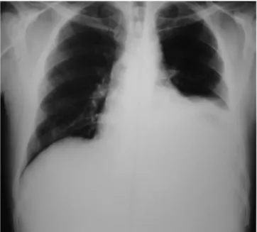
Figure 1 Figure 1 Figure 1 Figure 1
Figure 1 - Chest X-rays with marked elevation of left diaphragm and absence of the gastric air bubble.
relatively frequent, often preserves the spleen, making its involvement rather unusual, but of sometimes tragic consequences.
The low frequency and little experience with such cases do not allow definitive conclusions, but it is believed that the involvement of the spleen in the process would take place by three etiologic factors4,5: 1) vascular: thrombosis of the Splenic Vein is identified with possible complication in the evolution of chronic pancreatitis. It usually does not cause bleeding from the spleen, but may evolve with hypersplenism and even fugal type portal hypertension; 2) mechanical: this theory believes that perisplenic adhesions make the spleen more vulnerable to injuries, even minor ones; and 3) enzymatic: it occurs by direct action of pancreatic enzymes on splenic parenchyma or by invasion, through the hilum, along the blood vessels.
Although such patients may develop severe internal bleeding, the clinical presentation is usually not very different from those seen with uncomplicated pseudocysts. Uncharacteristic abdominal pain, eventual episodes of nausea, fever and weight loss are the most frequently reported complaints. Laboratory tests contribute little and a palpable mass is not very common, as the widespread use of CT and US make such pseudocysts diagnosed before appearing on physical examination, as was common in the past. Thus, some authors advocate early surgical indication due to the greater potential for complications that its evolution may result in4.
For most authors, the definitive treatment is splenectomy with distal pancreatectomy1,3, although prone to complications such as bleeding and infection. Heider3 presented a review of 238 pseudocysts and found only 14 (6%) of them affecting the spleen. This author believes that percutaneous drainage is not effective, since three of them required repetition of the procedure and many others ended up being operated. Likewise, he also pointed out flaws in the conservative management, as seven of the 10 other patients ended up requiring surgical treatment.
Unfortunately, the absence of pathological examination of the spleen itself rendered it difficult to characterize more clearly the type of splenic involvement, but the anatomical and radiological aspects similar to other cases, as well as a thick capsule surrounding the entire aggregate and leaving the entire convex surface of spleen in direct contact with the contents, prevent us to think of a commonplace pseudocyst.
In this case its long evolution may explain the extreme difficulty in finding safe plans that could allow surgical removal of the spleen and part of the pancreas. It is worth noting that the recommended splenectomy involves great risk of intraoperative hemorrhage and it is for the surgeon to establish the limits to accomplish it.
A c c e t t a A c c e t t a A c c e t t a A c c e t t a A c c e t t a
Pancreatic pseudocyst with splenic involvement. Case report 459
Rev. Col. Bras. Cir. 2010; 37(6): 457-459
A B S T R A C T A B S T R A C TA B S T R A C T A B S T R A C TA B S T R A C T
The authors present a case report of a pancreatic pseudocyst with an unusual spleen involvement. The aspects of this rare complication are discussed, as well as the probable etiologic factors. The outcome was satisfactory and the surgical treatment consisted of the resection of its thick capsule, since the local anatomic conditions would not permit a splenectomy with distal pancreatectomy, considered to be the ideal surgery.
Key words: Key words: Key words:
Key words: Key words: Pancreatic pseudocyst/complications. Pancreatic pseudocyst/surgery. Spleen.
REFERENCES
REFERENCES
REFERENCES
REFERENCES
REFERENCES
1. Sitzmann JV, Imbembo AL. Splenic complications of a pancreatic pseudocyst. Am J Surg. 1984;147(2):191-6.
2. McMahon NG, Norwood SH, Silva JS. Pancreatic pseudocyst with splenic involvement: a uncommon complication of pancreatitis. South Med J 1988; 81: 910-2.
3. Heider R, Behrns KE. Pancreatic pseudocysts complicated by splenic parenchymal involvement: results of operative and percutaneous management. Pancreas. 2001;23(1):20-5.
4. Scherer K, Kramann B. Rupture of the spleen by penetration of pancreatic pseudocysts. Eur J Radiol. 1987;7(1):67-9.
5. Yano T, Kaneda M, Yamamoto T, Suzuki T, Fujimori K, Itoh H. Pancreatic pseudocyst involving the spleen. Gastroenterol Jpn. 1988;23(1):73-7.
Received 06/10/2006
Accepted for publication 08/12/2006 Conflict of interest: none
Source of funding: none
How to cite this article: How to cite this article: How to cite this article: How to cite this article: How to cite this article:
Accetta P, Accetta I, Accetta R, Campos KB, Rodrigues MFO. Pancreatic pseudocyst with splenic involvement. Rev Col Bras Cir. [periódico na Internet] 2010; 37(6). Disponível em URL: http://www.scielo.br/rcbc
Correspondence to: Correspondence to: Correspondence to: Correspondence to: Correspondence to: Dr. Pietro Accetta