Lipoic acid increases glutathione
peroxidase, Na
+
, k
+
-atpase and
acetylcholinesterase activities
in rat hippocampus after
pilocarpine-induced seizures?
Geane Felix de Souza1, Gláucio Barros Saldanha1,Rivelilson Mendes de Freitas2
ABSTRACT
In the present study we investigated the effects of lipoic acid (LA) on acetylcholinesterase (AChE), glutathione peroxidase (GPx) and Na+, K+-ATPase activities in rat hippocampus during seizures. Wistar rats were treated with 0.9% saline (i.p., control group), lipoic acid (20 mg/kg, i.p., LA group), pilocarpine (400 mg/kg, i.p., P400 group), and the association of pilocarpine (400 mg/kg, i.p.) plus LA (20 mg/kg, i.p.), 30 min before of administration of P400 (LA plus P400 group). After the treatments all groups were observed for 1 h. In P400 group, there was a significant increase in GPx activity as well as a decrease in AChE and Na+, K+ -ATPase activities after seizures. In turn, LA plus P400 abolished the appearance of seizures and reversed the decreased in AChE and Na+, K+-ATPase activities produced by seizures, when compared to the P400 seizing group. The results from the present study demonstrate that preadministration of LA abolished seizure episodes induced by pilocarpine in rat, probably by increasing AChE and Na+, K+-ATPase activities in rat hippocampus.
Key words: glutathione peroxidase, Na+, K+-ATPase, acetylcholinesterase, hippocampus, seizures.
O ácido lipóico aumenta as atividades da glutationa peroxidase, da Na+, K+-ATPase
e da acetilcolinesterase no hipocampo de ratos após convulsões induzidas por pilocarpina?
RESUMO
No presente estudo nós investigamos os efeitos do ácido lipóico (AL) sobre as atividades da acetilcolinesterase (AChE), da glutationa peroxidase (GPx) e da Na+, K+-ATPase no hipocampo de ratos durante crises convulsivas. Ratos Wistar foram tratados com solução salina a 0,9% (i.p., grupo controle), ácido lipóico (20 mg/kg, i.p., grupo AL), pilocarpina (400 mg/kg, i.p., grupo P400), e a associação de AL (20 mg/kg, i.p.) com a pilocarpina (400 mg/kg, i.p.), 30 min antes da administração de pilocarpina (grupo AL + P400). Após os tratamentos todos os grupos foram observados durante 1 h. No grupo P400, houve um aumento significativo na atividade da GPx, assim como uma diminuição das atividades da AChE e Na+, K+-ATPase. Por sua vez, o pré-tratamento com AL aboliu o aparecimento de convulsões e reverteu a diminuição das atividades da AChE e da Na+, K+-ATPase causadas pelas convulsões, quando comparada com o grupo P400 sozinho. Os resultados do estudo demonstram que o pré-tratamento com AL aboliu os episódios de convulsão induzido pela pilocarpina em ratos, provavelmente por meio do aumento das atividades das enzimas AChE e Na+, K+-ATPase no hipocampo de ratos.
Palavras-chave: glutationa peroxidase, Na+, K+-ATPase, acetilcolinesterase, convulsões, hipocampo.
Correspondence
Rivelilson Mendes de Freitas Campus Sen. Helvídio Nunes de Barros Universidade Federal do Piauí Rua Cícero Eduardo s/n 64600-000 Picos PI - Brasil E-mail: [email protected]
Support
This work was supported in part by grants from the Brazilian National Research Council (CNPq), Brazil
Received 20 December 2009 Received in final form 12 February 2010 Accepted 19 February 2010
1Clinical Pathology Laboratory, Fortaleza CE, Brazil; 2Laboratory of Experimental Research in Biological Sciences of Federal
Oxidative stress is attractive as a possible mecha-nism for the pilocarpine-induced seizures for many rea-sons. he brain processes large amounts of O2 in
rela-tively small mass, and has a high content of substrates available for oxidation in conjunction with low antioxi-dant activities, making it extremely susceptible to oxida-tive damage1,2. In addition, certain regions of central
ner-vous system (CNS), such as the hippocampus, may be particularly sensitive to oxidative stress because of their low endogenous levels of antioxidants3. Such a depressed
defense system may be adequate under normal circum-stances. However, in pro-oxidative conditions, such as seizures, these low antioxidant defenses can predispose the brain to oxidative stress.
he mechanism behind seizures-induced oxidative stress is not well understood, but several explanations have been proposed. hese include excitotoxicity associ-ated with excessive neurotransmitter release and oxida-tive stress leading to free radical damage2,4. Recently,
sev-eral studies have examined the role of oxidative stress on pilocarpine-induced seizures whose underlying mecha-nisms are not yet fully established3.
Na+, K+-ATPase is a crucial enzyme responsible for
maintaining the ionic gradient necessary for neuronal ex-citability. It is present at high concentrations in brain cellu-lar membranes, consuming about 40-50% of the ATP gen-erated in this tissue5. It has been demonstrated that this
en-zyme is susceptible to free radical attack6. Besides, there are
some reports showing that Na+, K+-ATPase activity is
de-creased in various chronic neurodegenerative disorders6-8.
On the other hand, there is considerable evidence showing that oxidative stress is an important event occur-ring in various common acute and chronic neurodegener-ative pathologies9. his is understandable since the CNS is
potentially sensitive to oxidative damage due to its great oxygen consumption, high lipid content and poor antioxi-dant defenses10. We have recently shown that pretreatment
with lipoic acid (LA) induces alterations in antioxidant enzymatic activities in rat hippocampus, suggesting a di-rect efect of this antioxidant on this enzymatic activity11.
In addition, cholinergic transmission is mainly termi-nated by ACh hydrolysis by enzyme acetylcholinesterase (AChE)12,13. his enzyme substantially contributes to
syn-aptic transmission during seizures, thus, it would be im-portant to describe the efects of LA on this enzymatic activity. In the present study we investigated the LA ef-fects on AChE, glutathione peroxidase and Na+, K+
-AT-Pase activities in rat hippocampus after pilocarpine-in-duced seizures.
METHOD
Adult male Wistar rats (250-280 g) maintained in a temperature controlled room (26±1oC) with a 12-h light/
dark cycle with food and water ad libitum were used. All experiments were performed according to the Guide for the care and use of laboratory the US Department of Health and Human Services, Washington, DC14. he
re-search project was approved by the Ethics Committee of the Federal University of Piaui, Brazil (Protocol Number 038/09). he following substances were used: pilocarpine hydrochloride and alpha-lipoic acid (Sigma, Chemical USA). All doses are expressed in milligrams per kilogram and were administered in a volume of 10 ml/kg injected intraperitoneally (i.p.). In a set of experiments, the ani-mals were divided in four groups and treated with LA (20 mg/kg, i.p., n=36) or 0.9% saline (i.p., n=36) and 30 min later, they received pilocarpine hydrochloride (400 mg/ kg, i.p.), and in this 30-min interval rats were observed for the occurrence of any change in behavior. he treat-ments previously described represent the LA plus P400 and P400 groups, respectively. Other two groups received 0.9% saline (i.p., n=36, control group) or lipoic acid alone (20 mg/kg, i.p., n=36, LA group). After the treatments, the animals were placed in 30 cm x 30 cm chambers to record: latency to irst seizure (any one of the behavioral indices typically observed after pilocarpine administra-tion: wild running, clonus, tonus, clonic-tonic seizures)15,
number of animals that died after pilocarpine adminis-tration. Previous work have shown that the numbers of convulsions and deaths occurring within 1 h post-injec-tion always follow the same pattern, so we decided to ob-serve the animals for 1 h as pilocarpine-induced convul-sions occur in 1 h and deaths within 1 h after pilocarpine injection. he survivors were killed by decapitation and their brains dissected on ice to remove hippocampus for determinations AChE, glutathione peroxidase and Na+,
K+-ATPase activities. he pilocarpine group was
consti-tuted by those rats that presented seizures for over 30 min and that did not died within 1 h.
he drug dosages were determined from both dose-response studies, including pilocarpine (data not shown), and observations of the doses currently used in animals studies in the literature16,17. he doses used are not
equiv-alent to those used by humans because rats have difer-ent metabolic rates.
GPx was measured by method described by Sinet et al.18 using t-butyl-HPx as substrate. he protein
concen-tration was measured according to the method described by Lowry et al.19. he results expressed as mU per mg of
protein (mU/mg of protein).
Na+, K+-ATPase activity was determined by method
described by Wyse et al.20. Released inorganic phosphate
(Pi) was measured by method of Chan et al.21. Speciic
activity of the enzyme was expressed as nmol Pi released per min per mg of protein (nmol Pi/min/mg of protein).
al.22 with some modiications. he protein was measured
by the method of Lowry et al.19 using bovine serum
albu-min as standard. he results expressed as nmol acetyll-thiocholine hydrolyzed per min per mg protein (nmol/ min/mg of protein).
Results of latency to irst seizure and neurochemical alterations were compared using ANOVA and the Stu-dent-Newman-Keuls test as post hoc test, because these results show a parametric distribution. he number of an-imals that seized and the number that survived were cal-culated as percentages (percentage seizures and percent-age survival, respectively), and compared with a
nonpara-metric test (c2). In all situations statistical signiicance
was reached at p less-than-or-equals, slant 0.05. he sta-tistical analyses were performed with the software Graph-Pad Prism, Version 3.00 for Windows, GraphGraph-Pad Soft-ware (San Diego, CA, USA).
RESULTS
Animals studied showed generalized tonic-clonic con-vulsions (60%) with status epilepticus (SE), and 60% sur-vived the seizures. Pilocarpine induced the irst seizure at 35±0.70 min. Animals pretreated with the LA selected for this study were observed for 1 h before pilocarpine in-jection and its manifested alterations in behavior, such as peripheral cholinergic signs (100%), tremors (50%), star-ing spells, facial automatisms, wet dog shakes, rearstar-ing and motor seizures (25%), which develop progressively within 1-2 h into a long-lasting SE (25%) (Table). Results showed that when administered at the dose (20 mg/kg) before pilocarpine, LA reduced by 35% the percentage of animals that seized (p<0.0001), increased (126%) latency to the irst seizure (79.15±1.05 min) (p<0.0001) and in-creased (40%) the survival percentage (p<0.0001) as com-pared with the pilocarpine-treated group (Table). No ani-mal that received injections of isotonic saline (control) or LA alone showed seizure activity (Table).
Fig 1 shows the LA efects on glutathione peroxidase (GPx) and Na+, K+-ATPase activities in the hippocampus
during seizures induced by pilocarpine. Post hoc compar-ison of means indicated a signiicant (52%) increase in hippocampal GPx activity in the hippocampus during sei-zures (p<0.0003), when compared with the control group. he pretreatment with LA also produced a signiicant in-creases in hippocampal GPx activities (20%; p<0.0001), when compared with the P400 group. In addition, the pretreatment with LA, 30 min before administration of pilocarpine also produced a signiicant increased of 81% in GPx (p<0.0228) activities, when compared with corre-sponding values for the control group (Fig 1).
Na+, K+-ATPase activity in the hippocampus
dur-ing seizures showed a signiicant (17%) decrease in P400 group, when compared with corresponding values for the control group (p<0.0001). However, post hoc compari-son of means indicated that hippocampal Na+, K+
-AT-table. Efect of pretreatment with lipoic acid on pilocarpine-induced seizures and lethality in adult rats.
Groups Latency to irst seizures (min) percentage seizures percentage survival animals / groupNumber of
Pilocarpine 35±0.70 60 60 36
LA plus pilocarpine 79.15±1.05b 25a 100a 36
LA 0 0 100a 36
ap<0.0001 as compared with pilocarpine group (c2-test); bp<0.0001 as compared with pilocarpine group (ANOVA and Student-Newman-Keuls test).
Control
P400
LA plus P400 LA 0
10 20 30 40 50
a,b
GPx activity
(mU/mg of protein)
0 200 400 600 800 1000 1200
a b
Na
+ K + -ATPase activity
(nmol Pi/min/mg protein)
a: p<0.05 as compared to control animals (t-Student-Neuman-Keuls test); b: p<0.05 as compared to P400 group (t-Student-Neu-man-Keuls test).
Fig 1. Efect of lipoic acid in adult rats prior to pilocarpine-induced seizures on glutathione peroxidase (GPx) and Na+, K+-ATPase
Pase activity in the rat hippocampus pretreated with LA was not markedly altered during acute phase of seizures (p=0.1334), when compared with the control group (Fig 1). Post hoc comparison of means indicated a significant (23%) increase in hippocampal Na+, K+-ATPase
activi-ty of rats pretreated with LA (p<0.0001) when compared with the P400 group (Fig 1). However, no adult rats that received LA alone showed alterations in GPx (p=0.8913) and Na+, K+-ATPase activities (p=0.7039), when
com-pared with the control group (Fig 1).
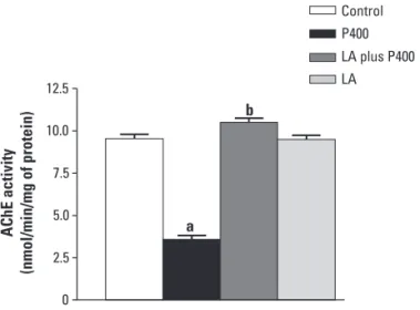
Fig 2 shows the LA efects in AChE activity in hip-pocampus during seizures induced by pilocarpine. Hip-pocampal AChE activity of rats in pilocarpine group was markedly decreased (63%) (p<0.0001), when compared with corresponding values for the control group. How-ever, post hoc comparison of means indicated a signif-icant (197%) increase in hippocampal AChE activity of rats pretreated with LA (p<0.0001), 30 min before ad-ministration of pilocarpine (LA plus P400 group), when compared with the P400 group. In addition, in LA plus P400 group it was observed no changes in AChE activi-ty (p=0.0534), when compared with corresponding val-ues for the control group (Fig 2). Moreover, AChE activi-ty in the hippocampus adult rats that received lipoic acid alone (LA group) was not markedly altered (p=0.9823), when compared with corresponding values for the con-trol group, but showed a signiicantly increased (169%) (p<0.0001), when compared with corresponding values for the P400 group (Fig 2).
DISCUSSION
he CNS contains some antioxidant enzymes, includ-ing superoxide dismutase (SOD) and GPx that are ex-pressed in higher quantities than catalase23. his
spec-trum of enzymatic defense suggests that the brain may ef-iciently metabolize superoxide but may have diiculties in eliminating the hydrogen peroxide produced by this re-action24. In the present study we have examined whether
the pretreatment with LA can reverse the alterations in the AChE, Na+, K+-ATPase and GPx activities in rat
hip-pocampus caused by seizures. Generation of reactive ox-ygen species (ROS) is currently viewed as one of the pro-cess through which epileptic activity exert their deleteri-ous efects on brain22. hese ROS in the absence of an
ef-icient defence mechanism cause peroxidation of mem-brane poly unsaturated fatty acids25. Brain is particularly
susceptible to peroxidation due to simultaneous presence of high levels of poly unsaturated fatty acids and iron24,
which is the target of free radical damage.
Previous studies conducted in our laboratory have shown that during seizures there are no alterations in hip-pocampal superoxide dismutase and catalase activities11.
Furthermore, other antioxidant systems such as
glutathi-one peroxidase can be responsible by inhibition of neu-rotoxicity induced by acute phase of seizures activity. It has been demonstrated that pretreatment with LA dur-ing acute phase of seizures induced by pilocarpine pro-duces increase in SOD, catalase activities11 and GPx in
rat hippocampus. he increase in antioxidant enzymes activities, after pretreatment with LA, is most readily ex-plained as a necessary consequence of inhibiting forma-tion of free radicals during convulsive process26-28.
LA plus P400 and P400 groups showed an increase in the GPx activities. hese data suggests that H2O2, which
is generated during superoxide dismutation, could be suf-iciently removed by GPx during seizures and after the pretreatment with lipoic acid. Previous studies showed an increased in hippocampal GPx activity after seizures26. In
addition, during the convulsive process, neuronal activi-ties changes are accompanied by alterations in the cere-bral metabolic rate29. Considering that an increased
met-abolic demand can be observed during the epileptic ac-tivity, we can suggest that GPx activity is modiied by sei-zures. his inding might suggest that pretreatment with LA produces an increase in this enzymatic activity. Its compensatory mechanisms against oxidative stress ob-served during seizures can explain the anticonvulsant ac-tions of LA. he seizures induced by pilocarpine are pre-vented by LA, suggesting a role of free radical in con-trolling seizures installation and propagation. In fact, we found that pretreatment with LA is able to inhibit pilo-carpine-induced seizures. In addition, the present data suggest evidence that free radical formation have a rele-vant role in the propagation and/or maintenance of con-vulsive activity. Meanwhile free radical formation
reduc-0 2.5 5.0 7.5 10.0 12.5
a b
AChE activity
(nmol/min/mg of protein)
Control P400
LA plus P400
LA
a: p<0.05 as compared to control animals (t-Student-Neuman-Keuls test); b: p<0.05 as compared to P400 group (t-Student-Neu-man-Keuls test).
es, an increase in antioxidant enzymes activities produced by LA produces a signiicant decrease in the susceptibil-ity to seizures induced by pilocarpine.
LA administration to convulsive animals has been shown to protect hippocampus against oxidative stress. LA has been observed to act as antioxidants towards hy-droxyl radicals and to inhibit the oxidation of lipids and protein4,9. Results of animal studies have demonstrated
that LA can reduce damage to neurons caused by free radicals that are produced in neurodegenerative diseases. he underlying mechanisms of brain dysfunction in seizures are poorly understood. Regarding this, it has been demonstrated that elevated free radical concentra-tions can be highly toxic, and that nitric oxide metabo-lites produced by the oxidative stress pathway such as ni-trite and nitrate might contribute to this toxicity30. It is
also known that hydroxyl radical has a synergistic efect on seizures elicited by pilocarpine.
Considering that Na+, K+-ATPase is decreased by
free radical formation6, lipid peroxidation31 and that –SH
groups of cell proteins are highly susceptible to oxidative stress32, we also investigated the LA efects on
inhibito-ry action of seizures on this enzyme activity. We veriied that seizures signiicantly inhibited this enzymatic activ-ity. On the other hand, we have shown in present work that LA increases this enzymatic activity during seizures in rat hippocampus11. hese observations may explain,
at least in part, the neuroprotective efects of LA against oxidative stress caused by seizures. Although the exact mechanism through which seizures inhibits Na+, K+
-AT-Pase activity is yet unknown, the present indings suggest the involvement of ROS probably by oxidizing SH groups of the enzyme and/or by peroxidation of membrane lip-ids, in which the enzyme is embedded. In this context, it should be noted that LA acts directly as a thiol-reducing agent, as well as a scavenger of free radicals and lipid per-oxidation products33. In turn, LA can be able to interact
with cell membranes, trapping ROS and interrupting the chain of oxidative reactions that damage cells. Further-more, there are studies in the literature showing that an-tioxidant compounds can efectively slow down the pro-gression of neurodegenerative diseases34-36.
Finally, we also evaluated the efect of LA on AChE activity in rat hippocampus. Our results show that this enzyme activity was decreased in seized rats. In order to conirm these indings, we veriied the efect of a single injection of LA on AChE activity. Results show that LA alone administration did not alter this enzyme activity in rat hippocampus killed 1 h after pilocarpine administra-tion. Moreover, a single injection of LA, 30 min before ad-ministration of pilocarpine produces increased on AChE activity. he increases in AChE may be due to the compen-satory mechanism of long-term administration with LA
may be due to the up-regulation of AChE activity. he re-sults obtained by AChE activities measurements could be further supported by Western blot analysis, which did not show higher protein contents of AChE (data not show).
Although it is diicult to extrapolate our animal model data to the human condition37-39, it is tempting to
specu-late that neurological symptoms observed in seizures may be related to high tissue concentrations of free radicals having an adverse efect on brain function through oxi-dative stress and inhibition of Na+, K+-ATPase and AChE
activities. However, whether these or other abnormalities are the main factors responsible for the brain damage in seizures remains to be elucidated. Furthermore, future studies should be carried out to provide additional infor-mation so as to clarify the action mechanisms of lipoic acid during the establishment of seizures.
ackNowLedGmeNts– We would like to thank Stenio Gardel Maia for her technical assistance.
REFERENCES
Bergamini CM, Gambetti S, Dondi A, Cervellati C. Oxygen, reactive oxygen 1.
species and tissue damage. Curr Pharm Des 2004;10:111-112.
Liang LP, Beaudoin ME, Fritz MJ, Fulton R, Patel M. Kainate-induced seizures, oxi-2.
dative stress and neuronal loss in aging rats. Neuroscience 2007;147:1114-1118. Henderson GI, Chen JJ, Schenker S. Ethanol, oxidative stress, reactive alde-3.
hydes, and the fetus. Frontiers Biosci 1999;4:541-550.
Andreoli SP, Mallett CP. Disassociation of oxidant-induced ATP depletion and 4.
DNA damage from early cytotoxicity in LLC-PK1 cells. Am J Physiol 1997; 272:729-735.
Erecinska M, Silver IA. Ions and energy in mammalian brain. Prog Neurobiol 5.
1994;43:37-71.
Lees GJ, Lehmann A, Sandberg M, Hamberg H. The neurotoxicity of oua-6.
bain, a sodium-potassium ATPase inhibitor, in the rat hippocampus. Neurosci Lett 1990;120:159-162.
Wyse ATS, Stefanello FM, Chiarani F, Delwing D, Wannmacher CMD, Wajner 7.
M. Arginine administration decreases cerebral cortex acetylcholinesterase and serum butyrylcholinesterase probably by oxidative stress induction. Neu-rochem Res 2004;29:385-389.
Grisar T, Guillaume D, Delgado-Escueta AV. Contribution of Na+, K+-ATPase 8.
to focal epilepsy: a brief review. Epilepsy Res 1992;12:141-149.
Reznick AZ, Packer L. Free radicals and antioxidants in muscular neurological 9.
diseases and disorders. In: Poli G, Albano E, Dianzani MU (Eds). Free radicals: from basic science to medicine. Birkhauser Verlag, Basel, 1993:425-437. Halliwell B, Gutteridge JMC. Free radicals in biology and medicine. London: 10.
Oxford Science Publications, 1999.
Freitas RM. The evaluation of efects of lipoic acid on the lipid peroxidation, 11.
nitrite formation and antioxidant enzymes in the hippocampus of rats after pilocarpine-induced seizures. Neurosci Lett 2009;455:140-144.
Massoulié J, Pezzementi L, Bon S, Krejci E, Vallette FM. Molecular and cellular 12.
biology of cholinesterases. Prog Neurobiol 1993;41:31-91.
Silver A. The biology of cholinesterases. New York: American Elsevier Pub-13.
lishing, 1974.
US Department of Health and Human Services, Institute of Laboratory Ani-14.
mal Resources. Guide for the care and use of laboratory animals. Washing-ton, DC: National Research Council, 1985.
Turski WA, Cavalheiro EA, Schwartz M, Czuczwar SJ, Kleinrok Z, Turski L. Lim-15.
bic seizures produced by pilocarpine in rats: a behavioural, electroencepha-lographic and neuropathological study. Behav Brain Res 1983;9:315-335. Ferreira PMP, Militão GCG, Freitas RM. Lipoic acid efects on lipid peroxida-16.
tion level, superoxide dismutase activity and monoamines concentration in rat hippocampus. Neurosci Lett 2009;464:131-134.
Militão GCG, Ferreira PMP, Freitas RM. Efects of lipoic acid on oxidative stress in 17.
Sinet PM, Michelson AM, Bazin A, Lejeune J, Jeerome H. Increase in glutathi-18.
one peroxidase activity in erythrocytes from trisomy 21 subjects. Biochem Biophys Res Com 1975;67:910-915.
Lowry H, Rosebrough NJ, Farr AL, Randall RJ. Protein measurements with the 19.
folin phenol reagent. J Biol Chem 1951;193:265-275.
Wyse ATS, Streck EL, Worm P, Wajner A, Ritter F, Netto CA. Preconditioning 20.
prevents the inhibition of Na+, K+-ATPase activity after brain ischemia. Neu-rochem Res 2000;25:971-975.
Chan K, Delfer D, Junger KD. A direct colorimetric assay for Ca2+-stimulated 21.
ATPase activity. Anal Biochem 1986;57:375-380.
Ellman GL, Courtney KD, Andres Jr V, Feather-Sotne RM. A new and rapid 22.
colorimetric determination of acetylcholinesterase activity. Biochem Phar-macol 1961;7:88-95.
Shivakumar BR, Ananndatheerthavarada HK, Ravindranath V. Free radical 23.
scavenging system in developing rat brain. Int J Dev Neurosci 1991;9:181-185. Castagne V, Gastschi M, Lefevre K, Posada A, Clarke PGH. Relationship be-24.
tween neuronal death and cellular redox status, focus on the developing nervous system. Prog Neurophysiol 1999;59:397-423.
Halliwell B, Gutteridge JMC. Lipid peroxidation: a radical chain reaction. Free 25.
radicals in biology and medicine. Oxford: Clarendon Press, 1989. Bellissimo MI, Amado D, Abdalla DSP, Ferreira E, Cavalheiro EA, Nafah-Maz-26.
zacoratti MG. Superoxide dismutase, glutathione peroxidase activities and the hydroperoxide concentration are modiied in the hippocampus of epi-leptic rats. Epilepsy Res 2001;46:121-128.
Kudin AP, Bimpong-Buta NY, Vielhaber S, Elger CE, Kunz WS. Characterization 27.
of superoxide-producing sites in isolated brain mitochondria. J Biol Chem 2004;279:4127-4135.
Maczurek A, Hager J, Kenklies M, et al. Lipoic acid as an anti-inlammatory 28.
and neuroprotective treatment for Alzheimer’s disease. Adv Drug Deliver Rev 2008;60:1463-1470.
Dymond AM, Crandall PH. Oxygen availability and blood low in the temporal 29.
lobes during spontaneous epileptic seizures in men. Brain Res 1976;102:191-196. Freitas RM, Souza FCF, Vasconcelos SMM, Viana GSB, Fonteles MMF. Oxidative stress 30.
in the hippocampus after status epilepticus in rats. FEBS J 2005;272:1307-1312. Viani P, Cervato G, Fiorilli A, Cestaro B. Age-related diferences in synaptosomal 31.
peroxidative damage and membrane properties. J Neurochem 1991;56: 253-258. Yufu K, Itho T, Edamatsu R, Mori A, Hirakawa M. Efect of hyperbaric oxy-32.
genation on the Na+, K+-ATPase and membrane luidity of cerebrocorti-cal membranes after experimental subarachnoid hemorrhage. Neurochem Res 1993;16:1033-1039.
Handelman GJ, Han D, Tritschler H, Packer L. Alpha-lipoic acid reduction by 33.
mammalian cells to the dithiol form, and release into the culture medium. Biochem Pharmacol 1994;47:1725-1730.
Barros DO, Xavier SM, Barbosa CO, et al. Efects of the vitamin E in catalase 34.
activities in hippocampus after status epilepticus induced by pilocarpine in Wistar rats. Neurosci Lett 2007;41:227-230.
Sano M, Ernesto C, Thomas RG, et al. A controlled trial of selegiline,
35. a-to-copherol,
or both as treatment for Alzheimer’s disease. N Engl J Med 1997;336:1216-1222. Satoh E, Nakazato Y. On the mechanism of ouabain-induced release of ace-36.
tylcholine from synaptosomes. J Neurochem 1992;58:1038-1044. Xavier SML, Barbosa CO, Barros DO, Silva RF, Oliveira RF, Freitas RM. Vitamin C 37.
antioxidant in hippocampus of adult Wistar rats after seizures and status ep-ilepticus induced by pilocarpine. Neurosci Lett 2007;420:76-79.
Freitas RM, Sousa FCF, Vasconcelos SMM, Viana GSB, Fonteles MMF. Alter-38.
ações agudas dos níveis de neurotransmissores em corpo estriado de ratos jovens após estado epiléptico induzido por pilocarpina. Arq Neuropsiquia-tr 2003;61:430-433.
Santos IMS, Freitas RLM, Saldanha GB, Tomé AR, Jordan J, Freitas RM. Altera-39.