ORIGIN
AL RESEAR
CH
Study in the field of Rheumatology of the Universidade Federal de São Paulo (Unifesp) – São Paulo (SP), Brazil.
1Universidade Federal do Amapá (Unifap) – Macapá (AP), Brasil. 2Universidade Federal de São Paulo (Unifesp) – São Paulo (SP), Brasil. 364
Corresponding address: Império Lombardi Junior – Rua Silva Jardim, nº 136 – Santos (SP), Brasil – Zip Code: 11015-020 – E-mail: [email protected] – Phone: +55 (13) 3878-3700 – Finance source: São Paulo Research Foundation (Fapesp) [grant 2012/20152-7] – Conflict of interest: Nothing to declare – Presentation: Apr. 6th, 2017 – Accepted for publication: Sept. 28th, 2017 – Approved by the Research Ethics Committee: Protocolo nº 66.320.
Lower-limb muscle strength in women with
rheumatoid arthritis and women without the disease:
is there a difference?
Força muscular de membros inferiores em mulheres com artrite reumatoide e mulheres sem a
doença: há diferença?
Fuerza muscular de los miembros inferiores entre las mujeres con artritis reumatoide y las
mujeres sin esta enfermedad: ¿habrá diferencias?
Ana Carolina Pereira Nunes Pinto1,2, Jamil Natour2, Império Lombardi Junior2
ABSTRACT | Rheumatoid arthritis (RA) is a systemic inflammatory disease, which is chronic and affects the joints’ synovial membrane. Among the physical aptitude qualities that can be impaired in individuals with RA, muscle strength deserves attention. It is directly associated with the capacity of performing all activities of daily living, from the simplest to the most complex. The aim of this study was to assess the muscle strength of the lower limbs of women with RA. Seventeen volunteers with RA (I, II and III functional classes) and 17 women without the disease, with mean age corresponding to 54.7+6.63 years, underwent the one-maximum repetition test to assess knee flexors and extensors, hip abductors and adductors. We used Student’s T test to analyze the data, considering significant p values < 0.05. In all assessments, the group of women with RA showed lower values when compared with women without the disease. However, there were no statistically significant differences between the groups. The descriptive levels obtained from the comparison between the groups’ muscle strength were: knee extensors, p=0.224; knee flexors, p=0.467; hip abductors, p=0.190 and hip adductors, p=0.127. The muscle strength of the lower limbs does not differ between women with RA (I, II and III functional classes) and women without the disease.
Keywords | Rheumatoid Arthritis; Muscle Strength; Lower
Limbs.
RESUMO | A artrite reumatoide (AR) é uma doença inflamatória sistêmica, crônica, que acomete preferencialmente a membrana sinovial das articulações. Dentre as qualidades de aptidão física que podem ser reduzidas em indivíduos com AR merece destaque a força muscular, que está diretamente relacionada à capacidade de realização de todas as atividades da vida diária, desde as mais simples até as mais complexas. O objetivo deste estudo foi avaliar a força muscular de membros inferiores de mulheres com AR. Métodos: 17 voluntárias com AR (das classes funcionais I, II e III) e 17 sem a doença, com idade média de 54,7+6,63 anos, foram submetidas ao teste de uma repetição máxima para avaliar a força muscular de flexores e extensores de joelho e de abdutores e adutores de quadril. Utilizou-se o teste τ para analisar os dados, sendo considerados estatisticamente significativos os níveis de α
<0,05. Em todas as avaliações o grupo de mulheres com AR apresentou valores inferiores quando comparados ao grupo sem a doença. No entanto não houve diferenças estatisticamente significativas entre os grupos. Os níveis descritivos obtidos da comparação entre a força muscular dos grupos foram: para extensores de joelho, p=0,224; flexores de joelho, p=0,467; abdutores de quadril, p=0,190; e adutores de quadril, p=0,127. A força muscular de membros inferiores não difere entre mulheres com AR (das classes funcionais I, II e III) e mulheres sem a doença.
Descritores | Artrite Reumatoide; Força Muscular;
RESUMEN | La artritis reumatoide (AR) es una enfermedad inflamatoria sistémica, crónica, que afecta específicamente a la membrana sinovial de las articulaciones. Entre las cualidades de aptitud física que pueden reducirse en individuos con AR, se señala la fuerza muscular, que está directamente relacionada con la capacidad para realizar todas las actividades de la vida diaria, desde las más simples hasta las más complejas. Este estudio propone evaluar la fuerza muscular de los miembros inferiores de mujeres con AR. Métodos: Se sometieron a 17 voluntarias con AR (de categorías funcionales I, II y III) y a 17 sin la enfermedad –con un promedio de edad de 54,7+6,63 años– a la prueba de repetición máxima para que se evalúe la fuerza muscular de los flexores y los extensores de rodilla, así como los extensores y abductores de cadera. Se utilizó la prueba τ
para analizar los datos, siendo considerados estadísticamente significativos los niveles de α <0,05. En todas las evaluaciones, el grupo de mujeres con AR presentó valores más bajos en comparación con el grupo sin la enfermedad. Sin embargo, no hubo ninguna diferencia estadísticamente significativa entre los grupos. Los niveles descriptivos obtenidos de la comparación entre la fuerza muscular de los grupos fueron: los extensores de rodilla, p=0,224; los flexores de rodilla, p=0,467; los abductores de cadera, p=,190; y los aductores de cadera, p=0,127. La fuerza muscular de los miembros inferiores no difirió entre las mujeres con AR (de categorías funcionales I, II y III) y las mujeres sin esta enfermedad.
Palabras clave | Artritis Reumatoide; Fuerza Muscular; Miembros
Inferiores.
INTRODUCTION
Rheumatoid arthritis (RA) is a systemic inflammatory disease, which is chronic and affects the joints’ synovial membrane. It is characterized by a progressive symmetrical polyarticular impairment that can lead to the joint’s destruction, with erosion of the bone and cartilage. Other
systems may also be damaged1.
It has an autoimmune character and although
its etiology is not known2, in recent decades there
have been relevant developments in the knowledge of the disease’s pathophysiology. Relevant changes in the approach and treatment of RA have been made, which emphasize the importance of early diagnosis
and treatment3
.
That is because, after its initial phase, the disease is characterized by persistent inflammation, which can lead to a significant loss of joint integrity. With its progression, patients develop impairments in the performance of their daily and professional activities. Due to its chronic nature and for affecting people in productive age, both for the sick individual as for society, RA represents a significant
socioeconomic burden4.
Among the physical fitness qualities that can be reduced in individuals with TA, muscle strength deserves attention. Its reduction is one of the most limiting losses and is directly related to mortality from all causes5. Muscle
strength is also directly related to the ability of carrying out all activities of daily living6, from the simplest, like the
ability to maintain one’s posture, balance and locomotion, to the most complex. Given this, the objective of this
study was to evaluate the muscle strength of the lower limbs of women with RA.
METHODOLOGY
Seventeen women with RA being followed up at the Rheumatology Clinic of the Federal University of São Paulo (Unifesp) were recruited to participate in the study, having been matched in relation to age and body mass index (BMI) to 17 volunteers from the community who made up the control group (CG). In the rheumatoid arthritis group (RAG), we included women classified according to the criteria of the American College of
Rheumatology (ACR)7 for RA and with stable medication
within the three months preceding the start of the study. Women with functional class IV RA according to the ACR’s criteria; engaged in some form of regular physical exercise program; with decompensated cardiac, respiratory, renal, coronary and/or liver failure; with decompensated systemic arterial hypertension and unable to carry out the proposed exercises were excluded from the study. Seventeen volunteers from the community were included in the CG. The exclusion criteria were the same ones adopted for the RAG’s participants.
The volunteers were evaluated in relation to functional
class according to the ACR criteria8. The participants’
usual practice of physical activity was evaluated via the International Physical Activity Questionnaire – Ipaq9, in
its short version. The Ipaq is one of the most important instruments to investigate the levels of physical activity and sedentarism of individuals, being composed of questions that evaluate the frequency, intensity and duration of physical activities performed by an individual in an ordinary week. With the analysis of the responses, it was possible to classify the participants in very active, active, irregularly active or sedentary9.
For the functional evaluation, we used the Health
Assessment Questionnaire (HAQ)10. This instrument
consists of 20 questions that measure eight areas of customary functional activities. The scores are grouped, the highest of each of the eight areas evaluated being considered for the calculation of the components’ scores. The arithmetic mean of the components’ scores is then calculated, and the end result is the HAQ score. Higher scores reflect a higher degree of functional impairment10.
The RAG group’s participants were also were evaluated for disease activity through the Disease Activity Score (DAS-28)11. It is considered that the higher the value of
the score, the greater the activity of the disease at the time of the evaluation. Cut-off points were defined for the DAS-28: ≤ 2.4 = remission; > 2.4 and 3.6 ≤ = low activity; > 3.6 and ≤ 5.5 = moderate activity; and > 5.5 = high activity11.
All volunteers underwent a session of familiarization with resistance exercises. This session was intended to make them familiar with the equipment and techniques of the exercises before the test to determine the one-repetition maximum load (1RM).
The familiarization session was composed of two sets of 10 repetitions for each exercise, with loads considered as light by the volunteers, one-minute rest intervals between sets and two-minute intervals between exercises having been adopted. A study indicates that, to make the application of the 1RM test more reliable in untrained individuals, one familiarization session is sufficient12.
Equipment with lever systems were used (Maxiflex Biodelta, Joinville, SC, Brazil), the following having been employed: seat extensor, seat flexor, seat abductor and seat adductor. The exercises performed were: knee extension, knee flexion, hip abduction and hip adduction. The volunteers performed the movements until the limit of their range or to the extent they were pain-free.
Muscle strength was evaluated via the 1RM test. This test started three minutes after the familiarization period, which also served as warm-up for performing the test. This procedure is intended to verify the maximum amount of force the individual can apply in a single movement, with maximum effort.
During the test, progressive loads were applied, based on the loads that had been previously considered light by the volunteers. At first, they were instructed to try to complete two repetitions. If more than one repetition was completed during this attempt, the load was increased. Then, the participant was allowed a three-minute rest interval so that another attempt could be made with the new load. The attempts were carried out progressively and successively, separated by the rest interval, until only one repetition could be performed.
If the load was increased and not a single repetition could be completed, a second attempt was performed with the same load with which the volunteer failed to complete the movement. If the participant was again not able to do it, a third and final attempt was carried out to ratify that she was performing the movement within the limits of her efforts. In case of new failure, the previous load was considered as the volunteer’s maximum load, i.e., the last load with which she managed to properly complete a repetition. The form and technique of execution of each exercise were continuously monitored to ensure the quality of the information.
Statistical analysis
To assess whether there was a difference between the muscle strength values observed in the 1RM test, Student’s t-test was employed. In all tests, we considered as statistically significant p-levels < 0.05.
The sample size was determined based on the work by Niehoff et al. (2011)13. Considering 0.05 significance level
and 90% power, we estimated that at least 17 participants would be needed for each group.
RESULTS
Sample’s characterization
Among the associated impairments observed in the RAG, we found: vasculitis, depression, systemic arterial hypertension (SAH), type II diabetes mellitus, carpal tunnel syndrome, bursitis in shoulders, arthrosis, osteoporosis and cardiac arrhythmia. As for physical activity level, when evaluated via the Ipaq, four participants of each group were considered abnormally active, and thirteen were considered active. No volunteer was classified as sedentary or very active.
The RAG was also assessed in relation to functional class. Among the volunteers, nine belonged to functional class III, five to functional class II, and three to class I. As for the other evaluations carried out in the RAG only, the data are described in Table 2.
Table 2. RAG’s characteristics in relation to health assessment, disease activity and time after the RA diagnosis
HAQ DAS-28 Time after the
diagnosis (in years)
Mean (SD) 1.24 (0.54) 6.24 (2.1) 11.7 (5.98)
Minimum 0.5 2.71 5
Maximum 2.25 9.39 26
HAQ: Health Assessment Questionnaire; DAS-28: Disease Activity Score
In relation to disease activity, nine volunteers had high disease activity, six had moderate, and two had low activity. No patient was in remission.
One-repetition maximum test
The values obtained in the 1RM are presented in Table 3. Despite the group of women with RA having showed lower values in the 1RM test in all evaluations, when compared with the group without the disease, there was no statistically significant difference between them.
Table 3. Mean load (in kg) obtained in the one-repetition maximum test in the RAG and in the CG
RAG* CG*
Percentage of difference between the
groups
P-value
Knee extensors 40.53 (16.83) 46.88 (12.70) 13.54% 0.224 Knee flexors 42 (11.13) 44.65 (9.80) 5.93% 0.467 Hip abductors 47.23 (7.36) 50.82 (8.23) 7.06% 0.190 Hip adductors 34.59 (8.77) 38.64 (6.04) 10.48% 0.127
RAG: rheumatoid arthritis group; CG: control group. *Values expressed in mean (standard deviation)
DISCUSSION
The results of our study show that there is no statistically significant difference between the muscle strength of the lower limbs of women with RA and of women without the disease, although there was a force reduction tendency in the former.
Ekdahl and Broman14 evaluated 67 functional class
II patients, aged between 23 and 65 years old and with 53 years of age on average. They also studied the strength of the lower limbs of people with RA and identified that the muscle strength of the knee and hip was 65% to 75% lower in the RA group when compared to the healthy group. However, the authors evaluated isometric force, and not dynamic force, like we did in our study.
Hakkinen et al.15 assessed 20 patients with RA and
20 without the disease and found that the dynamic force of the wrist and knee extensors was also lower in the RA group when compared to the healthy control group, but the groups did not differ among themselves in relation to the isometric strength of torso flexors and extensors and knee extensors.
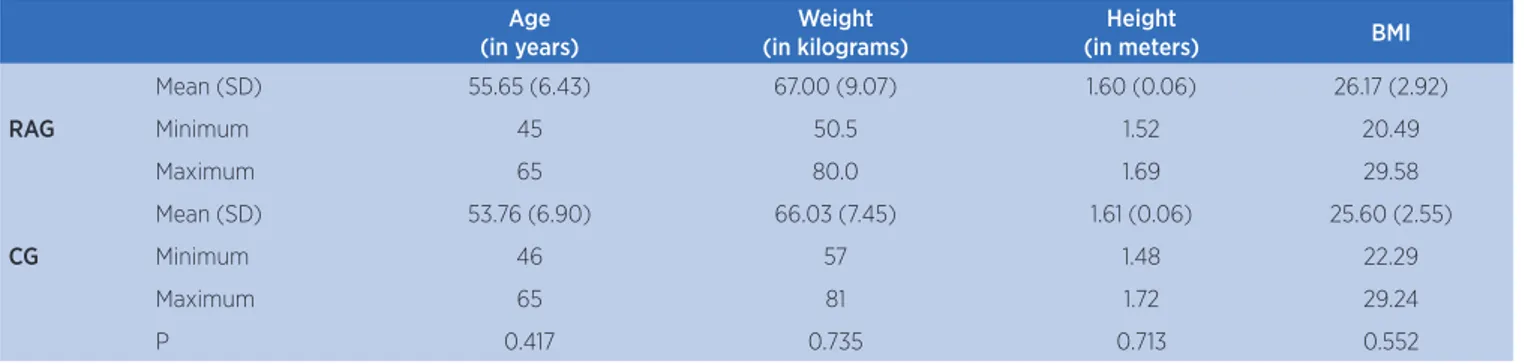
Table 1. General characteristics of the rheumatoid arthritis group and of the control group
Age (in years)
Weight (in kilograms)
Height
(in meters) BMI
Mean (SD) 55.65 (6.43) 67.00 (9.07) 1.60 (0.06) 26.17 (2.92)
RAG Minimum 45 50.5 1.52 20.49
Maximum 65 80.0 1.69 29.58
Mean (SD) 53.76 (6.90) 66.03 (7.45) 1.61 (0.06) 25.60 (2.55)
CG Minimum 46 57 1.48 22.29
Maximum 65 81 1.72 29.24
P 0.417 0.735 0.713 0.552
Madsen et al.16 evaluated 79 women with RA, who
had lived 10 years with the disease on average, and 67 women without the disease, matched in relation to age, having found that the women with RA had 20% less quadriceps strength when compared to the control group.
In our study, we observed no statistically significant difference between the muscle strength of the RA group and of the group without the disease. However, we did not evaluate functional class IV women, so we cannot rule out the hypothesis that they have reduced muscle strength when compared to their non-diseased peers. It is also possible that there is a reduction of the muscle strength of the upper limbs, which have not been assessed in this study.
It is known that the loss of mass and muscle strength may promote the increased inactivity of patients with RA, which contributes to the reduction of their functional capacity, physical independence and quality of life. As non-pharmacological strategy of approach to several diseases, including RA, exercise has been proposed in recent years for providing muscle mass and strength gains and for improving functional capacity, without exacerbating disease activity or joint damages17.
The studies found which compared the muscle strength of people with RA and of people without the disease date back to more than a decade. We believe that the evolution of the treatment of people with RA over the past few years certainly contributed to the delayed onset of physical impairments, such as loss of muscle strength. Studies with a higher number of participants and with possibility of stratification of the functional classes may assist the better identification of differences in muscle strength between the functional classes and the ratification of our hypothesis that the advance of therapies contributed to the reduction of differences over time.
CONCLUSION
The muscle strength of the lower limbs does not differ between women with functional class I, II and III rheumatoid arthritis and women without arthritis.
REFERENCES
1. Aletaha D, Neogi T, Silman AJ, Funovits J, Felson DT, Bingham CO 3rd, et al. 2010 rheumatoid arthritis classification criteria: an American College of Rheumatology/European League Against Rheumatism collaborative initiative. Ann Rheum Dis. 2010;69(9):1580-8. doi: 10.1136/ard.2010.138461
2. Goeldner I, Skare T, Reason I, Utiyama S. Artrite reumatoide: uma visão atual. J Bras Patol Med Lab. 2011;47(5):495-503. doi: 10.1590/S1676-24442011000500002
3. Emery P. Treatment of rheumatoid arthritis. BMJ. 2006;332:152-5. doi: 10.1136/bmj.332.7534.152
4. American College of Rheumatology, Subcommittee on rheumatoid arthritis guidelines. Guidelines for the management of rheumatoid arthritis: 2002 Update. Arthritis Rheum. 2002;46(2):328-46. doi: 10.1002/art.10148
5. Ruiz J, Sui X, Lobelo F, Morrow J, Jackson A, Sjöström M, et al. Association between muscular strength and mortality in men: prospective cohort study. BMJ. 2008;337(7661):92-5. doi: 10.1136/bmj.a439
6. Bjarnason-Wehrens B, Mayer-Berger W, Meister ER, Gielen S. Recommendations for resistance exercise in cardiac rehabilitation. Recommendations of the German Federation for Cardiovascular Prevention and Rehabilitation. Eur J Cardiovasc Prev Rehabil. 2004;11(4):352-61. doi: 10.1097/01. hjr.0000137692.36013.27
7. Arnett FC, Edworthy SM, Bloch DA, McShane DJ, Fries JF, Cooper NS, et al. The American Rheumatism Association 1987 revised criteria for the classification of rheumatoid arthritis. Arthritis Rheum. 1988;31(3):315-24. doi: 10.1002/art.1780310302 8. Hochberg MC, Chang RW, Dwosh I, Lindsey S, Pincus T, Wolfe
F. The American College of Rheumatology 1991 revised criteria for the classification of global status in Rheumatoid Arthritis. Arthritis Rheum. 1992;35(5):498-502. doi: 10.1002/art.1780350502 9. Matsudo SM, Araújo T, Matsudo VR, Andrade D, Andrade E,
Oliveira LC, et al. Questionário Internacional de Atividade física (IPAQ): estudo de validade e reprodutibilidade no Brasil. Rev Ativ Fís Saúde. 2001;6(2):5-18. doi: 10.12820/rbafs.v.6n2p5-18 10. Bruce B, Fries JF. The Health Assessment Questionnaire (HAQ).
Clin Exp Rheumatol. 2005 [cited 2018 Oct 9].;23(5 Suppl 39):S14-8. Available from: https://bit.ly/2OhJEEU
11. Aletaha D, Smolen J. The Simplified Disease Activity Index (SDAI) and the Clinical Disease Activity Index (CDAI): a review of their usefulness and validity in rheumatoid arthritis. Clin Exp Rheumatol. 2005;23(5 Suppl 39):S100-8.
12. Levinger I, Goodman C, Hare DL, Jerums G, Toia D, Selig S. The reliability of the 1RM strength test for untrained middle-aged individuals. J Sci Med Sport. 2009;12(2):310-6. doi: 10.1016/j. jsams.2007.10.007
13. Niehoff A, Müller M, Brüggemann L, Savage T, Zaucke F, Eckstein F, et al. Deformational behavior of knee cartilage and changes in serum cartilage oligomeric matrix protein (COMP) after running and drop landing. Osteoarthr Cartil. 2011;19(8):1003-10. doi: 10.1016/j.joca.2011.04.012
14. Ekdahl C, Broman G. Muscle strength, endurance, and aerobic capacity in rheumatoid arthritis patients: a comparative study with healthy subjects. Ann Rheum Dis. 1992;51(1):35-40. 15. Hakkinen A, Hannonen P, Hakkinen K. Muscle strength in
healthy people and in patients suffering from recent-onset inflammatory arthritis. Br J Rheumatol. 1995;34(4):355-60. 16. Madsen OR, Egsmose C, Hansen B, Sorensen OH. Soft tissue
composition, quadriceps strength, bone quality and bone mass in rheumatoid arthritis. Clin Exp Rheumatol. 1998;16(1):27-32. 17. Lemmey AB, Marcora SM, Chester K, Wilson S, Casanova F,