Case Report
Keywords
Echocardiography, three-dimensional; heart failure; left ventricular reverse remodeling; cardiomyopathy, dilated.
Cardiac resynchronization therapy consists of a promising treatment for patients with severe heart failure, but about 30% of patients do not exhibit clinical improvement with this procedure. However, approximately 10% of patients undergoing this therapy may have hyperresponsiveness, and three-dimensional echocardiography can provide an interesting option for the selection and evaluation of such patients.
“Hyper-response” Evaluated by 3D Echocardiography after Cardiac
Resynchronization Therapy
Viviane Tiemi Hotta
1,2, Marcelo Luiz Campos Vieira
1,3, Daniela do Carmo Rassi
1, Silvana Angelina D’orio Nishioka
1,
Martino Martinelli Filho
1, Wilson Mathias Jr
1Instituto do Coração (InCor) da Faculdade de Medicina da Universidade de São Paulo1; Fleury Medicina e Saúde2; Hospital Israelita Albert
Einstein3, São Paulo, SP - Brazil
Mailing address: Viviane Tiemi Hotta •
Av. Dr. Enéas de Carvalho Aguiar, 44 - 05403-000 - Cerqueira César - São Paulo, SP - Brazil
E-mail: [email protected]
Manuscript received August 06, 2009; revised manuscript received January 26, 2010; accepted March 25, 2010.
Introduction
Cardiac resynchronization therapy (CRT) has been used to treat patients with severe heart failure (left ventricular ejection fraction ≤ 0.35), intraventricular conduction disturbance (QRS ≥ 120 ms) and NYHA functional class III or IV despite an optimized medical treatment1. CRT leads to a reduction of ventricular volumes and improvement of the diastolic pattern and left ventricular ejection fraction, reducing mitral regurgitation resulting in clinical and functional improvement. It also reduces mortality2,3.
Three-dimensional echocardiography (3D Echo) has proven very useful in evaluating left ventricular volumes and ejection fraction (LVEF), and especially in the evaluation of cardiac dyssynchrony4,5. This is the case of a patient treated with CRT who developed significant improvement of LVEF, reduction of left ventricular volumes and dyssynchrony indexes assessed by 3D echocardiography.
Case report
Female patient, 64 years old, diagnosed with heart failure five years ago has evolved with progressive worsening of symptoms (NYHA functional class III at baseline) despite
regular use of optimized medication. She reports a history of hypertension and dyslipidemia under regular treatment. The patient was taking the following drugs: enalapril 20 mg 2x/day, carvedilol 25 mg 2x/day, spironolactone 25 mg/day, furosemide 40 mg/day and digoxin 0.25 mg ½ pill/day.
During the investigation, serology for Chagas (nonreactive) and cineangiocardiography (coronary arteries) were performed. On physical examination, the patient was in regular condition, eupneic at rest, BP 122 x 68 mmHg and HR of 68 bpm. It also found: two-stroke rhythmic sounds with mitral regurgitation murmur, lung auscultation with no abnormalities and edema + / 4 + in the legs.
Electrocardiogram (figure 1A) revealed sinus rhythm, first degree atrioventricular block, left anterior divisional block and left bundle branch block. Transthoracic echocardiogram revealed moderate dilation of the left chambers, severe reduction of LVEF reduced by a significant degree (LVEF estimated at 28% on 3D Echo, movie clip 1) at the expenses of diffuse hypokinesis and significant dyssynchrony assessed by 3D echocardiography (dyssynchrony index: 13.38% in the analysis of 16 segments; VN < 5%) (Figure 2A and 2B).
The patient received an atriobiventricular pacemaker for CRT, evolving to significant improvement in symptoms and clinical parameters. Six months after CRT, there was improvement in NYHA functional class and quality of life (assessed by the Minnesota Living with Heart Failure Questionnaire), and the electrocardiogram (EKG) of the patient (Figure 1B) revealed reduction of QRS duration in relation to preoperative findings. 3D Echo revealed a significant reduction in left ventricular diastolic and systolic volumes, significant increase in LVEF and improved dyssynchrony indexes (Figure 2C and 2D; movie clip 2).
Discussion
To date, clinical studies regarding the CRT reported rates around 30% of non-responders by assessing NYHA functional class. Considering echocardiographic parameters or imaging methods for assessment of ventricular volumes and LVEF, this rate increases, reaching up to 50%. There has been much discussion about the causes for the “non response” of these patients, but little is said about patients with “hyper-response”, i.e., patients who have virtually normalization of LVEF after CRT.
In a recent study to assess the prevalence of hyper-responders after CRT, 13% of patients were found to present standardization
Case Report
Hotta et al “Hyper-response” evaluated by 3D echocardiography after CRTArq Bras Cardiol 2011;96(6):e119-e122
Figure 1 -(A) Basal ECG: sinus rhythm, HR: 76 bpm, PR interval: 204 ms, anterior superior divisional block and left bundle branch block (QRS: 189 ms). (B) Post-CRT ECG: pacemaker rhythm, HR: 60 bpm, and QRS: 135 ms.
Movie clip 1 Movie clip 2
Case Report
Hotta et al“Hyper-response” evaluated by 3D echocardiography after CRT
Arq Bras Cardiol 2011;96(6):e119-e122
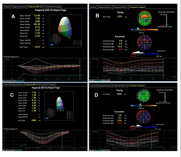
Figure 2 -(A) Dyssynchrony index on 3D Echo (T msv%). Volumetric variation curves are presented for each of the 16 left ventricular segments. The standard deviation (SD) of the duration of systolic contraction of each segment is calculated in relation to the cardiac cycle duration (Tmsv 16-SD: 13.38%, VN < 5%).( B) Parametric image of the left ventricular segments. In green color, the segments with normal contraction time, and in red, segments with conduction delay (greater delay in the apical anterior segment). (C) Normalization of dyssynchrony index on 3D after CRT (Tmsv 16-SD: 1.53%).(D) Parametric image of the left ventricle after CRT showing improvement in contraction times of myocardial segments.
(LVEF: 0.55 ± 2SD) in LVEF with CRT. In this study, the only factor associated with hyper-responsive patients was the etiology of cardiomyopathy. All hyper-responsive patients had nonischemic dilated cardiomyopathy. No patient with ischemic cardiomyopathy presented normalization of LVEF6.
This case illustrates an uncommon situation after CRT. The patient had significant improvement of LVEF, normalization of ventricular volumes and dyssynchrony indexes on 3D Echo after CRT. Such patient, probably had dilated cardiomyopathy secondary to hypertension, moderate dilation of the left ventricle, as well as preserved right ventricular systolic function
and no signs of pulmonary hypertension. These features, besides a major intraventricular conduction disturbance (QRS > 150 ms) and a significant cardiac dyssynchrony evaluated by 3D echocardiography may be associated with this significant response after CRT.
Anyway, in light of current knowledge, the predictors of good response to CRT are still under discussion, and 3D Echo emerged as a promising and useful method to evaluate dyssynchrony in the setting of patients undergoing CRT, as well as an accurate method in assessing ventricular volumes and LVEF.
Case Report
References
1. American College of Cardiology (ACC), American Heart Association (AHA). Heart faliure: 2009 focused update: ACC/AHA guidelines for the diagnosis and management of heart failure in adults heart failure focused update. J Am Coll Cardiol 2009; 53:1343-82.
2. Cleland JGF, Daubert JC, Erdmann E, Freemantle N, Gras D, Kappenberger L, et al. Cardiac resynchronization - heart failure (CARE-HF) study investigators. The effect of cardiac resynchronization on morbidity and mortality in heart failure. N Engl J Med. 2005; 352 (15): 1539-49.
3. Kapetanakis A, Kearney MT, Siva A, Gall N, Cooklin M, Monaghan MJ. Real-time three-dimensional echocardiogra¬phy: a novel technique to
quantify global left ventricular mechanical dyssynchrony. Circulation. 2005; 112 (7): 992-1000.
4. Sonne C, Sugeng L, Takeuchi M, Weinert L, Childers R, Watanabe N, et al. Real-time 3-dimensional echocardiographic assessment of left ventricular dyssynchrony: pitfalls in patients with dilated cardiomyopathy. JACC Cardiovasc Imaging. 2009; 2 (7): 802-12.
5. Castellant P, Fatemi M, Bertault-Valls B. Cardiac resynchronization therapy: “nonresponders” and “hyperresponders”. Heart Rhytm. 2008; 5 (2): 193-7.
Hotta et al “Hyper-response” evaluated by 3D echocardiography after CRT
Arq Bras Cardiol 2011;96(6):e119-e122
Potential Conflict of Interest
No potential conflict of interest relevant to this article was reported.
Sources of Funding
There were no external funding sources for this study.
Study Association
This article is part of the thesis of doctoral submitted by Viviane Tiemi Hotta, from Faculdade de Medicina da USP.