Arq Bras Cardiol 2002; 78: 267-70.
Sacramento et al Electrocardiographic alterations in patients with leptospirosis
267
Hospital Universitário Professor Edgard Santos da Faculdade de Medicina-UFB and Hospital Couto Maia of the State Health Care Authority of Bahia Mailing address: Edilson Sacramento - Rua Moacir Leão, 360/1503 B - 40080-160 - Salvador, BA, Brazil - e-mail: [email protected]
Received for publication on 1/2/01 Accepted on 2/7/01
English version by Stela Maris C. e Gandour
Objective - To report the frequency and types of elec-trocardiographic alterations in patients with leptospi-rosis in the first 24 hours of hospitalization.
Methods - We analyzed the electrocardiograms of 157 patients admitted to the Hospital Couto Maia in the city of Salvador, in the State of Bahia, Brazil, from March 1998 to June 1999. The electrocardiograms were per-formed in the first 24 hours after hospital admission, in-dependent of the clinical manifestations of the patients.
Results - The mean ± SD for patients’ age was 35.5± 13.7 (median = 32) years, and jaundice was present in 95.5% of them. Alterations in the electrocardiogram were detected in 68.2% (107/157) of the patients (95% confi-dence interval = 60.6% - 75.1%). Atrial fibrillation was the most frequent arrhythmia, occurring in 10.8% (17/157) of the patients. Other frequent findings were alterations in ventricular repolarization detected in 38.9% (61/157) of patients and first-degree atrioventricular block in 10.2% (16/157). The patients with atrial fibrillation were older and had higher levels of creatinine and aminotransferases.
Conclusion - In this sample, approximately 2/3 of the patients had electrocardiographic alterations after hospi-tal admission. Of the major arrhythmias, atrial fibrillation was the most frequent, and the patients with this arrhythmia had evidence of more severe disease. The relation between the presence and type of electrocardiographic alteration and the prognosis of leptospirosis is yet to be assessed.
Key words: electrocardiography, leptospirosis
Arq Bras Cardiol, volume 78 (nº 3), 267-70, 2002
Edilson Sacramento, Antonio Alberto Lopes, Everaldo Costa, Olaivio Lima Passos, Yara Aragão Costa, Eliana Dias Matos
Salvador, BA - Brazil
Electrocardiographic Alterations in Patients Hospitalized
with Leptospirosis in the Brazilian City of Salvador
Original Article
Leptospirosis is a zoonosis of universal distribution, even though more frequent in countries with a tropical climate 1. The disease is endemic in several Brazilian
regions, and epidemics occur in the rainy season 2,3.
Experi-mental studies in animals and autopsies in human beings have shown that cardiac involvement in leptospirosis is frequent, even though it may occur without clinical mani-festations 4-7. Despite this, few studies exist that report
electrocardiographic alterations in patients with leptos-pirosis.
The estimates of the prevalence of electrocardiogra-phic alterations in leptospirosis have been reported to reach 80% 7-10. Nevertheless, the influence of bias in these
findings is difficult to eliminate due to the preferential selec-tion of patients with clinical manifestaselec-tions of cardiac disea-se. Apparently, Escosteguy et al 9 conducted the unique
study properly designed to assess the electrocardiographic alterations occurring with leptospirosis. This study compri-sed only 56 patients, and the estimate of prevalence (71.4%) had little accuracy. Our study was conducted in the city of Salvador, in the Brazilian state of Bahia, where the incidence of leptospirosis is high 11. The study aimed at reporting the
frequency and types of electrocardiographic alterations in patients with leptospirosis right after hospital admission.
Methods
268
Sacramento et al
Electrocardiographic alterations in patients with leptospirosis
Arq Bras Cardiol 2002; 78: 267-70.
findings. A standardized questionnaire was used for reporting the cardiac alterations, including the presence of arrhythmias, hypertrophy of the cardiac chambers, disor-ders of cardiac conduction, and alterations in ventricular repolarization.
The diagnosis of leptospirosis was established based on clinical and epidemiological data, and on the ma-croscopic agglutination (n=157) and mima-croscopic agglu-tination (n=19) tests for leptospire. The microscopic agglutination tests were performed in the Centro de Pes-quisas da Fundação Osvaldo Cruz – FIOCRUZ, and the macroscopic agglutination tests were performed in the La-boratório Central do Estado da Bahia (LACEN). The microscopic agglutination test was considered positive when the titer increased more than 4 times in paired serums or when the titer was greater than 1:800 in a single sample. The macroscopic agglutination test was classified as positive or negative.
The Fisher exact and Kruskal-Wallis tests were used for comparing categorical and quantitative variables, respectively. The accuracy of the prevalence of electro-cardiographic alterations was estimated using the 95% confidence interval (CI), an exact method (mid-p), and the CONFINT module of Computer Programs for Epidemio-logists 12. For the Fisher exact test, the Exact 2xk module of
the same software was used. The other analyses were performed with the Statistical Package for Social Science (SPSS) software 13.
Results
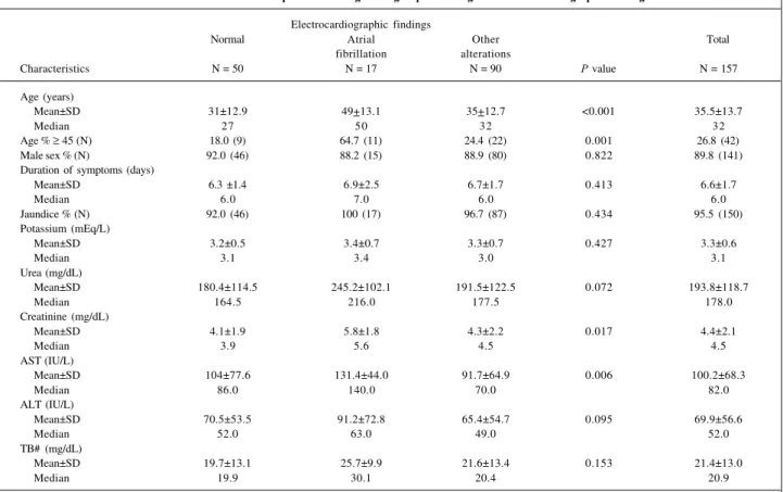
All patients had positive macroscopic agglutination tests for leptospirosis. The characteristics of the 157 patients in general and stratified into 3 levels according to the type of electrocardiographic findings are shown in table I. The electrocardiogram was initially classified as normal (n=50), with atrial fibrillation (n=17), and with other altera-tions (n=90). The mean (±SD) age in the general group was 35.5±13.7 (median = 32) years. Predominance (89.8%) of the male sex was observed; 95.5% of the patients were icteric. The mean (±SD) duration of the symptoms was 6.6±1.7 (median=6) days. The mean (±SD) value of serum potassium was 3.27±0.65 mEq/L (median=3.1 mEq/L). The mean (±SD) levels of urea and creatinine were 193.8±118.7 mg/dL (median=178 mg/dL) and 4.43±2.12 mg/dL (median=4.5 mg/ dL), respectively. AST, ALT, and total bilirubin had the following means (±SD): 100.2±68.3 IU/L (median=82 IU/L), 69.9±56.6 IU/L (median=52 IU/L), and 21.4±13 IU/L (me-dian=20.9 IU/L), respectively. The AST and creatinine levels were significantly higher in the group with atrial fibrillation (P=0.006 and P=0.017, respectively).
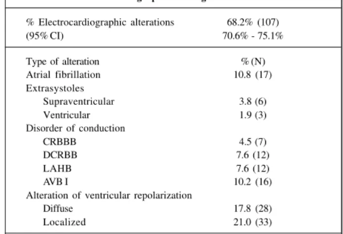
The prevalence of electrocardiographic alterations in the patients studied was 68.2% (107/157), 95% CI = 60.6%-75.1%. Table II shows in detail the percentage distribution of the types of electrocardiographic alterations. Of the ma-jor arrhythmias, atrial fibrillation was the most frequent, being present in 10.8% (17/157) of all electrocardiograms.
Table I - Characteristics of the patients in the general group according to the electrocardiographic findings
Electrocardiographic findings
Normal Atrial Other Total
fibrillation alterations
Characteristics N = 50 N = 17 N = 90 P value N = 157
Age (years)
Mean±SD 31±12.9 49+13.1 35+12.7 <0.001 35.5±13.7
Median 27 50 32 32
Age % ≥ 45 (N) 18.0 (9) 64.7 (11) 24.4 (22) 0.001 26.8 (42)
Male sex % (N) 92.0 (46) 88.2 (15) 88.9 (80) 0.822 89.8 (141)
Duration of symptoms (days)
Mean±SD 6.3 ±1.4 6.9±2.5 6.7±1.7 0.413 6.6±1.7
Median 6.0 7.0 6.0 6.0
Jaundice % (N) 92.0 (46) 100 (17) 96.7 (87) 0.434 95.5 (150)
Potassium (mEq/L)
Mean±SD 3.2±0.5 3.4±0.7 3.3±0.7 0.427 3.3±0.6
Median 3.1 3.4 3.0 3.1
Urea (mg/dL)
Mean±SD 180.4±114.5 245.2±102.1 191.5±122.5 0.072 193.8±118.7
Median 164.5 216.0 177.5 178.0
Creatinine (mg/dL)
Mean±SD 4.1±1.9 5.8±1.8 4.3±2.2 0.017 4.4±2.1
Median 3.9 5.6 4.5 4.5
AST (IU/L)
Mean±SD 104±77.6 131.4±44.0 91.7±64.9 0.006 100.2±68.3
Median 86.0 140.0 70.0 82.0
ALT (IU/L)
Mean±SD 70.5±53.5 91.2±72.8 65.4±54.7 0.095 69.9±56.6
Median 52.0 63.0 49.0 52.0
TB# (mg/dL)
Mean±SD 19.7±13.1 25.7±9.9 21.6±13.4 0.153 21.4±13.0
Median 19.9 30.1 20.4 20.9
Arq Bras Cardiol 2002; 78: 267-70.
Sacramento et al Electrocardiographic alterations in patients with leptospirosis
269
Atrial fibrillation was also associated with low voltage (3 patients), complete right bundle-branch block (2 patients), and alterations in ventricular repolarization (10 patients). Frequencies of supraventricular and ventricular extrasys-toles were 3.8% (6/157) and 1.9% (3/157), respectively. Alterations in ventricular repolarization were observed in 38.9% (61/157) of the tracings, being the most frequent finding in the electrocardiograms. First-degree atrio-ventricular block was the most common disorder of cardiac conduction, accounting for 10.2% (16/157) of the electro-cardiographic tracings. Left anterior hemiblock (12/157) and disorder of conduction of the right bundle-branch (12/157) had the same 7.6% frequency. Complete right bundle-branch block was reported in 4.5% (7/157) of the patients. Left ventricular hypertrophy was observed in 1.3% (2/157) of the patients. In 5.1% (8/157) of the patients, the electro-cardiogram was compatible with low voltage (tab. II).
Discussion
According to the results obtained, alterations in the electrocardiographic tracing could be observed in the first 24 hours of hospitalization in approximately 2/3 of the pa-tients with leptospirosis in Salvador. This finding is in ac-cordance with previous studies also carried out in Brazil 6-9.
However, the present study used a sample almost 3 times greater than those used in previous studies, which resulted in a greater accuracy in the estimate of prevalence of elec-trocardiographic alterations. Of the major arrhythmias, atrial fibrillation was the most frequent (10.8%). Alterations in ventricular repolarization (38.9%) and first-degree atrioventricular block (10.2%) were other frequent findings. It is worth noting that more than 70% of the patients were younger than 45 years, an age bracket in which the elevated frequency of arrhythmias, particularly atrial fibrillation, found in our study is not expected 14,15.
The-refore, most alterations observed should have been caused by leptospirosis. The mechanisms determining the electro-cardiographic alterations were not specifically assessed in the present study. We observed, however, that patients with atrial fibrillation had evidence of greater systemic impairment due to the disease. They had higher levels of
Table II – Distribution of the findings in the 157 electrocardio-graphic tracings
% Electrocardiographic alterations 68.2% (107)
(95% CI) 70.6% - 75.1%
Type of alteration % (N)
Atrial fibrillation 10.8 (17) Extrasystoles
Supraventricular 3.8 (6)
Ventricular 1.9 (3)
Disorder of conduction
CRBBB 4.5 (7)
DCRBB 7.6 (12)
LAHB 7.6 (12)
AVB I 10.2 (16)
Alteration of ventricular repolarization
Diffuse 17.8 (28)
Localized 21.0 (33)
↓CI- confidence interval; CRBBB- complete right bundle-branch block; DCRBB- disorder of conduction of the right bundle-branch; LAHB- left anterior hemiblock; AVB I- first-degree atrioventricular block.
AST and creatinine. It is reasonable to believe that patients with more severe clinical findings have a tendency towards more severe metabolic alterations and greater hemodynamic impairment. It is worth noting that myocarditis is a frequent autopsy finding in patients with leptospirosis, probably constituting an important cause of arrhythmias in these patients. Even though, in the present study the mean level of potassium was similar in patients with and without elec-trocardiographic alterations, we cannot discount the possi-bility that hypokalemia and other alterations contribute to arrhythmias in these patients.
According to our results, the prevalence of electrocar-diographic alterations in patients with leptospirosis right after hospitalization is high. Of the major arrhythmias, atrial fibrillation was the most frequent, and patients with this arrhythmia had more severe disease. New studies are required for a better understanding of the mechanisms of the electrocardiographic alterations in leptospirosis. The assessment of whether the presence and type of the elec-trocardiographic alterations are associated with the prog-nosis of leptospirosis is also important.
1. Farr R. Leptospirosis. Clin Infect Dis 1995; 21: 1-6.
2. Azevedo R, Corrêa M. Considerações em torno da epidemia de leptospirose na cidade de Recife em 1966. Aspectos epidemiológicos, laboratoriais e clínicos. Rev Inst Adolfo Lutz 1968; 28: 85-111.
3. Ko AI, Galvao Reis M, Ribeiro Dourado CM, Johnson WD, Jr., Riley LW. Urban epidemic of severe leptospirosis in Brazil. Salvador Leptospirosis Study Group. Lancet 1999; 354: 820-5.
4. de Brito T, Bohm GM, Yasuda PH. Vascular damage in acute experimental leptospirosis of the guinea-pig. J Pathol 1979; 128: 177-82.
5. de Brito T, Morais CF, Yasuda PH, et al. Cardiovascular involvement in human and experimental leptospirosis: pathologic findings and
immunohis-tochemical detection of leptospiral antigen. Ann Trop Med Parasitol 1987; 81: 207-14.
6. Herdy GVH, Assis SM, Marins AB, Silva JJP, Ferrari AH. Miocardite na leptospirose: correlação clínico-patológica de 14 casos. Arq Bras Med 1993; 67: 79-84. 7. Meira DA, Wainman JT, Pileggi F, Salles JC, Meira JA, Decourt LV.
Comprome-timento miocárdico na leptospirose: estudo eletrocardiográfico e anatomo-pato-lógico. Arq Bras Cardiol 1965; 18: 177-94.
8. Machado R, Pondé A, Viana YC, Oliveira G. Alterações cardíacas na doença de Weil. O Hospital 1966; 4.
9. Escosteguy CC, Mansur EM, Alves MLM, et al. Análise do envolvimento car-díaco na leptospirose. Arq Bras Med 1991; 65: 42-8.
270
Sacramento et al
Electrocardiographic alterations in patients with leptospirosis
Arq Bras Cardiol 2002; 78: 267-70.
10. Rajiv C, Manjuran RJ, Sudhayakumar N, Haneef M. Cardiovascular involvement in leptospirosis. Indian Heart J 1996; 48: 691-4.
11. Relatório da Avaliação Epidemiológica da Leptospirose no Estado da Bahia. Salvador (Bahia, Brazil): Departamento de Vigilância da Saúde. Secretaria de Saúde do Estado da Bahia, 1996.
12. Abramson JH, Gahlinger PM. Computer Programs for Epidemiologists. Stone Mountain, GA: PEPI: USD Inc., 1999.
13. Norusis MJ. Statistical Package for Social Science (SPSS) for Windows: Base system user’s. Chicago, IL: SPSS Inc., 1993.
14. Savioli Neto F, Batlouni M, Guedes MCS, Armaganijan D, Faludi AA. Arritmias cardíacas em idosos saudáveis: detecção através da eletrocardiografia dinâmica. Arq Bras Cardiol 1988; 51: 373-5.
15. Carvalho Filho ET, Miotta ST, Alves ATR, Curati JAE, Alencar YMG. Fibrilação atrial crônica no idoso. Arq Bras Cardiol 1991; 57: 109-14.
Aldeia dos Índios Waiwai - PA Fernando Kawai - SP
Editor da Seção de Fotografias Artísticas: Cícero Piva de Albuquerque