Arq Bras Cardiol 2001; 76: 393-4.
Fernandes et al Cholesterol pericarditis
393 393
Instituto do Coração do Hospital das Clínicas - FMUSP
Mailing address: Fábio Fernandes – InCor - Unidade Clínica de Cardiopatias Gerais - Av. Dr. Enéas C. Aguiar, 44 - 05403-900- São Paulo, SP, Brazil – E-mail: [email protected]
English version by Stela Maris C. Gandour
Fábio Fernandes, Glacy Sabra Vieira, Edmundo Arteaga, Barbara Maria Ianni, Paulo Pêgo-Fernandes, Charles Mady
São Paulo, SP - Brazil
Cholesterol Pericarditis. A Specific but Rare Cause of
Pericardial Disease
Case Report
During a diagnostic investigation in a 40-year-old male with pericardial effusion associated with hypothyroi-dism, cholesterol pericarditis was detected. We report a brief review on the etiopathogeny, clinical findings, and therapeutical possibilities of this entity.
Pericardial diseases have diverse causes, which deter-mine the different types of morphological involvement. A rare but specific entity is cholesterol pericarditis. This term is used in cases of chronic pericardial effusion with the pre-sence of cholesterol crystals or an elevated concentration of cholesterol, or both 1. Cases associated with systemic di-seases, such as tuberculosis, rheumatoid arthritis, and hy-pothyroidism have been reported, as have idiopathic cases. The objective of this study was to report a case of cholesterol pericarditis and review the medical literature.
Case Report
The patient is a 40-year-old male from the city of Reci-fe, in the Brazilian state of Pernambuco, who was referred to the Instituto do Coração with a diagnosis of pericardial effu-sion of unknown etiology. The patient reported that 6 mon-ths prior to hospital admission he began to feel progressi-vely tired on exertion, and also noted edema in the lower limbs and enlargement of the abdominal volume. A chest X-ray depicted an enlarged cardiac silhouette. An electrocar-diogram showed low-voltage QRS complexes. An echocar-diogram revealed a significant pericardial effusion with no signs of restriction. The laboratory test results were as fol-lows: total cholesterol of 322mg/dL; triglycerides of 386mg/ dL; antinuclear factor, LE cells, and rheumatoid factor were negative. Measurements of thyroid hormones were as
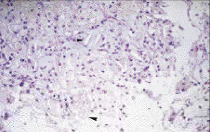
fol-lows: T3 = 0.2 ng/mL (0.8-2.0); T4 = 1.2 µg/dL (4.5-12.5); and TSH >47 µIU/mL (0.3-4.5). The diagnostic hypothesis of pericardial effusion secondary to hypothyroidism was esta-blished and treatment with 50 µg of levothyroxine (Puran T4®) per day was started. As the patient remained clinically symptomatic in NYHA functional class IV, we decided to perform a pericardial drainage via video pericardioscopy on 9/5/98. An anterior pericardiotomy was performed, and 2000 mL of pericardial fluid of a gold-yellow color were with-drawn. Microscopic examination revealed the presence of multinucleated giant cells compatible with a chronic inflam-matory process. The search for neoplastic cells, bacteria, and fungi was negative. Results of the biochemical analysis of the pericardial fluid were as follows: amylase, 65; choles-terol, 63 (normal up to 70mg/dL); glucose, 83mg/dL; trigly-cerides, 10mg/dL; total protein, 7.1; albumin, 5; and lactic dehydrogenase, 218 U/L. The anatomicopathological stu-dy revealed mild fibrosis, lymphocytic and plasmacytic in-filtrate, and proliferation of capillary vessels. The material adhered to the pericardium consisted of a large amount of xanthomatous macrophages (fig. 1) and foreign body giant cells engulfing cholesterol crystals (fig. 2). Deposits of intracytoplasmic hemosiderin in the macrophages were also found. On the basis of these findings, the diagnosis of cho-lesterol pericarditis was made. After a 1-year ambulatory fol-low-up, no recurrence of the pericardial effusion was obser-ved, and the patient remained asymptomatic.
Discussion
In 1919, Alexander 2 reported the first case of pericar-dial effusion with a sparkling gold paint appearance secon-dary to the presence of cholesterol. Since then, several ca-ses of cholesterol pericarditis have been reported in the lite-rature. The incidence of cholesterol pericarditis has not been progressively increasing due to the diagnosis and treatment of the associated diseases 1. Pericarditis has been reported associated with systemic diseases, such as tuber-culosis, rheumatoid arthritis, and hypothyroidism, and idio-pathic pericarditis has been reported as well.
Cholesterol pericarditis is not properly a pericarditis. It
394 394
Fernandes et al
Cholesterol pericarditis
Arq Bras Cardiol 2001; 76: 393-4.
is a special type of pericardial effusion rich in LDL-cholesterol, which may form masses along with debris and fibrin, crys-tallizing over time and depositing in the pericardium, leading to pericarditis. LDL-cholesterol levels in the pericardial fluid are higher than those in the plasma, while in healthy individu-als, they are lower 3. Cholesterol pericarditis is secondary to recurring outbreaks of acute pericarditis, which in many si-tuations are subclinical, resulting in inflammation with effusi-on, pericardial thickening, and change in the absorption ca-pacity of the pericardium. The increase in cholesterol con-centration, particularly of the LDL fraction, causes precipita-tion of the crystals, which, located in the pericardial membra-ne, trigger a granulomatous response of the foreign body type. The result is exudation of the pericardial fluid, which leads to effusion 4,5.
Clinically, cholesterol pericarditis may manifest as pericardial effusion, cardiac tamponade 5, and constrictive pericarditis6. Usually, the patient remains asymptomatic for a long time, because pericarditis is secondary to chronic diseases, and, consequently, accumulation of the pericar-dial fluid is slow.
The diagnosis of cholesterol pericarditis is made by the presence of crystals of cholesterol or an elevated concentra-tion of cholesterol in the pericardial fluid (above 70mg/dL).
Treatment of cholesterol pericarditis consists of the treatment of the underlying disease. In recurring cases, a pleuropericardial window is indicated, and, in more refractory cases, pericardiectomy may be a good therapeutical option. Cholesterol pericarditis is a specific disease, even though rare, that should be considered in the course of chronic diseases involving the pericardium.
1. Torres S, Carvalho H, Moreira I, Gomes L, Pimenta A. Pericardite de colesterol. A propósito de um caso clínico. Rev Port Cardiol 1989; 8: 715-19.
2. Alexander JS. A pericardial effusion of gold paint appearance due to the presence of cholesterin. Br Med J 1919; 2: 463.
3. El Maraghi, NRH. Diseases of the pericardium. In: Silver MD (ed.). Cardiovascu-lar Pathology. New York: Churchil Livingstone, 1983: 125-70.
Fig. 1 - Cholesterol pericarditis: xanthomatous macrophages containing fat and crystals of cholesterol (HE, 10x).
Fig. 2 - Multinucleated giant cells engulfing a large amount of crystals of choleste-rol. (HE, 10x).
References
4. Brawley RK, Vasko JS, Morrow AG. Cholesterol pericarditis: considerations of its pathogenesis and treatment. Am J Med 1966; 41: 235-48.
5. Ford EJ, Bear PA, Adams RW. Cholesterol pericarditis causing cardiac tampona-de. Am Heart J 1991; 122: 877-9.