BACCI MR ETAL.
58 REV ASSOC MED BRAS 2015; 61(1):58-60
ORIGINAL ARTICLE
Inflammatory biomarker kinetics after mechanical and
bioprosthetic valve replacement
MARCELO RODRIGUES BACCI1*, NEIF MURAD2, JOÃO ROBERTO BREDA3, AMANDA VOLTARELI CESARDE OLIVEIRA4, NATHAN HERSZKOWICS4,
NATHALIA KITAMOTO CARDOSO4 , FERNANDO LUIZ AFFONSO FONSECA5
1Assistant Professor, Department of Internal Medicine, ABC Medical School (FMABC), Santo André, SP, Brazil 2Adjunct Professor of Cardiology, ABC Medical School (FMABC), Santo André, SP, Brazil
3Assistant Professor of Cardiac Surgery, Federal University of São Paulo (Unifesp), São Paulo, SP, Brazil 4Physician, Department of General Practice, ABC Medical School (FMABC), Santo André, SP, Brazil 5Vice Director, ABC Medical School (FMABC), Santo André, SP, Brazil
S
UMMARYStudy conducted at ABC Medical School
Article received: 6/9/2014
Accepted for publication: 6/15/2014
*Correspondence:
Address: Av. Príncice de Gales, 821 Postal code: 09060-650
Vila Príncipe de Gales Santo André – SP – Brazil [email protected]
http://dx.doi.org/10.1590/1806-9282.61.01.058
Conflict of interest: none
Introduction: valve disease is an important cause of heart failure. There is a di-rect relationship between valve deterioration and the patient’s inflammatory sta-tus and cytokines: interleukin-6, interleukin-1, tumor necrosis factor, and C-re-active protein, involved in this major state of inflammation.
Objective: to report a series of cases of valve replacement, using a bioprosthetic or mechanical valve, and the inflammatory profile of them.
Methods: patients older than 18 years and with bioprosthetic or mechanical valve placed for a minimum of 6 months and maximum of 2 years were included. In addition to the demographic characteristics of each patient, inflammatory mark-ers were measured and a comparison was made of echocardiographic results be-fore (based on medical records) and after surgery. A total of 46 patients were en-rolled, 23 with mechanical valve and 23 with bioprosthetic valve.
Results: of the 46 patients, 20 presented complete data were included, 12 with bioprosthetic and 8 with mechanical valve. There was no difference between types of prosthesis or implant site for the values of inflammatory markers although they were all above reference range.
Discussion: patients undergoing aortic mechanical valve implant benefited more than those undergoing bioprosthetic implant and both with much better results than those of valve replacements performed on mitral valve. In short, there was no difference in relation to inflammatory biomarkers.
Keywords: interleukin-6, heart failure, aortic valve stenosis, heart valve pros-thesis.
I
NTRODUCTIONHeart diseases that affect valve leaflets are the main cau-se of heart failure.1
Recent data indicate that the number of deaths cau-sed by heart valve diseases in the United States reaches a rate of 20,000 individuals per year, i.e., 7/100,000 people in the general population.2 Aortic and mitral valves are
most often affected, although dysfunctional pulmonary and tricuspid valves are indicators of adverse evolution. The durability of biological valves is limited to 10-15 years,3 and around 50% of the patients develop
compli-cations within 10 years.4
A recent meta-analysis revealed that in male patients a biological valve lasts 15.1 years when implantation oc-curs at the age of 55, 16.8 years at the age of 65, and 18.8 years when individuals are 75 years of age. Structural valve degeneration was not observed in mechanical val-ve models.3
INFLAMMATORYBIOMARKERKINETICSAFTERMECHANICALANDBIOPROSTHETICVALVEREPLACEMENT
REV ASSOC MED BRAS 2015; 61(1):58-60 59
We reported the relation of a series of cases of aortic and mitral valve replacements (mechanical and biologi-cal) with the inflammatory markers IL-1, IL-6, usCRP and TNF-α in a hospital environment. The aim of this study, however, was to report not only the cases, but also the in-flammatory profile of these patients.
M
ETHODSThis is a report of a series of cases of patients with valve diseases who underwent surgery between January 2008 and March 2010.
Inclusion criteria were: adult individuals, aged 18 or over, with bioprosthetic or mechanical valve replacement over a period of no less than 6 months and no more than 2 years. Exclusion criteria were: body mass index (BMI) ≥25, diabe-tes, the presence of chronic kidney disease in patients with glomerular filtration rate below 60 mL/min/1.73m2, the
pre-sence of hepatic disease, Aids, patients undergoing immu-nosuppressant therapy, patients who routinely made use of non-steroidal anti-inflammatory drugs, patients who were hospitalized within a period of 6 months prior to the scree-ning interview for problems unrelated to valve replacement, loss to follow-up and death.
Within this period there were 46 patients who under-went valve replacement: 23 with St. Jude Epic heart valve bioprostheses (St. Jude Medical Inc., Minneapolis, MN, USA) and 23 with mechanical valves.
During a second phase of the study the measurement of usCRP, IL-1, IL-6 and TNF-α was performed using che-miluminescent immunoenzymatic assay with an Immu-lite 2000® analyzer (Siemens®, Frankfurt, Germany).
The echocardiographic parameters established for the sake of comparison of pre- and postoperative values were: Troy equation for the calculation of left ventricular mass; Bernoulli equation for the calculation of transval-vular flow velocity (mean transvaltransval-vular gradient); ejection fraction and left atrial diameter for patients with mitral prosthesis.
All individuals who were included in this study, pre-viously approved by the Ethics and Research Committee of the ABC Medical School under number 012/2011, sig-ned an informed consent form.
The results of the continuous variables were presen-ted as their mean value and respective standard deviation.
R
ESULTSThe analysis of 46 medical records (23 patients in the bio-prosthetic valve group and 23 in the mechanical valve group) was carried out in the period between August-Sep-tember 2011.
Of these, 26 patients were excluded due to loss to fol-low-up, mortality or incomplete data supplied. A total of 20 patients were left, 12 with bioprosthetic valve and 8 with mechanical prosthesis valve (Table 1).
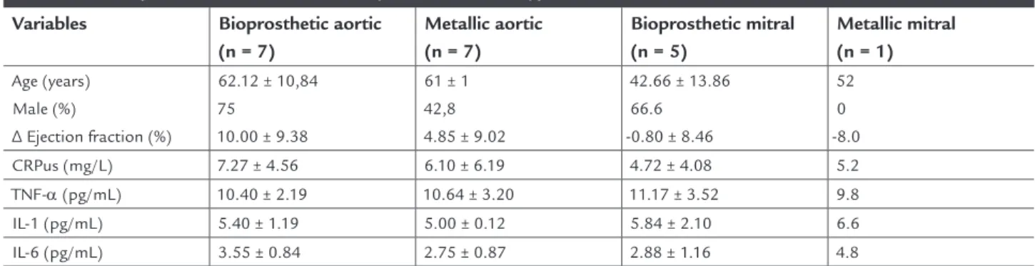
Table 1 shows a higher prevalence of male patients. The mean age was of 42.66 years for the bioprosthetic mi-tral valve replacement group and 61 years for the mecha-nical aortic valve replacement group.
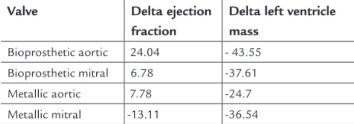
An increased differential in the ejection fraction could be observed only in patients submitted to aortic valve re-placement with a value of 10% in the biological group and of 4.85% in the mechanical valve group. Regarding the group submitted to mitral valve replacement, there was a subtle decrease in the postoperative ejection frac-tion value (Table 2).
As to the inflammatory markers, no differences between the types of prosthesis and implant sites could be observed in regard to the measured values of 1, IL-6, usCRP and TNF-α. However, their overall values were increased.
D
ISCUSSIONA case-by-case approach suggests that, in this series, pa-tients who underwent aortic mechanical valve replace-ment benefited more than those who were submitted to biological implant, and both procedures showed much
TABLE 1 Comparison between inflammatory biomarkers and type of valve used
Variables Bioprosthetic aortic (n = 7)
Metallic aortic (n = 7)
Bioprosthetic mitral (n = 5)
Metallic mitral (n = 1)
Age (years) Male (%)
∆ Ejection fraction (%)
62.12 ± 10,84 75
10.00 ± 9.38
61 ± 1 42,8 4.85 ± 9.02
42.66 ± 13.86 66.6 -0.80 ± 8.46
52 0 -8.0 CRPus (mg/L) 7.27 ± 4.56 6.10 ± 6.19 4.72 ± 4.08 5.2
BACCI MR ETAL.
60 REV ASSOC MED BRAS 2015; 61(1):58-60
better results than the ones found in the performance of mitral valve replacements.
As time passes by, after the implantation of biologi-cal and mechanibiologi-cal valves, the onset of a deposit of extra-cellular matrix and calcification of the leaflets can be ob-served.5,6 Many reports point to the existence of alterations
in the serum levels of cytokines, namely IL-1, IL-6, usCRP and TNF-α. These variations are not exclusive to valve re-placement procedures. Nevertheless, they are present, and are likely to cause valve degeneration.7-9
The individual analysis of each patient revealed that the levels of serum inflammatory markers had no direct re-lation with their clinical and echocardiographic data, sin-ce high serum concentrations of cytokines could be obser-ved in hemodynamically and clinically stable patients and, on the other hand, lower serum values were found in tho-se with higher functional class and lower ejection fraction. In their study, Kastellanos et al.4 showed a decrease in
usCRP and TNF-αserum levels in the medium term (6 months). However, the decrease in TNF-α values proved to be a more reliable marker of inflammation recovery than usCRP. Although no significant differences in cytokine va-lues could be observed regarding sex, there was a stronger tendency for women to present higher levels of usCRP when they were submitted to aortic valve replacement.4
Taking into consideration the differences in the ejec-tion fracejec-tion before and after the replacement procedu-re, the conclusion is that there was a better response in patients who had the aortic rather than the mitral valve replaced. Moreover, no differences in the values of IL-1, IL-6, TNF-α and usCRP could be observed regardless of the type of valve used or the implant site.
R
ESUMOCinética dos marcadores inflamatórios após troca valvar protética mecânica e bioprótese: série de casos.
Introdução: doença valvar é importante causa de insufi-ciência cardíaca. Existe relação direta entre a
deteriora-ção valvar e o estado inflamatório do paciente, sendo as citocinas interleucina-6, interleucina-1, fator de necrose tumoral e a proteína C reativa as principais envolvidas nesse estado de estimulação.
Objetivo: relatar uma série de casos de troca valvar, biopró-tese ou mecânica e seu perfil inflamatório.
Métodos: pacientes maiores de 18 anos e portadores de bioprótese ou protética mecânica, com período mínimo de 6 meses e máximo de 2 anos, foram incluídos. Além das características demográficas de cada paciente, colhe-ram-se os marcadores inflamatórios e comparou-se o eco-cardiograma conforme registro de prontuário antes e de-pois da cirurgia. Um total de 46 pacientes foi incluído, tendo sido 23 com valva mecânica e 23 de bioprótese. Resultados: dos 46 pacientes, chegamos ao total de 20 pacientes com dados completos, sendo 12 com biopróte-se e 8 com protética mecânica. Não houve diferença en-tre tipo de prótese ou local de implante para os valores dos marcadores inflamatórios, contudo, na média, seus valores estavam aumentados.
Discussão: pacientes submetidos ao implante de valva pro-tética mecânica aórtica beneficiaram-se mais do que os sub-metidos ao implante de bioprótese e ambos com resultado bem superior às trocas realizadas na valva mitral. Não hou-ve diferença em relação aos biomarcadores inflamatórios.
Palavras-chave: insuficiência cardíaca, estenose da valva aórtica, interleucina-6, bioprótese.
R
EFERENCES1. Wilson PW. An epidemiologic perspective of systemic hypertension, ischemic heart disease and heart failure. Am J Cardiol 1997;80(9B):3J-8J.
2. Thom T, Haase N, Rosamond W, et al. Heart disease and stroke statistics 2006 update: a report from the American Heart Association Statistical Committee and Stroke Statistics Subcommittee.Circulation 2006;113(6):85-121. 3. Puvimanasinghe JP, Takkenberg JJ, Edwards MB, Eijkemans MJ, Steyerberg
EW, Van Herwerden LA, et al. Comparison of outcomes after aortic valve replacement with a mechanical valve or a bioprosthesis using microsimulation. Heart 2004;90(10):1172-8.
4. Kastellanos SS, Toumpoulis IK, Aggeli C, et al. Time Course of C-Reactive Protein, Tumour Necrosis Factor-Alpha and Monocyte Chemoattractant Protein-1 Following the Surgical Treatment of Patients with Aortic Valve Stenosis. Hellenic J Cardiol 2007; 48:5-14.
5. Skowasch D, Schrempf S, Wernert N, Steinmetz M, Jabs A, Tuleta I, et al. Cells of primarily extravalvular origin in degenerative aortic valves and bioprotheses. Eur Heart J. 2005;26(23):2576-80
6. Srivatsa SS, Harrity PJ, Maercklein PB, Kleppe L, Veinot J, Edwards WD, et al. Increased cellular expression of matrix proteins that regulate mineralization is associated with calcification of native human and porcine xenograft bioprosthetic heart valves. J Clin Invest. 1997;99(5):996-1009
7. Heinrich PC, Behrmann I, Haan S, Hermanns HM, Müller-Newen G, Schaper F. Principles of interleukin (IL)-6-type cytokine signalling and its regulation. Biochem J 2003; 374(1):1-20.
8. Wajant H, Pfizenmaier K, Scheurich P. Tumor necrosis factor signaling. Cell Death Differ 2003; 10 (1): 45-65.
9. Pepys MB, Hirschfield GM. C- reactive protein: a critical update. J Clin Invest 2003; 111(12):1805-12.
TABLE 2 Difference in echocardiographic parameters compared to pre-operative measures
Valve Delta ejection fraction
Delta left ventricle mass
Bioprosthetic aortic 24.04 - 43.55 Bioprosthetic mitral 6.78 -37.61