Short Communication
Corresponding author:Dr.Márcio Bezerra Santos.
e-mail: [email protected]
Received 4 October 2016
Accepted 1 February 2017
Spatial analysis of viral hepatitis and schistosomiasis
coinfection in an endemic area in Northeastern Brazil
Márcio Bezerra Santos
[1],[2], Allan Dantas dos Santos
[2],[3], Patrícia Pereira da Silva
[4],
Aline Silva Barreto
[2], Erlon Oliveira dos Santos
[5], Alex Vianey Callado França
[6],
Constança Simões Barbosa
[7], and Karina Conceição Gomes Machado de Araújo
[2],[8][1]. Departamento de Educação em Saúde, Universidade Federal de Sergipe, Campus Prof. Antônio Garcia Filho, Lagarto, SE, Brasil. [2]. Programa de Pós-Graduação Stricto Sensu em Ciências da Saúde, Universidade Federal de Sergipe, Campus Prof. João Cardoso Nascimento, Aracaju, SE, Brasil. [3]. Departamento de Enfermagem, Universidade Federal de Sergipe, Campus Prof. Antônio Garcia Filho, Lagarto, SE, Brasil. [4]. Programa de Pós-graduação
Stricto Sensu em Biologia Parasitária, Universidade Federal de Sergipe, Cidade Universitária Prof. José Aloísio de Campos, São Cristóvão, SE, Brasil. [5]. Departamento de Biologia, Universidade Estadual de Alagoas, Arapiraca, AL, Brasil. [6]. Departamento de Medicina, Universidade Federal de Sergipe,
Campus Prof. João Cardoso Nascimento, Aracaju, SE, Brasil. [7]. Laboratório de Referência em Esquistossomose, Centro de Pesquisa Aggeu Magalhães, Fundação Oswaldo Cruz, Recife, PE, Brasil. [8]. Departamento de Morfologia, Universidade Federal de Sergipe, Cidade Universitária
Prof. José Aloísio de Campos, São Cristóvão, SE, Brasil.
Abstract
Introduction: This cross-sectional study analyzed the spatial distribution of hepatitis B or C virus (HBV/HBC) and schistosomiasis coinfection. Methods: Serum samples were collected from patients with Schistosoma mansoni infection. These were tested for serological markers of HBV/HCV infection. The spatial distribution of coinfection was analyzed using intensity kernel estimation. Results: Overall, 9.4% of individuals had contact with HBV and 1.7% of samples tested positive for anti-HCV antibodies. We identiied clusters of risk located in the central region. Conclusions: Spatial analysis allowed visualization of high-risk areas, leading to a deinition of priority areas to be targeted for intensiication of control interventions.
Keywords: Schistosoma mansoni. Hepatitis B virus. Hepatitis C virus. Distribution mapping.
Schistosomiasis, a serious parasitic disease, is prevalent in tropical and subtropical regions, especially in communities with inadequate water and sanitation services. It is caused by trematode worms of the genus Schistosoma, and both acute and chronic infection occurs. The disease is found in 78 countries across Latin America, Africa, and Asia1. Brazil is the worst affected country in the Americas2, with 42.9 million people exposed to the risk of infection and approximately 6.8 million infected individuals3-5.
Another important infection, especially in schistosomiasis endemic areas, is viral hepatitis, particularly that caused by hepatitis B virus (HBV) and hepatitis C virus (HCV)6. Viral hepatitis is a serious public health problem, not only in Brazil but also in many other countries worldwide7,8. Despite the controversies in the literature about the prevalence of schistosomiasis and viral hepatitis coinfection, some reports have indicated that the presence of both infections accelerates and aggravates the process of ibrosis in the liver and can lead to the development of cirrhosis and hepatocellular carcinoma3.
A high prevalence of Schistosoma mansoni and hepatitis virus coinfection has been reported, mainly in Egypt where 19.6-64.0% of individuals infected with S. mansoni are HBV coinfected and 10.3-67.0% are HCV coinfected9. In Brazil, most surveys have been conducted in referral centers that manage patients with chronic schistosomiasis3,10,11. Because the treatment of chronic schistosomiasis often requires transfusion of blood products and the use of parenterally administered drugs, there is an increased risk of infection with HBV/HCV. However, few studies have investigated the associations between these infections in areas endemic for schistosomiasis6.
TABLE 1
Frequency of HBV and HCV serological markers.
Serological marker Number Percentage
No markers 120 70.6
Anti-HBc only 1 0.6
HBsAg and anti-HBc 3 1.7
Anti-HBs only 32 18.8
Anti-HBs and anti-HBc 11 6.5
Anti-HCV only 2 1.2
Anti-HCV and anti-HBc 1 0.6
HBV: hepatitis B virus; HCV: hepatitis C virus; Anti-HBc: hepatitis B virus core antigen; HBsAg: hepatitis B surface antigen; Anti-HBs: hepatitis B surface antibody; Anti-HCV: hepatitis C virus antibody.
positive for S. mansoni (using the Kato-Katz method) from an inquiry conducted from March 2010 to July 2011. These patients were invited to participate in the study and were included in the study if they signed an informed consent form.
A total of 187 individuals agreed to participate in the study. Data were collected using a questionnaire that included identifying variables and variables pertaining to epidemiological risk factors for HBV/HCV infection. A 10mL sample of peripheral blood, collected from each participant by venipuncture, was sent to the Health Foundation Parreiras Horta [Fundação de Saúde Parreiras Horta (FSPH)], Central Laboratory of the State of Sergipe [Laboratório Central de Saúde Pública de Sergipe (LACEN/SE)] for serological testing to detect the following markers of HBV and HCV infection: total immunoglobulin G (IgG), hepatitis B core antibody (anti-HBc), hepatitis B surface antibody (anti-HBs), hepatitis B surface antigen (HbsAg), and hepatitis C virus antibody (anti-HCV), using chemiluminescence immunoassays. Of the 187 serum samples collected, 170 were considered suitable for laboratory testing; 17 had hemolyzed and were discarded.
The spatial analysis was performed using the participants’ data and the municipal grid. To determine the spatial location of each participant’s address, the absolute method of instantaneous positioning was used. This was done using the Global Positioning System navigation receiver that captures geographical coordinates on the Universal Transverse Merchant projection system and the ellipsoidal reference geodectic datum, South American Datum 1969. To construct the maps, we used the Aracaju cartographic base obtained from the GIS service of Aracaju. The maps were constructed and analyzed using the TerraView program (http://www.dpi.inpe.br/terraview_eng/ index.php). Data availability was determined by means of a GIS-based query system. The GIS enabled the stored data to be connected to the corresponding geographical features, allowing visualization and spatial analysis13; results were obtained in the form of thematic maps. We used the Kernel method with an adaptive radius of inluence; this system is able to describe how the density at one point can inluence a neighborhood. All statistical analyses were performed using BioEstat, version 5.0.
To test the hypothesis of no association between two categorical variables, we used the χ2 test of equality (for two categories) or the G-test (for more than two categories). We used multiple logistic regression to study the relationship between serological positivity for HBV infection and risk factors for HBV infection.
Overall, 12.8% of participants or their relatives had had hepatitis before the study period. The majority (66.8%) of participants were aged 15-42 years. Half originated from other cities. In total, 16 (9.4%) participants tested anti-HBc positive (Table 1), suggesting that they had had contact with HBV. Three (1.7%) serum samples were anti-HCV positive. Moreover, 32 serum samples were anti-HBs positive (18.8%), indicating a low proportion of HBV-vaccinated individuals in this population. One (0.6%) serum sample presented isolated reactivity to anti-HBc. Eleven (6.5%) serum samples were positive for anti-HBc and anti-HBs, indicating that these participants had had contact with HBV and had developed spontaneous disease remission and immunity. One (0.6%) participant tested positive for both anti-HBc and anti-HCV antibodies. Furthermore, acute HBV infection was found in 1.7% of participants; these patients were only positive for HBsAg and anti-HBc.
50
40
30
20
10
0
0-14 15-28 28-42 42-56 >56 Positive
Anti-HBc isolated or associated to HBsAg (%)
Age in years
50
40
30
20
10
0
Anti-HBc isolated or associated to HBsAg (%)
0 1-4 5-8 9-11 >12 Positive
Years of schooling
100
80
60
40
20
0
<1 1-2 >3
Family income (minimum wage) Vaccine for HBV Positive
Anti-HBc isolated or associated to HBsAg (%)
100
80
60
40
20
0
Anti-HBc isolated (%)
Yes No
A
B
C
D
FIGURE 1 - (A): Association between patient age and HBV infection (anti-HBc positive) (*p < 0.0001). (B): Association between years of schooling and HBV infection (anti-HBc positive) (*p < 0.0001). (C): Association between monthly income and HBV infection (anti-HBc positive) (*p = 0.007). (D): Percentage of patients that were anti-HBs positive because of being vaccinated against HBV (*p < 0.0001). HBV: hepatitis B virus; anti-HBc: hepatitis B virus core antigen
Using a multiple logistic regression model, we identiied the following important risk factors for HBV infection: having undergone surgery [p = 0.002; odds ratio (OR), 4.39; conidence interval (CI), 1.51 - 12.74], having received a blood transfusion (p = 0.02; OR, 2.66; CI, 0.51 - 13.87), having required sutures (p = 0.01; OR, 2.66; CI, 1.09 - 12.05), having been exposed to blood (p = 0.02; OR, 4.84; CI, 0.61 - 1.17), and not using condoms (p = 0.04; OR, 3.04; CI, 0.09 - 1.37).
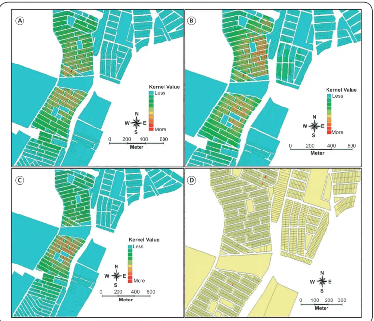
To understand the distribution dynamic of these infections, we analyzed the spatial distribution of cases of schistosomiasis and HBV/HCV coinfection in the study area. Figure 2A shows the spatial analysis of the density of schistosomiasis cases in the neighborhood of Santa Maria, estimated by the Kernel estimator. The regions marked in blue are areas where schistosomiasis is rare. A proportional increase in the number
of schistosomiasis cases is represented by the change from green to yellow and from orange to red. The hottest areas are in the most central neighborhood region, close to the Unidade de Saúde Celso Daniel. We observed a similar spatial distribution for schistosomiasis and HBV coinfection (Figure 2B) and for participants with a positive anti-HBs serum marker alone (indicative of having been vaccinated against HBV) (Figure 2C). The red areas in Figure 2D indicate the domiciles of participants who tested positive for anti-HCV antibodies.
Our data revealed a high prevalence of HBV infection (9.4% anti-HBc positive) in the population with S. mansoni
of participants tested positive for anti-HCV antibodies; this prevalence is higher than the estimated prevalence of HCV infection in the blood donor population (0.49–0.69%)3,10,11. Although our findings are slightly above the estimated prevalences for the population, we cannot infer that individuals with schistosomiasis are more susceptible to HBV or HCV infection. Moreover, the statistical analysis showed that in this study population, the risk factors for HBV transmission were related both to health services and to behavioral factors (no condom use). Silva et al.3 identiied a high (11.9%) prevalence of anti-HCV positivity among patients with hepatosplenic illness who were managed at a schistosomiasis referral center at the
Hospital das Clínicas, Universidade Federal de Pernambuco/ Kernel Value
Less
More
0 200 400 600
Meter N
W E
S
Kernel Value
Less
More
Meter N
W E
S
0 200 400 600
Kernel Value
Less
More
N
W E
S
0 200 400 600
Meter
N
W E
S
0 100 200 300
Meter
A B
C D
FIGURE 2 - Kernel maps of the neighborhood of Santa Maria: (A): Spatial analysis of individuals with schistosomiasis. (B): Spatial analysis of anti-HBc/HBsAg positive individuals. (C): Spatial analysis of individuals positive for anti-HBs alone (indicative of having been vaccinated against HBV). (D): Spatial distribution of place of residence (in red) of anti-HCV positive individuals. anti-HBc: hepatitis B virus core antigen; HBsAg: hepatitis B surface antigen; anti-HBs: hepatitis B surface antibody HBV: hepatitis B virus; HCV: hepatitis C virus.
UFPE. In that study, the authors also reported high levels of coinfection, with a prevalence of 30% for anti-HBc and/or HBsAg markers and 7.4% for anti-HCV. Although the work conducted in schistosomiasis-endemic areas mainly involved patients with the acute or hepatointestinal form schistosomiasis, correlations between schistosomiasis and the viral hepatitis were signiicantly lower than those observed among patients receiving treatment for severe forms of schistosomiasis6; this
corroborates our indings.
between HBV positivity and older people6,10. Our results showed that engaging in sex without using a condom was a risk factor for HBV infection. We did not detect any cases of anti-HBc positivity in the group of participants aged 0-14 years.
A history of surgery, sutures, blood transfusion, and blood exposure were statistically signiicant risk factors for HBV transmission. Some authors have suggested that blood transfusion is the most eficient viral transmission pathway14. The higher prevalence of schistosomiasis and HBV/HCV coinfection in Egypt was related to the indiscriminate use of unsterilized injections with tartar emetic15.
Our study was conducted during an HBV vaccination campaign. In the study interview, 37.9% of participants reported having been vaccinated against HBV. However, serological testing revealed that only 18.8% of all participants were anti-HBs (only) positive (indicating previous vaccination). Hence, participants either did not actually know whether they had been vaccinated or they had not completed the vaccination schedule. These indings highlight a serious public health problem – that most of those people residing in Santa Maria were not immunized against HBV.
There is much published research on the spatial distribution of schistosomiasis and other diseases12,13. However, the present study is the irst to determine the spatial distribution of schistosomiasis and viral hepatitis coinfection in northeastern Brazil. The spatial analysis of schistosomiasis revealed a concentration of cases in central neighborhood areas and a progressive decrease in the suburbs. There are several water reservoirs that provide conditions suitable for S. mansoni
transmission12. A similar spatial distribution pattern was observed for schistosomiasis – HBV coinfection. Given that the central region has a greater population density, the spatial analysis most likely relects this distribution proile.
The Kernel analysis showed that the majority of vaccinated individuals resided close to the Unidade de Saúde Celso Daniel, indicating those living within close proximity of a health unit have better access to health services, such as vaccination. The spatial distribution scenario showing a low (18.8%) proportion of HBV vaccinated individuals in the population indicates that a large proportion of individuals in this neighborhood is susceptible to HBV infection. These spatial distribution studies can support public health programs to control, prevent, and monitor infections. Finally, understanding the prevalence of viral hepatitis in areas endemic for schistosomiasis can be used to inform disease control programs, thereby potentially preventing clinical conditions that carry a poor prognosis, such as chronic hepatitis, liver cirrhosis, and hepatocellular carcinoma.
Ethical considerations
The study was approved by the Research Ethics Committee of the Universidade Federal de Sergipe (UFS) (Number 0168.0.107.000-11; report 188/2011). Individuals who tested positive for HBV/HCV were referred to the Department of Hepatology, Hospital Universitário (HU)/UFS for appropriate medical care.
Acknowledgments
The authors would like to thank the Unidade de Saúde Celso Daniel, the
Fundação de Saúde Parreiras Horta of the Laboratório Central de Saúde Pública de Sergipe (FSPH-LACEN/SE), Tiago Pinheiro Vaz de Carvalho, and Renata Dultra Torres Moura.
Conlict of interest
The authors declare that have no conlicts of interest.
REFERENCES
1. World Health Organization. Schistosomiasis. February, 2014. Available at: http://www.who.int/mediacentre/factcheets/fs115/en/ index.html.
2. Ministério da Saúde. Secretaria de Vigilância em Saúde. Guia de Vigilância Epidemiológica. Brasília: Ministério da Saúde; 2010. 3. Silva JLA, Souza VSB, Villela TAS, Domingues ALC, Coêlho
MRCD. HBV and HCV serological markers in patients with the hepatosplenic form of mansonic schistosomiasis. Arq Gastroenterol. 2011;48(2):124-30.
4. Souza FPC, Vitorino RR, Costa AP, Faria-Júnior FC, Santana LA, Gomes AP. Esquistossomose mansônica: aspectos gerais, imunologia, patogênese e história natural. Rev Soc Bras Clín Méd. 2011;9(4):300-7.
5. Sarvel AK, Oliveira AA, Silva AR, Lima ACL, Katz N. Evaluation of a 25-year-program for the control of schistosomiasis mansoni in an endemic area in Brazil. PLoS Negl Trop Dis. 2011;5(3):e990. 6. Tavares-Neto J, Prata A, Paraná R, Valente BR, Vitvitski L,
Figueiredo JFC. Very low prevalence of hepatitis C virus infection in rural communities of northeastern Brazil with a high prevalence of Schistosoma mansoni. Rev Soc Bras Med Trop. 2005;38(4):290-3. 7. Santos BFO, Santana NO, França AVC. Prevalence, genotypes
and factors associated with HCV infection among prisoners in Northeastern Brazil. World J Gastroenterol. 2011;17(25):3027-34. 8. Scaraveli NG, Passos AM, Voigt AR, Livramento A, Tonial G,
Treitinger A, et al. Soroprevalence of hepatitis B and hepatitis C markers in adolescents in Southern Brazil. Cad Saude Publica. 2011;27(4):753-58.
9. El-Sayed HF, Abaza SM, Mehanna S, Winch PJ. The prevalence of hepatitis B and C infections among immigrants to a newly reclaimed area endemic for Schistosoma mansoni in Sinai, Egypt. Acta Tropica. 1997;68(2):229-37.
10. Silva JLA, Coêlho MRCD, Souza VSB, Domingues ALC. Soroprevalência da Hepatite C em pacientes com Esquistossomose. Rev Para Med.2008;22(1):27-32.
11. Morais CNL, Carvalho BM, Melo WG, Melo FL, Lopes EPA, Domingues ALC, et al. Correlation of biological serum markers
with the degree of hepatic ibrosis and necroinlammatory activity
in hepatitis C and schistosomiasis patients. Mem Inst Oswaldo Cruz. 2010;105(4):460-6.
12. Barbosa CS, Araújo KC, Sevilla MAA, Melo F, Gomes ECS, Souza-Santos R. Currente epidemiological status of schistosomiasis in the state of Pernambuco, Brazil. Mem Inst Oswaldo Cruz. 2010;105(4):549-54.
13. Araújo KCGM, Rezendes APC, Souza-Santos R, Silveira Jr JC, Barbosa CS. Análise espacial dos focos de Biomphalaria glabrata e de casos humanos de esquistossomose mansônica em Porto de Galinhas, Pernambuco, no ano de 2000. Cad Saude Publica. 2007;23(2):409-18. 14. Khouri ME, Cordeiro Q, Luz DABP; Duarte LS, Gama, MEA,
Corbett CEP. Endemic hepatitis B and C virus infection in a Brazilian eastern Amazon region. Arq Gastroenterol. 2010;47(1):35-41. 15. Mohamed MK, El-Hoseiny M, Arafa N, Hassan A, Ismail S, et al.